. 2022 Dec 2;12(1):20815.
doi: 10.1038/s41598-022-24636-1.

Genetic epidemiology of inherited retinal diseases in a large patient cohort followed at a single center in Italy

Affiliations

Genetic epidemiology of inherited retinal diseases in a large patient cohort followed at a single center in Italy

Marianthi Karali et al. Sci Rep. .

Abstract

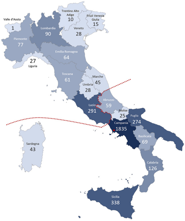
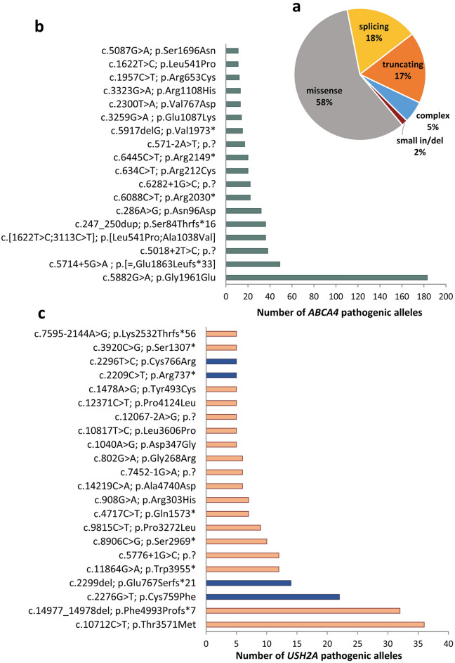
Inherited retinal diseases (IRDs) are the leading cause of vision loss in the working-age population. We performed a retrospective epidemiological study to determine the genetic basis of IRDs in a large Italian cohort (n = 2790) followed at a single referral center. We provided, mainly by next generation sequencing, potentially conclusive molecular diagnosis for 2036 patients (from 1683 unrelated families). We identified a total of 1319 causative sequence variations in 132 genes, including 353 novel variants, and 866 possibly actionable genotypes for therapeutic approaches. ABCA4 was the most frequently mutated gene (n = 535; 26.3% of solved cases), followed by USH2A (n = 228; 11.2%) and RPGR (n = 102; 5.01%). The other 129 genes had a lower contribution to IRD pathogenesis (e.g. CHM 3.5%, RHO 3.5%; MYO7A 3.4%; CRB1 2.7%; RPE65 2%, RP1 1.8%; GUCY2D 1.7%). Seventy-eight genes were mutated in five patients or less. Mitochondrial DNA variants were responsible for 2.1% of cases. Our analysis confirms the complex genetic etiology of IRDs and reveals the high prevalence of ABCA4 and USH2A mutations. This study also uncovers genetic associations with a spectrum of clinical subgroups and highlights a valuable number of cases potentially eligible for clinical trials and, ultimately, for molecular therapies.


Conflict of interest statement

The authors declare no competing interests.

Figures

Figure 1
Figure 1
Overview of the geographical representation of the clinic’s cohort. Map of Italy, divided by administrative districts (Regions; delimited by a grey line), depicting the origin of patients followed at the Reference Center for Rare Eye Diseases at the Eye Clinic of the University of Campania ‘Luigi Vanvitelli’ (Naples, Campania Region; red dot). Color intensity indicates the relative number of patients originating from each administrative region. The geographic area corresponding to the Southern Italy and Islands is delimited by a dotted line.
Figure 2
Figure 2
Distribution of different types of alleles in the solved cohort. (a) Pie chart showing the relative contribution of each causal IRD gene in disease pathogenesis of the genetically defined cohort. Genes (n = 99) implicated in less than 15 cases are plotted as a single group. (bd) Pie chart depicting the prevalence and relative contribution of causative genes implicated in autosomal recessive (b), autosomal dominant (c) and X-linked (d) retinal dystrophy forms. Genes that are responsible for IRD pathogenesis in less than 10 cases with autosomal recessive (or less than 4 cases with dominant) forms are depicted as a single group. (e) Contribution of the most recurrent IRD-associated genes involved in syndromic forms. Genes implicated in less than 3 cases are plotted as a single group.
Figure 3
Figure 3
Most frequent variants identified in ABCA4- and USH2A-associated cases. (a) Pie-chart showing the relative abundance of the different classes of variants across the ABCA4 pathogenic alleles. (b) Histogram showing the most frequent ABCA4 alleles. (c) Histogram of occurrence of the most recurrent USH2A variants. Blue bars show variants located in exon 13.

References

    1. Sahel JA, Marazova K, Audo I. Clinical characteristics and current therapies for inherited retinal degenerations. Cold Spring. Harb. Perspect. Med. 2014;5:a017111. doi: 10.1101/cshperspect.a017111. - DOI - PMC - PubMed
    1. Berger W, Kloeckener-Gruissem B, Neidhardt J. The molecular basis of human retinal and vitreoretinal diseases. Prog. Retin. Eye Res. 2010;29:335–375. doi: 10.1016/j.preteyeres.2010.03.004. - DOI - PubMed
    1. Tatour Y, Ben-Yosef T. Syndromic inherited retinal diseases: Genetic, clinical and diagnostic aspects. Diagnostics (Basel) 2020 doi: 10.3390/diagnostics10100779. - DOI - PMC - PubMed
    1. Schatz P, et al. Fundus albipunctatus associated with compound heterozygous mutations in RPE65. Ophthalmology. 2011;118:888–894. doi: 10.1016/j.ophtha.2010.09.005. - DOI - PubMed
    1. Aoun M, et al. Inherited retinal diseases due to RPE65 variants: From genetic diagnostic management to therapy. Int. J. Mol. Sci. 2021 doi: 10.3390/ijms22137207. - DOI - PMC - PubMed

Publication types

Supplementary concepts