. 2023 Apr;32(4):425-435.
doi: 10.1111/exd.14722. Epub 2022 Dec 19.

Crisaborole efficacy in murine models of skin inflammation and Staphylococcus aureus infection

Affiliations

Crisaborole efficacy in murine models of skin inflammation and Staphylococcus aureus infection

Christine Youn et al. Exp Dermatol. 2023 Apr.

Abstract

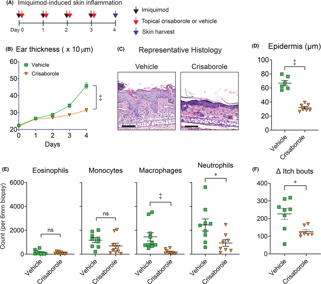
Phosphodiesterase 4 (PDE4) is highly expressed in keratinocytes and immune cells and promotes pro-inflammatory responses upon activation. The activity of PDE4 has been attributed to various inflammatory conditions, leading to the development and approval of PDE4 inhibitors as host-directed therapeutics in humans. For example, the topical PDE4 inhibitor, crisaborole, is approved for the treatment of mild-to-moderate atopic dermatitis and has shown efficacy in patients with psoriasis. However, the role of crisaborole in regulating the immunopathogenesis of inflammatory skin diseases and infection is not entirely known. Therefore, we evaluated the effects of crisaborole in multiple mouse models, including psoriasis-like dermatitis, AD-like skin inflammation with and without filaggrin mutations, and Staphylococcus aureus skin infection. We discovered that crisaborole dampens myeloid cells and itch in the skin during psoriasis-like dermatitis. Furthermore, crisaborole was effective in reducing skin inflammation in the context of filaggrin deficiency. Importantly, crisaborole reduced S. aureus skin colonization during AD-like skin inflammation. However, crisaborole was not efficacious in treating S. aureus skin infections, even as adjunctive therapy to antibiotics. Taken together, we found that crisaborole reduced itch during psoriasis-like dermatitis and decreased S. aureus skin colonization upon AD-like skin inflammation, which act as additional mechanisms by which crisaborole dampens the immunopathogenesis in mouse models of inflammatory skin diseases. Further examination is warranted to translate these preclinical findings to human disease.

Keywords: Staphylococcus aureus; atopic dermatitis; crisaborole; inflammation; psoriasis.


Conflict of interest statement

CONFLICT OF INTEREST

The remaining authors state no conflict of interest.

Figures

FIGURE 1
FIGURE 1
Efficacy of crisaborole in an imiquimod model of psoriasis-like dermatitis. Wt mice ears were treated daily for 4 days with topical imiquimod followed by topical application with either 2% crisaborole ointment or vehicle ointment 4 h later. Ear skin was harvested on day 4 for histological and flow analysis (n ≥ 7/group). (A) Model timeline. (B) Mean ± SEM ear thickness (0.01 × mm). (C) Representative skin histology (haematoxylin and eosin stain). Scale bar = 100 μm. (D) Mean ± SEM epidermal thickness (μm). (E) Mean ± SEM number of cells from skin. (F) Mean ± SEM itch bouts on Day 4 normalized to Day 0. ns = not significant, *p < 0.05; ‡p < 0.001, vehicle versus crisaborole-treated mice, as calculated by two-way ANOVA (B) or two-tailed Student’s t-test (E, F). Results are combined from or representative of at least two independent experiments
FIGURE 2
FIGURE 2
Efficacy of crisaborole in a skin injury model of AD-like skin inflammation. Skin injury was performed on ft/ft mice and on Day 21 post-injury, ft/ft mice were treated topically daily for 10 days with either 2% crisaborole ointment or vehicle (n ≥ 7/group). (A) Model timeline. (B) Representative skin photographs. (C) Mean ± SEM area of skin inflammation (cm2) on Day 31 normalized to Day 21. ns = not significant, *p< 0.05, vehicle versus crisaborole-treated mice, as calculated by two-tailed Student’s t-test. Data are representative of at least two independent experiments.
FIGURE 3
FIGURE 3
Efficacy of crisaborole in a model of epicutaneous Staphylococcus aureus exposure-induced AD-like skin inflammation. Wt mice were epicutaneously challenged with Staphylococcus aureus (1 × 10⁸ CFU) on the dorsal skin for 7 days. On Day 7, mice were treated for 3 days with 2x/day topical 2% crisaborole ointment (Crisa), 1×/day oral 10 mg/ml linezolid (Lin), crisaborole and linezolid (Crisa + Lin), or vehicle (Veh) as specified in (A) (n ≥ 5/group). (A) Model timeline. (B) Representative skin photographs. (C) Mean ± SEM disease score. (D) Mean ± SEM ex vivo skin CFUs. ns = not significant, *p < 0.05; ‡p < 0.001, as calculated by one-way ANOVA with Tukey’s correction (C) and Kruskal–Wallis with Benjamini, Krieger and Yekutieli correction for multiple comparisons (D). Results are combined from or representative of at least two independent experiments
FIGURE 4
FIGURE 4
Efficacy of crisaborole in a Staphylococcus aureus intradermal skin infection model. The S. aureus i.d. skin infection was performed on wt mice. After 4-h post-infection, mice were treated for 7 days with 2×/day topical 2% crisaborole ointment (Crisa), 1×/day oral 10 mg/ml linezolid (Lin), crisaborole and linezolid (Crisa + Lin), or vehicle (Veh), as specified in (A) (n ≥ 7/group). (A) Model timeline. (B) Representative S. aureus in vivo bioluminescent signals. (C) Mean ± SEM total flux (photons/s). (D) Mean ± SEM ex vivo skin CFUs on Day 3. (E) Representative photographs of skin lesions. (F) Mean ± SEM total lesion size (cm2). ns = not significant, *p < 0.05; ‡p < 0.001, as calculated by two-way ANOVA test with Tukey’s correction (C, F) and Kruskal–Wallis with Benjamini, Krieger and Yekutieli correction for multiple comparisons (D). Results are combined from or representative of at least two independent experiments

References

    1. Chiricozzi A, Caposiena D, Garofalo V, Cannizzaro MV, Chimenti S, Saraceno R. A new therapeutic for the treatment of moderate-to-severe plaque psoriasis: apremilast. Expert Rev Clin Immunol. 2016;12(3):237–249. doi:10.1586/1744666X.2016.1134319 - DOI - PubMed
    1. Li H, Zuo J, Tang W. Phosphodiesterase-4 inhibitors for the treatment of inflammatory diseases. Front Pharmacol. 2018;9:1048. doi:10.3389/fphar.2018.01048 - DOI - PMC - PubMed
    1. Guttman-Yassky E, Hanifin JM, Boguniewicz M, et al. The role of phosphodiesterase 4 in the pathophysiology of atopic dermatitis and the perspective for its inhibition. Exp Dermatol. 2019;28(1):3–10. doi:10.1111/exd.13808 - DOI - PubMed
    1. Milakovic M, Gooderham MJ. Phosphodiesterase-4 inhibition in psoriasis. Psoriasis (Auckl). 2021;11:21–29. doi:10.2147/PTT.S303634 - DOI - PMC - PubMed
    1. Phillips JE. Inhaled phosphodiesterase 4 (PDE4) inhibitors for inflammatory respiratory diseases. Front Pharmacol. 2020;11:259. doi:10.3389/fphar.2020.00259 - DOI - PMC - PubMed

Publication types

MeSH terms

Substances