. 2022 Dec 3;33(12):79.
doi: 10.1007/s10856-022-06701-3.

Lyophilized bovine acellular tendon linear fiber material for the reconstruction of attachment structure of paraspinous muscles: an animal in vivo study

Affiliations

Lyophilized bovine acellular tendon linear fiber material for the reconstruction of attachment structure of paraspinous muscles: an animal in vivo study

Bo Yuan et al. J Mater Sci Mater Med. .

Abstract

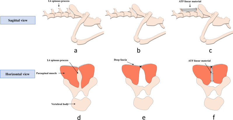
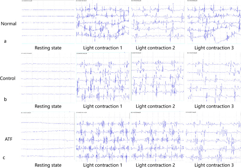
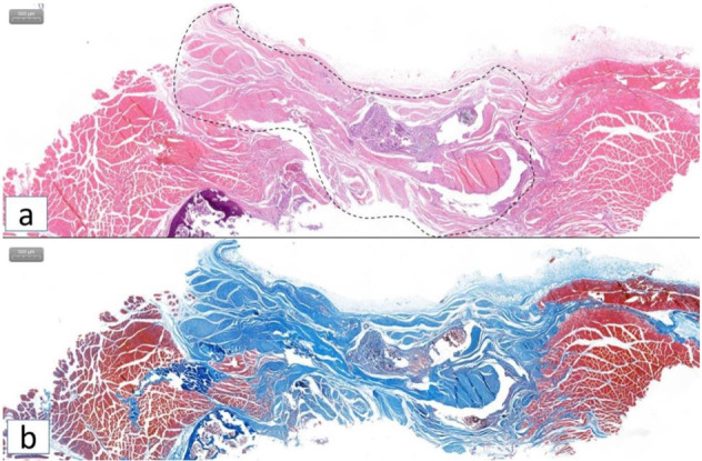
Low back pain is common after lumbar spine surgery and the injury from extensive detachment of paraspinal muscles during the surgery may play a vital role. Previously, we prepared a bovine acellular tendon fiber (ATF) material through lyophilization and proved that it could retain its original fibrillar structure and mechanical properties. The objective of this study is to evaluate the effectiveness of this new fiber material used for attachment structure reconstruction of paraspinal muscle. Defect of spinous process, interspinous and supraspinous ligament was established on lumbar spine in rabbit and rat and ATF linear material was implanted to reconstruct the attachment structure. Ultrasound showed the cross-sectional area of the paraspinal muscle in ATF group was larger than that of control group in rats. MRI showed the irregular shape and high signal changes in control group, but regular shape and uniform signal in the ATF group in rabbit. For Electromyogram, the frequency of evoked potential in control group was lower than ATF group and normal rats. HE and Masson staining showed good tissue healing, and immunohistochemical results showed the immune rejection of ATF is significantly lower than that of suture. Reconstruction of the attachment structure of paraspinous muscles with ATF linear material could maintain the morphology, volume and function of paraspinal muscle. ATF material has the potential to be used to manufacture personalized ligaments and other tissue engineering scaffolds. Graphical abstract.


Conflict of interest statement

The authors declare no competing interests.

Figures

None
Graphical abstract
Fig. 1
Fig. 1
Rabbit model establishment and material implantation. a Lumbar sacral bone structure; b Resection of L6 spinous process; c Use AFT linear material to fix between L5 and L7; d cross-section of paraspinal muscle near lumbosacral spinous process; e Direct suture of bilateral deep fascia after resection of the interspinous ligament; f Bilateral paraspinal muscles was sutured to fix on the AFT linear material between L5 and L7 spinous process
Fig. 2
Fig. 2
MRI scan results of paraspinal muscle for volume evaluation in rabbits. a Magnified figure of MRI on lumbosacral spine of normal New Zealand white rabbit; b MRI structure of normal L5; c MRI results showed regular shape and basically uniform signal of paraspinous muscle in ATF group; d MRI results showed the irregular shape and uneven signal of the boundary of paraspinous muscle in control group. The white arrow indicates the paraspinous muscle, and the white transparent arrow indicates the space between the paraspinous muscle and the lamina
Fig. 3
Fig. 3
EMG examination results of SD rats for paraspinal muscle function test. a EMG results of normal SD rats in resting state and awake light contraction state; b Control group EMG results showed more interference waves on the baseline without pathological wave and no low amplitude or shortened phase, but frequency of evoked potential in control group was lower than ATF group; c ATF group EMG examination showed straight baseline without pathological wave manifestations
Fig. 4
Fig. 4
Immunohistochemical staining results of SD rats for Collagen I antibody of specimens in ATF and control group. a, b Histological test showed obvious gaps between the bilateral paraspinal muscles in control group. Red arrow indicated the tissue gap (×62 and ×124); c, d The bilateral paraspinal muscle and the collagen fibers between them were well healed, and the tissue structure of the bilateral muscle was intact in ATF group. Red arrow indicated the ATF linear material (×74 and ×148)
Fig. 5
Fig. 5
HE and Masson staining of specimens in ATF group in SD rats. a HE staining and b Masson stating showed that the collagen fiber structure and the muscle tissue structure were intertwined, and there was no obvious tissue gap, suggesting good tissue healing. Dashed line area indicates the ATF linear material
Fig. 6
Fig. 6
Immunohistochemical staining results for immune inflammation factor of specimens in ATF and control group in SD rats. af Immunohistochemical test of neutrophils, monocytes, macrophages, NK cell markers (CD11B and CD11C). Suture used in the surgery showed strongly positive (a, d × 700), ATF material showed positive (b, e × 500), and normal low back fascia collagen fibers showed positive (c, f × 500); gi Immunohistochemical test of T cell markers (CD3). Suture used in the surgery showed strongly positive (g × 700), ATF material showed negative (h × 500), and normal low back fascia collagen fibers showed negative (i × 500)

Similar articles

References

    1. Huang Q, Zhang Y, Li D, Yang D, Huo M, Maruyama H. The evaluation of chronic low back pain by determining the ratio of the lumbar multifidus muscle cross-sectional areas of the unaffected and affected sides. J Phys Ther Sci. 2014;26:1613–4. doi: 10.1589/jpts.26.1613. - DOI - PMC - PubMed
    1. Shafaq N, Suzuki A, Matsumura A, Terai H, Toyoda H, Yasuda H, et al. Asymmetric degeneration of paravertebral muscles in patients with degenerative lumbar scoliosis. Spine (Philos Pa 1976) 2012;37:1398–406. doi: 10.1097/BRS.0b013e31824c767e. - DOI - PubMed
    1. Cornwall J, John Harris A, Mercer SR. The lumbar multifidus muscle and patterns of pain. Man Ther. 2006;11:40–5. doi: 10.1016/j.math.2005.02.002. - DOI - PubMed
    1. Fritzell P, Hägg O, Wessberg P, Nordwall A, Swedish Lumbar Spine Study Group. Chronic low back pain and fusion: a comparison of three surgical techniques: a prospective multicenter randomized study from the Swedish lumbar spine study group. Spine (Philos Pa 1976) 2002;27:1131–41. doi: 10.1097/00007632-200206010-00002. - DOI - PubMed
    1. Fan S, Hu Z, Zhao F, Zhao X, Huang Y, Fang X. Multifidus muscle changes and clinical effects of one-level posterior lumbar interbody fusion: minimally invasive procedure versus conventional open approach. Eur Spine J. 2010;19:316–24. doi: 10.1007/s00586-009-1191-6. - DOI - PMC - PubMed

LinkOut - more resources