. 2022 Dec 3;20(1):559.
doi: 10.1186/s12967-022-03780-2.

Monocytes educated by cancer-associated fibroblasts secrete exosomal miR-181a to activate AKT signaling in breast cancer cells

Affiliations

Monocytes educated by cancer-associated fibroblasts secrete exosomal miR-181a to activate AKT signaling in breast cancer cells

Katayoon Pakravan et al. J Transl Med. .

Abstract

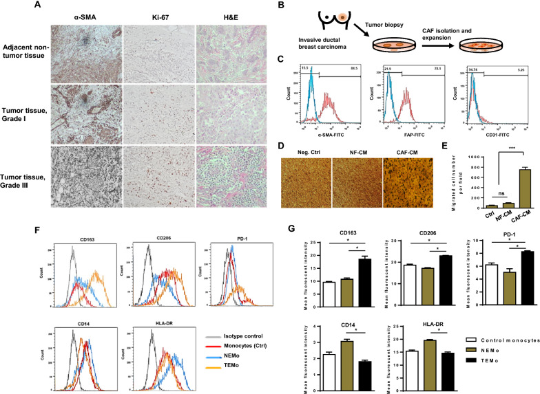
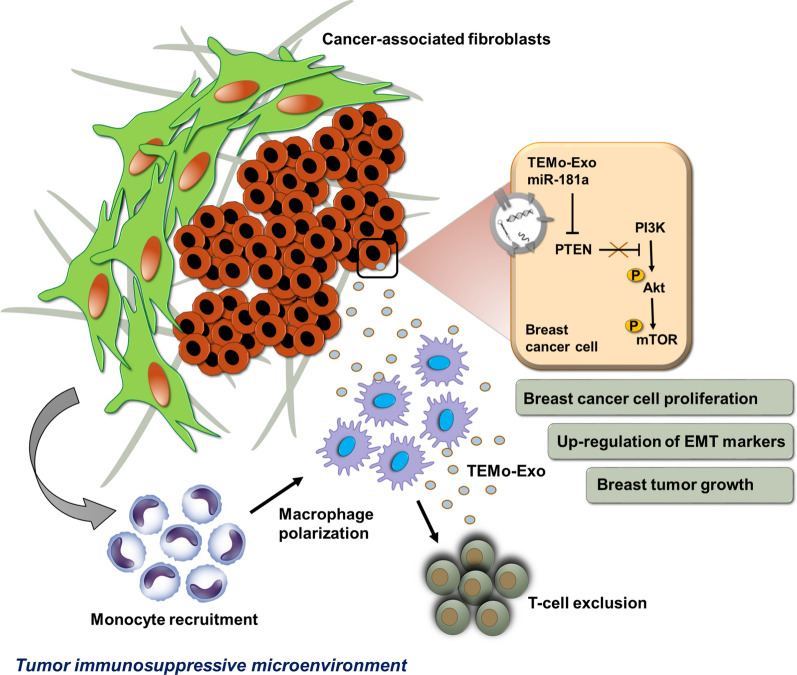
Background: Cancer-associated fibroblasts (CAFs), one of the major components of the tumor stroma, contribute to an immunosuppressive tumor microenvironment (TME) through the induction and functional polarization of protumoral macrophages. We have herein investigated the contribution of CAFs to monocyte recruitment and macrophage polarization. We also sought to identify a possible paracrine mechanism by which CAF-educated monocytes affect breast cancer (BC) cell progression.

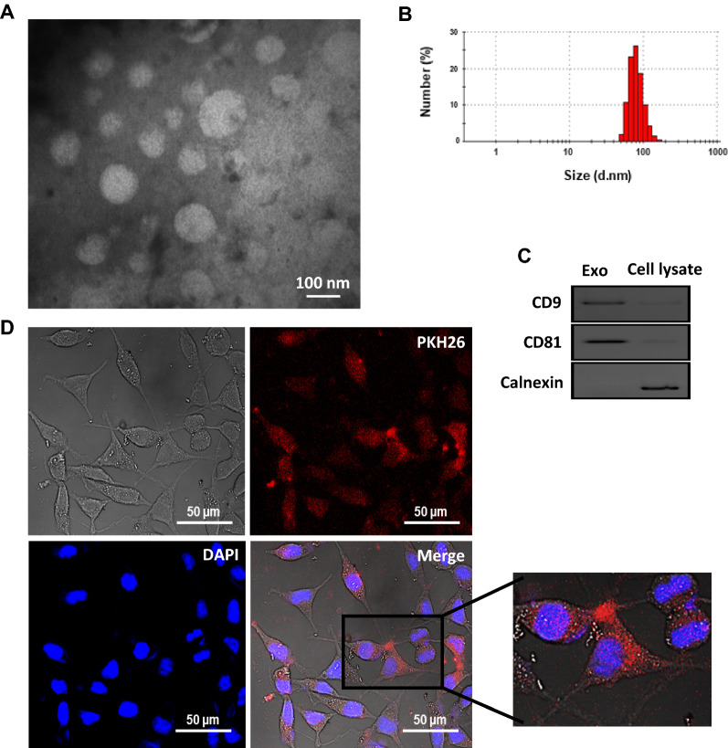
Methods: Monocytes were educated by primary CAFs and normal fibroblast (NF); the phenotypic alterations of CAF- or NF-educated monocytes were measured by flow cytometry. Exosomes isolated from the cultured conditioned media of the educated monocytes were characterized. An in vivo experiment using a subcutaneous transplantation tumor model in athymic nude mice was conducted to uncover the effect of exosomes derived from CAF- or NF-educated monocytes on breast tumor growth. Gain- and loss-of-function experiments were performed to explore the role of miR-181a in BC progression with the involvement of the AKT signaling pathway. Western blotting, enzyme-linked immunosorbent assay, RT-qPCR, flow cytometry staining, migration assay, immunohistochemical staining, and bioinformatics analysis were performed to reveal the underlying mechanisms.

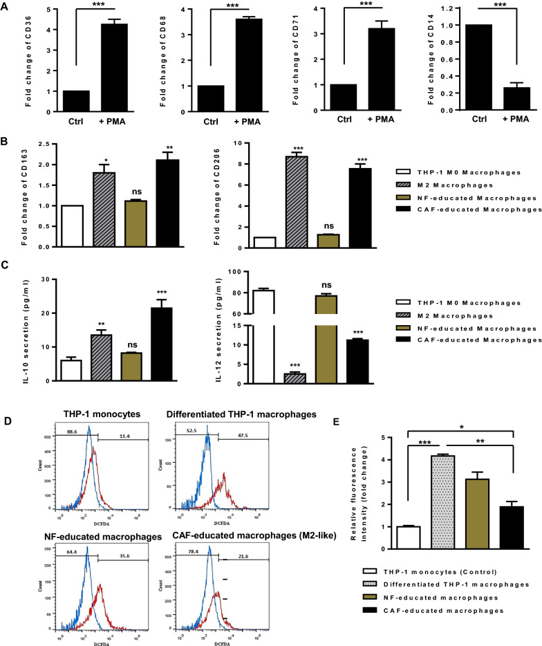
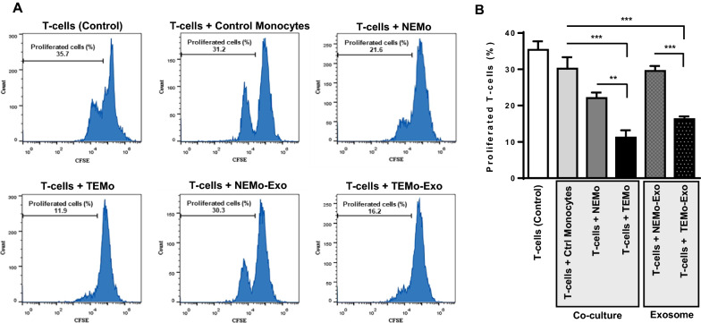
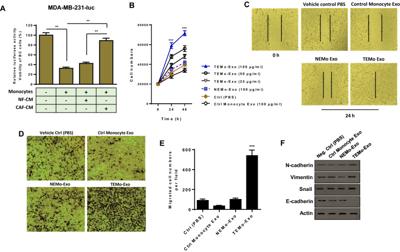
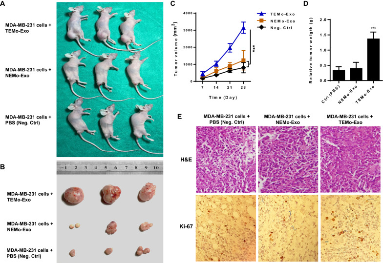
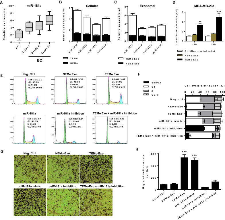
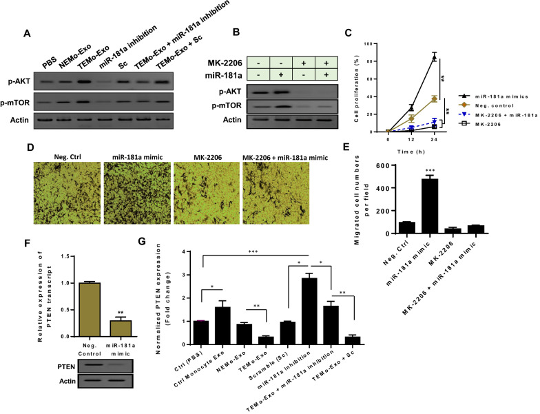
Results: We illustrated that primary CAFs recruited monocytes and established pro-tumoral M2 macrophages. CAF may also differentiate human monocyte THP-1 cells into anti-inflammatory M2 macrophages. Besides, we revealed that CAFs increased reactive oxygen species (ROS) generation in THP-1 monocytes, as differentiating into M2 macrophages requires a level of ROS for proper polarization. Importantly, T-cell proliferation was suppressed by CAF-educated monocytes and their exosomes, resulting in an immunosuppressive TME. Interestingly, CAF-activated, polarized monocytes lost their tumoricidal abilities, and their derived exosomes promoted BC cell proliferation and migration. In turn, CAF-educated monocyte exosomes exhibited a significant promoting effect on BC tumorigenicity in vivo. Of clinical significance, we observed that up-regulation of circulating miR-181a in BC was positively correlated with tumor aggressiveness and found a high level of this miRNA in CAF-educated monocytes and their exosomes. We further clarified that the pro-oncogenic effect of CAF-educated monocytes may depend in part on the exosomal transfer of miR-181a through modulating the PTEN/Akt signaling axis in BC cells.

Conclusions: Our findings established a connection between tumor stromal communication and tumor progression and demonstrated an inductive function for CAF-educated monocytes in BC cell progression. We also proposed a supporting model in which exosomal transfer of miR-181a from CAF-educated monocytes activates AKT signaling by regulating PTEN in BC cells.

Keywords: AKT signaling; Breast cancer; Cancer-associated fibroblasts; Exosomes; Immunosuppressive tumor microenvironment; Tumor-associated macrophages.


Conflict of interest statement

The authors declare that they have no competing interests.

Figures

Fig. 1
Fig. 1
Stromal fibroblasts isolated from BC tissues exhibit characteristics of CAFs, effectively recruit monocytes and subsequently affect their polarization states. A Representative photomicrographs of H&E staining and IHC staining of α-SMA (a CAF-specific marker) and Ki67 (a proliferation marker) positive cells in breast tumor and adjacent non-tumor tissues (×100). Immunohistochemically, the presence of α-SMA demonstrated the presence of stromal myofibroblasts surrounding the cancer nests, which were distributed among the invasive cancer cells in the heterogeneous cancer tissue. B A schematic illustration of primary CAF isolation and expansion in vitro. C Flow cytometry staining of primary cultured CAFs with anti-α-SMA, anti-FAP, and anti-CD31. CAFs were characterized by the expression of α-SMA and FAP but lacked the expression of endothelial cell marker CD31 by flow cytometry. D, E To elucidate that monocytes are functionally recruited by CAFs, serum-starved monocytes were allowed to migrate for 12 h toward CMs from CAFs or NFs and compared to the negative control. D Representative photomicrographs of the migration potential of monocytes in different conditions assessed using the transwell assay. E Quantitative assessment of migrated cells showed that monocytes were effectively recruited by CAF-CM. Representative flow cytometry histograms (F) and bar graphs of mean fluorescent intensity (G) show that the expression levels of CD163, CD206, and PD-1 were considerably higher in TEMo than in NEMo or control monocytes, while the expression levels of HLA-DR and CD14 were significantly lower in TEMo compared to NEMo. Columns, mean of three different experiments; bars, SD. *P-value < 0.05, ***P-value < 0.001
Fig. 2
Fig. 2
CAFs induce the polarization of THP-1 cells into anti-inflammatory M2-like macrophages. A After 24 h of incubation with 150 nM PMA, the monocyte-to-macrophage differentiation was confirmed by the elevated transcript levels of recognized macrophage markers CD36, CD68, and CD71. The decreased transcript level of CD14 in PMA-treated THP-1 cells further confirmed that the THP-1 monocytes were differentiated into macrophage-like cells. B Transcript expression levels of M2 macrophage markers CD163 and CD206 in THP-1 macrophages incubated with CAF-CM were significantly increased compared with those in THP-1 M0 macrophages alone or incubated with NF-CM after 48 h. As a positive control, THP-1 macrophages were stimulated with 20 ng/mL of IL-4 for 48 h. C Protein secretion levels of the anti-inflammatory cytokine IL-10 and the pro-inflammatory cytokine IL-12 were assessed by ELISA. THP-1 macrophages incubated with CAF-CM as well as positive control M2 macrophages were identified by IL-10high IL-12low phenotype when compared to THP-1 M0 macrophages alone or incubated with NF-CM, confirming the potential of CAFs in promoting the polarization of THP-1 cells into anti-inflammatory M2-like macrophages after 48 h. D, E The relative levels of intracellular ROS production during macrophage polarization. The amount of ROS produced by differentiated THP-1 macrophages increased noticeably when monocytes were treated with PMA. Importantly, ROS generation in CAF-educated macrophages was lower than that in macrophages educated by NF-CM after 48 h. Even though the level of ROS diminishes during M1/M2 polarization, CAF-induced M2-like macrophages were found to produce higher levels of ROS than THP-1 control monocytes. Representative flow cytometry histograms (D) and bar graphs of relative DCFDA fluorescence intensity (E) imply that CAFs contribute to M2 polarization in part by increasing ROS production, as M2 macrophages require a level of ROS for proper polarization. Columns, mean of three different experiments; bars, SD. ns: non-significant, *P-value < 0.05, **P-value < 0.01, ***P-value < 0.001
Fig. 3
Fig. 3
Characterization and cellular uptake of purified exosomes derived from monocytes. The morphology and size of exosomes were observed using (A) transmission electron microscopy. B Representative dynamic light scattering (DLS) number distribution measurement of purified exosomes showed a single peak at ~ 90 nm. C Exosome-specific surface markers CD9 and CD81 were detected in exosomes by western blotting. The cytoplasmic protein marker Calnexin was expressed in the whole cell lysate but was undetectable in the isolated exosomes, indicating that the exosome preparations were not contaminated with other vesicles such as endoplasmic reticulum ones. D Cellular internalization of PKH26-labeled exosomes by MDA-MB-231 BC cells was visualized and imaged under a confocal microscope. The red fluorescence in the cytoplasm showed that exosomes were uptaken by MDA-MB-231 BC cells. The nuclear staining was performed by DAPI
Fig. 4
Fig. 4
CAF-educated monocytes and their derived exosomes suppress T-cell proliferation. TEMo, NEMo, their corresponding exosomes as well as control monocytes were co-cultured with autologous peripheral T-cells which were labeled with CFSE for 72 h. Representative flow cytometry histograms (A) and bar graphs of proliferated T-cells (B) illustrated that TEMo and their derived exosomes (100 µg/mL) had a greater inhibitory effect on T-cell proliferation when compared with control monocytes, NEMo, or derived exosomes. Effector cells (i.e., monocytes) were co-cultured with target cells (labeled T-cells) at the Effector: target (E:T) ratio of 1:4. Columns, mean of three different experiments; bars, SD. **P-value < 0.01, ***P-value < 0.001
Fig. 5
Fig. 5
CAF-activated, polarized monocytes lose their tumoricidal properties and their derived exosomes enhance the proliferation and migration of BC cells and up-regulate the expression of EMT markers. A The cytotoxic activity of monocytes alone or incubated with either CAF- or NF-CM was evaluated by measuring the luciferase activity of MDA-MB-231 BC cells expressing luciferase (MDA-MB-231-luc) in a co-culture condition. Bar graphs represent the relative luciferase activity, indicative of BC viability. The reduced luciferase activity of BC cells co-cultured with control monocytes pointed to the effective anti-tumoral functions of monocytes. In contrast, when monocytes were treated with CAF-CM, these cells lost their tumoricidal properties and instead promoted BC proliferation. Columns, mean of three different experiments; bars, SD. **P-value < 0.01. B MDA-MB-231 BC cells were incubated with exosomes derived from all differentiated monocytes. Results showed that different concentrations of TEMo-Exo (25, 50, and 100 μg/mL) have promoting effects on the proliferation rate of MDA-MB-231 BC cells in a dose- and time-dependent manner. In contrast, BC cell proliferation was not substantially affected by incubation with 100 µg/mL NEMo-Exo as compared to PBS-treated BC cells. As expected, BC cells displayed the lowest rate of proliferation when incubated with 100 µg/mL control monocyte exosomes at the indicated time points. Points, mean of three different experiments, bars, SD. ***P-value < 0.001. C Representative photomicrographs of MDA-MB-231 BC cells cultured with 100 μg/mL exosomes derived from TEMo, NEMo, or control monocytes as well as in standard medium containing PBS at 24 h after scratch wounding. BC cells incubated with 100 μg/mL TEMo-Exo exhibited a considerably higher migration potential compared with those incubated with exosomes derived from control monocytes or NEMo. D, E Enhanced migration of MDA-MB-231 BC cells incubated with 100 μg/mL of TEMo-Exo, compared with the corresponding control cells. D Representative photomicrographs of the migration potential of MDA-MB-231 BC cells in different conditions after 24 h of incubation, assessed using the transwell assay. E Quantitative assessment of migrated cells showed that MDA-MB-231 BC cells treated with 100 μg/mL TEMo-Exo exhibited a significantly higher migration potential compared with cells incubated with exosomes derived from control monocytes or NEMo as well as cells cultured in standard medium containing PBS. Columns, mean of three different experiments; bars, SD. ***P-value < 0.001. F Western blot analysis showed up-regulation of EMT protein markers (N-cadherin, Vimentin, and Snail) and down-regulation of epithelial marker E-cadherin in MDA-MB-231 BC cells, 48 h after treatment with 100 μg/mL TEMo-Exo compared with the corresponding control cells. Actin was used as an endogenous loading control. Western blot images are representative of at least three independent experiments
Fig. 6
Fig. 6
Exosomes derived from TEMo promote breast tumor growth in vivo. A Representative images of tumor-bearing nude mice subcutaneously implanted with MDA-MB-231 cells alone or mixed with exosomes derived from TEMo or NEMo. B Representative photographs of xenograft tumors obtained from mice at day 28 post-implantation. C Tumor volume at days 7, 14, 21, and 28 after subcutaneous implantation of nude mice with MDA-MB-231 cells alone or mixed with 200 µg/mL exosomes derived from TEMo or NEMo. Co-implantation of MDA-MB-231 BC cells and TEMo-Exo together resulted in a faster growth rate of tumors and a larger tumor diameter than that of mice injected with either BC cells alone or BC cells mixed with NEMo Exo. ***P-value < 0.001. D Tumor weight in mice implanted with MDA-MB-231 BC cells mixed with 200 µg/mL TEMo-Exo was significantly higher than in those implanted with BC cells alone or mixed with NEMo Exo at day 28 post-implantation. ***P-value < 0.001. E Representative photographs of H&E and IHC staining for Ki-67 on formaldehyde-fixed, paraffin-embedded MDA-MB-231-derived xenograft tumor sections from different treatment groups. IHC analysis of the cell proliferation marker Ki67 showed much higher immunoreactivity for nuclear Ki67 in the TEMo-Exo co-implantation group compared to the cell-only group or cells implanted with NEMo-Exo. **P-value < 0.01, ***P-value < 0.001
Fig. 7
Fig. 7
The pro-oncogenic effect of exosomes derived from TEMo on BC cell progression is partly dependent on miR-181a. A The relative expression level of plasma-derived circulating miR-181a in breast ductal carcinoma patients compared to healthy controls. A higher level of miR-181a was detected in BC plasma samples as compared with healthy controls, and, importantly, its expression level was correlated positively with tumor aggressiveness, as grade III tumors showed the highest expression of miR-181a with respect to grade I and II tumors. B, C Differential expression of miR-181 family members in TEMo and NEMo (B) as well as their corresponding exosomes (C). RT-qPCR results revealed a higher level of miR-181a in TEMo and TEMo-Exo compared to their normal counterparts. D The mean normalized ratio for miR-181a levels was assessed by RT-qPCR in MDA-MB-231 BC cells at 12 and 24 h time points. MDA-MB-231 BC cells were pre-treated with RNA polymerase inhibitor α-amanitin for 8 h before incubation with 100 μg/mL TEMo-Exo. The cells incubated with PBS and α-amanitin were used as a control. RT-qPCR results revealed that TEMo-secreted exosomal miR-181a is transferred to MDA-MB-231 BC cells in a time-dependent manner. E, F Representative flow cytometry histograms (E) and bar graphs (F) of cell cycle distribution in MDA-MB-231 BC cells in different conditions after 48 h of incubation. Flow cytometry results indicated transfection of miR-181a mimic caused BC cell progression similar to the cell cycle distribution observed in TEMo-Exo-treated BC cells. Additionally, inhibition of miR-181a resulted in the reduced distribution in the S and G2/M phases but induced an increased accumulation of cells in the sub G1 phase. Importantly, the TEMo-Exo-induced increases in S and G2/M proportions were partly prevented by reintroducing the miR-181a inhibitor, indicating that the promoting effect of TEMo-Exo on BC cell progression depends on miR-181a. G, H Representative photomicrographs (G) and bar graphs (H) illustrating the migration potential of MDA-MB-231 BC cells in different conditions after 24 h of incubation, assessed using a transwell assay. Overexpression of miR-181a recapitulated the promoting effect of TEMo-Exo on BC cell migration. However, miR-181a inhibition dramatically suppressed the phenotypes induced by TEMo-Exo in BC cells. Columns, mean of three different experiments; bars, SD. **P-value < 0.01, ***P-value < 0.001
Fig. 8
Fig. 8
TEMo-secreted miR-181a activates AKT signaling partly by inhibiting PTEN in BC cells. A Western blot analysis showed up-regulation of the phosphorylated levels of AKT and mTOR in MDA-MB-231 BC cells, 48 h after treatment with 100 μg/mL TEMo-Exo compared with the corresponding control cells. Results indicated that the promoting effects of TEMo-Exo on Akt–mTOR signaling were partially abolished when the cells were transfected with the miR-181a inhibitor. B Western blot analysis revealed that when MDA-MB-231 BC cells were incubated with the AKT inhibitor MK-2206, the activation of the Akt–mTOR signaling caused by miR-181a was suppressed. Actin was used as an endogenous loading control. Western blot images are representative of at least three independent experiments. C BC cells displayed the lowest rate of proliferation when treated with MK-2206 at the indicated time points. Although transfection of miR-181a mimic considerably increased the proliferation rate of MDA-MB-231 BC cells compared to control cells, miR-181a did not increase BC cell proliferation in the cell group treated with MK-2206. Points, mean of three different experiments; bars, SD. **P-value < 0.01. D, E Representative photomicrographs (D) and bar graphs (E) illustrated that MK-2206 abrogated the promoting effect of miR-181a on BC cell migration. F Transfection of miR-181a mimic caused a significant reduction of PTEN expression at both mRNA and protein levels in MDA-MB-231 BC cells, suggesting that miR-181a may regulate PTEN in BC cells. G The mean normalized ratio for PTEN mRNA levels were measured by RT-qPCR in different conditions, 48 h after incubation. Results demonstrated that PTEN mRNA levels were considerably decreased in MDA-MB-231 BC cells incubated with 100 μg/mL TEMo-Exo compared to those cells incubated with exosomes derived from control monocytes or NEMo. In contrast, inhibition of miR-181a in MDA-MB-231 BC cells partially prevented the inhibitory effect of TEMo-Exo on PTEN expression. Columns, mean of three different experiments; bars, SD. *P-value < 0.05, **P-value < 0.01, ***P-value < 0.001
Fig. 9
Fig. 9
A proposed model illustrating how CAFs drive a more aggressive phenotype in BC by recruiting monocytes in a paracrine manner. CAFs contribute to preparing an immunosuppressive TME by recruiting monocytes and educating them into a distinct population of macrophages exhibiting an M2-like phenotype. As a plausible mechanism, exosomal transfer of miR-181a from CAF-educated monocytes is partly associated with activating Akt signaling and up-regulating EMT markers, thereby promoting breast tumor progression and growth

Similar articles

Cited by

References

    1. Anderson NM, Simon MC. The tumor microenvironment. Curr Biol. 2020;30:R921–R925. doi: 10.1016/j.cub.2020.06.081. - DOI - PMC - PubMed
    1. Costa A, Kieffer Y, Scholer-Dahirel A, Pelon F, Bourachot B, Cardon M, Sirven P, Magagna I, Fuhrmann L, Bernard C. Fibroblast heterogeneity and immunosuppressive environment in human breast cancer. Cancer Cell. 2018;33(463–479):e410. - PubMed
    1. Liu T, Han C, Wang S, Fang P, Ma Z, Xu L, Yin R. Cancer-associated fibroblasts: an emerging target of anti-cancer immunotherapy. J Hematol Oncol. 2019;12:1–15. doi: 10.1186/s13045-019-0770-1. - DOI - PMC - PubMed
    1. Gunaydin G. CAFs interacting with TAMs in tumor microenvironment to enhance tumorigenesis and immune evasion. Front Oncol. 2021;11:2669. doi: 10.3389/fonc.2021.668349. - DOI - PMC - PubMed
    1. Cassetta HT-AM. Monocyte transcriptional landscapes reveal cancer-specific reprogramming, biomarkers, and therapeutic targets. Cancer Cell. 2019;35:588–602. doi: 10.1016/j.ccell.2019.02.009. - DOI - PMC - PubMed

Publication types