. 2023 Jan 17;122(2):374-385.
doi: 10.1016/j.bpj.2022.12.003. Epub 2022 Dec 5.

A hemifused complex is the hub in a network of pathways to membrane fusion

Affiliations

A hemifused complex is the hub in a network of pathways to membrane fusion

Jason M Warner et al. Biophys J. .

Abstract

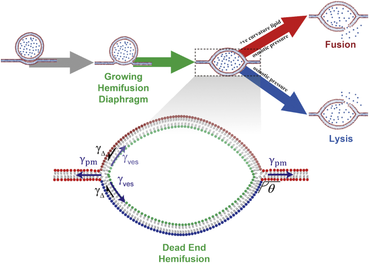
Membrane fusion is a critical step for many essential processes, from neurotransmission to fertilization. For over 40 years, protein-free fusion driven by calcium or other cationic species has provided a simplified model of biological fusion, but the mechanisms remain poorly understood. Cation-mediated membrane fusion and permeation are essential in their own right to drug delivery strategies based on cell-penetrating peptides or cation-bearing lipid nanoparticles. Experimental studies suggest calcium drives anionic membranes to a hemifused intermediate that constitutes a hub in a network of pathways, but the pathway selection mechanism is unknown. Here we develop a mathematical model that identifies the network hub as a highly dynamic hemifusion complex. Multivalent cations drive expansion of this high-tension hemifusion interface between interacting vesicles during a brief transient. The fate of this interface determines the outcome, either fusion, dead-end hemifusion, or vesicle lysis. The model reproduces the unexplained finding that calcium-driven fusion of vesicles with planar membranes typically stalls at hemifusion, and we show the equilibrated hemifused state is a novel lens-shaped complex. Thus, membrane fusion kinetics follow a stochastic trajectory within a network of pathways, with outcome weightings set by a hemifused complex intermediate.


Conflict of interest statement

Declaration of interests The authors declare no competing interests.

Figures

Figure 1
Figure 1
Ca2+ boosts membrane tension, induces membrane adhesion, and mediates the outcomes in the network of pathways to membrane fusion. (A) Ca2+ increases membrane tension and adheres negatively charged vesicles. Ca2+ interacts with anionic or zwitterionic lipids in the outer leaflet of the vesicle membrane to contract the vesicle membrane by a factor ε to increase tension. A second effect is to adhere the vesicles, with adhesion energy W per unit area. (B) Network of pathways to Ca2+-mediated membrane fusion. Calcium and other divalent cations adhere phospholipid bilayer vesicle membranes (state A) and provoke hemifusion, fusion of the outer phospholipid monolayers only. The initial hemifusion connection is thought to be a minimal stalk (state S) that is metastable and yields to an expanding hemifusion diaphragm, HD (state HD). HD expansion is driven by high calcium-induced membrane tension and outer monolayer contraction. The HD bilayer tension is greatest and may generate a pore (state HD) that could reseal or dilate and rupture the HD (fusion outcome); or the vesicle membrane could nucleate a pore (state V) that dilates and causes rupture (lysis outcome); or the HD may survive the high-tension transient unscathed, expanding to full equilibrium (dead-end hemifusion outcome). Inset: the transient hemifused state (HD) selects the pathway. The HD tension γHD(t) is greatest, as it balances two vesicle tensions γves (t), but its area is least, AHDt<Avest. These effects compete to set the ratio of fusion to lysis. Since tension rapidly decays as the HD expands, the HD may escape to low-tension equilibrium (dead-end hemifusion). To see this figure in color, go online.
Figure 2
Figure 2
Calcium concentration governs outcome distribution in Ca2+-mediated GUV-GUV fusion. Model predictions, conditions as in experiments of (11) (see Table 1 for parameters). GUVs are made of phospholipid with PS/PE/PC = 1/1/3. (A) Predicted outcome distributions versus calcium concentration. In the low-concentration regime, dead-end hemifusion is most probable; fusion is maximized in the intermediate regime; lysis dominates at high concentrations. (B) Predicted outcome distributions for the two specific cation concentrations used in (11). D, dead-end hemifusion; L, lysis; F, fusion. (C and D) Predicted time evolution of HD area and tension and vesicle tension at 2 mM Mg2+ (C) and 6 mM Ca2+(D). HD growth decays the vesicle and HD tensions to below their respective rupture thresholds. More fusion occurs at the higher concentration because the HD tension exceeds its rupture threshold for longer. Calcium only interacts with the PS component in the non-HD region to switch its spontaneous curvature to a negative pore-hating value. Compared with HDs, non-HD membranes are less likely to rupture. (E) Model predicted rates of Ca2+-mediated GUV-GUV fusion and lysis versus time for the conditions of the experiments of ref (11). To see this figure in color, go online.
Figure 3
Figure 3
Lipid composition regulates outcome distribution in Ca2+-mediated GUV-GUV fusion. GUVs are made of PS and PE with different PE fractions. Model predictions, conditions as in experiments of (9,10) (see Table 1 for parameters). The negative-curvature lipid PE disfavors pore formation and suppresses fusion. (A) Predicted outcome distributions for the compositions of (9) (pure PS) and (10) (75% PE, 25% PS) in the presence of 5 mM Ca2+. (B) Predicted outcome distributions versus PS/PE composition in the presence of 5 mM Ca2+. (C and D) Predicted time evolution of HD area, vesicle and HD tension for pure PS (C) and 75% PE, 25% PS (D). (E) Mechanism that protects pure PS vesicles from lysis. As PS has positive spontaneous curvature, nucleated pores have low line tension and will likely be expanded by the HD membrane tension γHD (left). By contrast, the pore line tension τ in the vesicle membrane is large, as PS is thought to have negative spontaneous curvature in the presence of Ca2+, and pores close (right). Were it not for the PS curvature sign reversal, almost no fusion would occur as the pore nucleation rate is much greater in the far bigger vesicle membrane. To see this figure in color, go online.
Figure 4
Figure 4
Calcium cannot typically fuse vesicles with a plasma membrane (PM). Model predicted sequence is shown (schematic). At high [Ca2+], the vesicle has high tension and adheres strongly to the PM. Following hemifusion and HD growth the present model predicts that the vesicle tension is dissipated and a lens-shaped equilibrium hemifused complex is attained. Additional forces are required to drive fusion. Application of pore-promoting positive-curvature lipids can selectively activate fusion or lysis. To see this figure in color, go online.

References

    1. Martens S., McMahon H.T. Mechanisms of membrane fusion: disparate players and common principles. Nat. Rev. Mol. Cell Biol. 2008;9:543–556. doi: 10.1038/nrm2417. - DOI - PubMed
    1. Wickner W., Schekman R. Membrane fusion. Nat. Struct. Mol. Biol. 2008;15:658–664. doi: 10.1038/nsmb.1451. - DOI - PMC - PubMed
    1. Brunger A.T., Choi U.B., et al. Zhou Q. Molecular mechanisms of fast neurotransmitter release. Annu. Rev. Biophys. 2018;47:469–497. doi: 10.1146/annurev-biophys-070816-034117. - DOI - PMC - PubMed
    1. Südhof T.C. Neurotransmitter release: the last millisecond in the life of a synaptic vesicle. Neuron. 2013;80:675–690. doi: 10.1016/j.neuron.2013.10.022. - DOI - PMC - PubMed
    1. Ginsberg L. Does Ca2+ cause fusion or lysis of unilamellar lipid vesicles? Nature. 1978;275:758–760. - PubMed

Publication types

LinkOut - more resources