. 2022 Dec 9;50(22):13063-13082.
doi: 10.1093/nar/gkac1119.

The multivalency of the glucocorticoid receptor ligand-binding domain explains its manifold physiological activities

Affiliations

The multivalency of the glucocorticoid receptor ligand-binding domain explains its manifold physiological activities

Alba Jiménez-Panizo et al. Nucleic Acids Res. .

Abstract

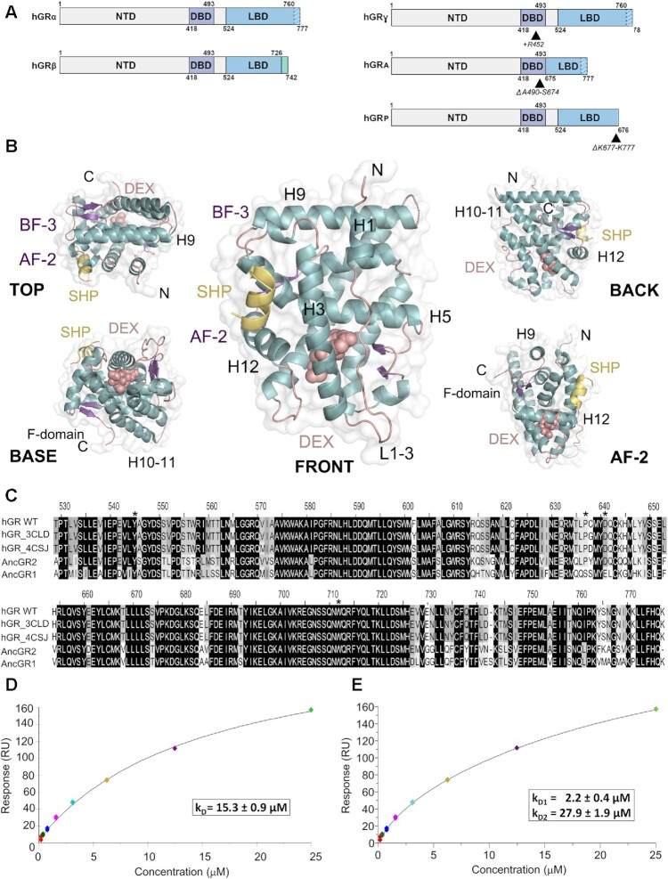
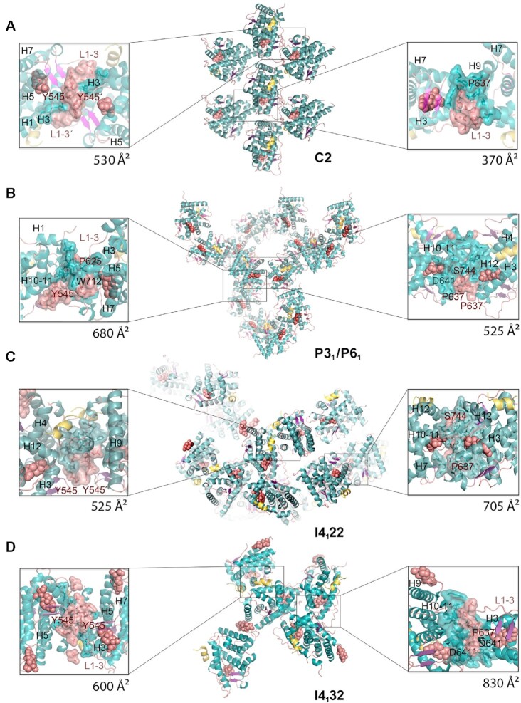
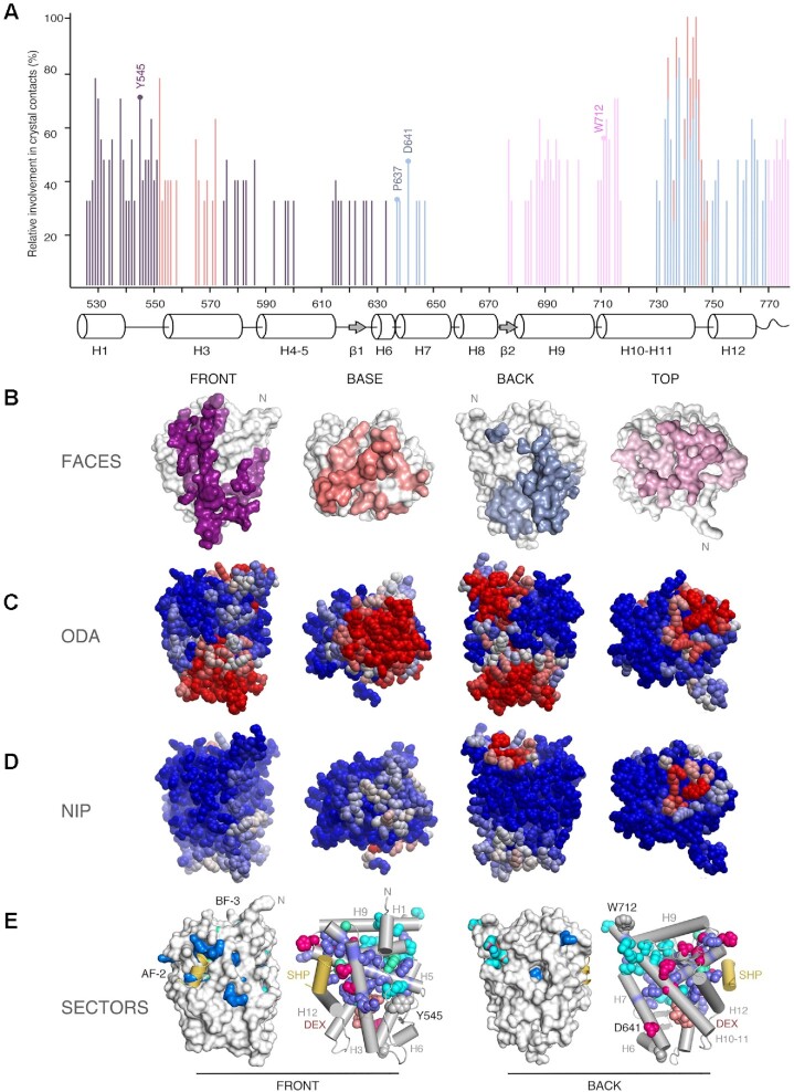
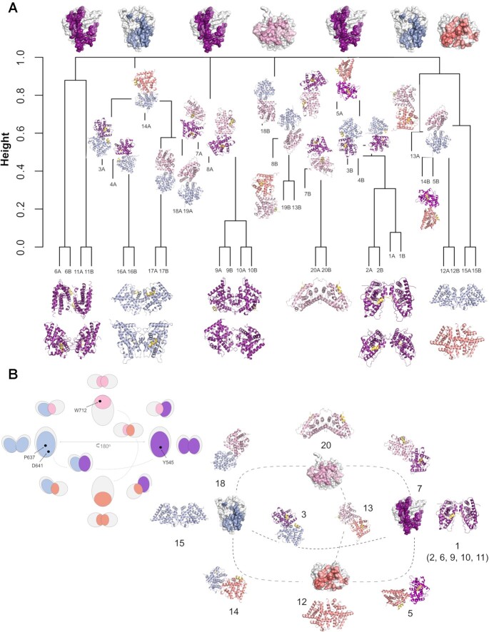
The glucocorticoid receptor (GR) is a ubiquitously expressed transcription factor that controls metabolic and homeostatic processes essential for life. Although numerous crystal structures of the GR ligand-binding domain (GR-LBD) have been reported, the functional oligomeric state of the full-length receptor, which is essential for its transcriptional activity, remains disputed. Here we present five new crystal structures of agonist-bound GR-LBD, along with a thorough analysis of previous structural work. We identify four distinct homodimerization interfaces on the GR-LBD surface, which can associate into 20 topologically different homodimers. Biologically relevant homodimers were identified by studying a battery of GR point mutants including crosslinking assays in solution, quantitative fluorescence microscopy in living cells, and transcriptomic analyses. Our results highlight the relevance of non-canonical dimerization modes for GR, especially of contacts made by loop L1-3 residues such as Tyr545. Our work illustrates the unique flexibility of GR's LBD and suggests different dimeric conformations within cells. In addition, we unveil pathophysiologically relevant quaternary assemblies of the receptor with important implications for glucocorticoid action and drug design.


Figures

Figure 1.
Figure 1.
The ligand-binding domain of GR self-associates in solution. (A) Schematic representation of GR domain organization. GRα and GRβ are identical up to residue 727 (H10/11), but the last 50 (in GRα) and 15 residues (in GRβ, green box) are unrelated. Other GR isoforms are shown (right). (B) Overall structure of the GR-LBD monomer. The module is shown in standard orientation in the middle of the panel (i.e. with H1 and H3 displayed facing the viewer and the AF-2 pocket on the left). Four additional views are shown to present other LBD areas. Models are depicted as cartoons with helices (blue), loops (pink) and beta-sheets (purple). DEX (salmon spheres) and SHP peptide (yellow cartoon) are shown. The BF-3 pocket is also labeled. (C) Sequence alignment of LBDs between WT human GR, two engineered variants used in several structures (PDBs 3CLD and 4CSJ), and the ancGR1 and 2 forms. Strictly conserved residues are white with black shading; other conservatively replaced residues are shaded gray. Residues mutated in the current study are marked with asterisks. (D, E) SPR analysis of GR-LBD self-association according to (D) 1:1 or (E) multisite models. The results of experiments conducted in duplicate are shown along with the calculated affinity constants (kD). Data were fitted to the 1:1 and multisite models with χ2 values of 4.52 and 1.25, respectively.
Figure 2.
Figure 2.
New crystal structures of GR-LBD·DEX reveal a variety of quaternary assemblies. For all structures, the overall crystal packing is shown in the central panels. Monomers are depicted as cartoons with helices colored blue and loops colored salmon. DEX molecules are represented as salmon spheres and SHP peptides as yellow ribbons. Details of intermonomer interfaces are given in the lateral panels, in which the side chains of interacting residues are shown as sticks. (A) C2 crystals. Note that major contacts are centered on L1–3 with stacked Tyr545 phenol rings from two neighboring molecules. A monoclinic structure of ancGR2-LBD bound to another synthetic GC, triamcinolone acetonide, and complexed to a shorter SHP peptide had been previously reported (5UFS; (38)). Interestingly, 5UFS features a Tyr545-centered parallel dimer almost identical to the topologically equivalent arrangement in our current C2 crystals. (B) Related P31 and P61 crystals. Tyr545 engages in heterologous contacts with a neighboring molecule in these crystals (see the position of the Trp712’ side chain). Structures of ancGR2-LBD bound to either DEX or a different GC (mometasone furoate) and complexed to a TIF-2 peptide had been previously reported in a similar hexagonal crystal form (3GN8 and 4E2J; a and b axes are ∼5% longer in our crystals, while the c axis is ∼4% shorter). These relatively small differences in the cell constants compared to the current P61 structure result in a markedly different small intermonomer interface, however, which is asymmetric in 3GN8/4E2J. (C, D) Common packing of I4122 and I4132 crystals. Note that the phenol rings of two Tyr545 residues stack as in the C2 crystals, although the two interacting monomers are fully differently oriented relative to each other. Note also that the largest interface in these crystals features abutting Asp641 side chains from three monomers organized around a local (I4122) or exact 3-fold axis (I4132; right side panel in D).
Figure 3.
Figure 3.
Experimental structures and bioinformatics analyses unveil four major homodimerization surfaces on GR-LBD. (A) Relative frequencies of residue involvement in GR-LBD homodimer formation. Bar height indicates how often a given residue engages in crystal contacts in all available structures of GR-LBD, normalized to the residue most frequently found in homodimer interfaces, Leu741. Secondary structure elements given below the plot correspond to the crystal structure of human GR-LBD resolved at the highest resolution, 6NWL. (B) Residues involved in GR-LBD homodimerization cluster in continuous patches on its front (colored purple), base (coral), back (blue) and top (pink) faces. The association of these four faces yields the catalog of GR-LBD dimers represented in Figure 4. Models are shown in the same orientation and at the same magnification in panels (C)–(E). (C) Predicted protein-protein interaction ODA. ODA ‘hotspots’ (residues with favorable docking energy; ODA < –10.0 kcal/mol) are colored red, residues with ODA > 0 kcal/mol are shown in blue, and intermediate values are scaled accordingly. ODA hotspots form continuous surface patches that essentially overlap with the four protein-protein interaction interfaces shown in panel (B). (D) Hotspot interface residues predicted from docking experiments. Surface residues are colored according to their NIP. Residues with NIP > 0.4 and < 0 are colored red and blue, respectively; intermediate values are scaled accordingly. (E) SCA identifies two sectors of clustered, physically connected residues in GR-LBD. The front and back orientations of GR-LBD are depicted, and in both cases the module is represented as a solid surface and as a cartoon, with helices shown as rods and labeled. Residues belonging to sectors I and II are shown with their side chains atoms as spheres, colored cyan and dark blue, respectively. Residues belonging to both sectors are colored pink. SHP peptide is colored yellow, and DEX is shown as salmon spheres.
Figure 4.
Figure 4.
An integrated catalog of GR-LBD homodimers. The four distinct GR-LBD protein-protein interfaces associate to generate 20 topologically different homodimers. (A) A dendrogram based on a hierarchical analysis of protein-protein contacts using Jaccard's index groups the 20 unique GR-LBD assemblies into six different clusters. (B) Relationships between the different GR-LBD homodimers. For orientation, monomers highlighting the four interacting surfaces are placed at the cardinal points in this panel (top, nord; front, east; base, south; and back, west), colored-coded as in Figure 3. Monomers in 10 representative homodimers are depicted as cartoons; each monomer is colored according to the face used to associate with its partner. Dimers are placed closest to the generating monomers. An equivalent schematic representation of GR-LBD homodimerization potential is shown at the upper left corner, with the position of major interacting residues indicated.
Figure 5.
Figure 5.
Several GR-LBD homodimers are populated in solution. (A) SDS-PAGE analysis of GR-LBD after incubation with EDC. Notice bands with relative molecular masses corresponding to GR-LBD dimers (D) and tetramers (T) in all the lanes except control lane 1 (no EDC added). Lanes 2 and 3, protein incubated at about 0.37 mg/ml; lanes 4 and 5, protein incubated at about 1.5 mg/ml. Samples in lanes 2 and 4 were treated at RT; those in lanes 3 and 5 at 30°C. Bands corresponding to co-purified DnaK and GroEL E. coli chaperones are marked with (*) and (**), respectively. (B) Schematic representation of GR-LBD structure, with side side chains of residues identified using EDC shown as spheres (Asp in green, Lys in lilac, and Glu in yellow). (C) Crosslink map of EDC-treated GR-LBD showing all intermonomer crosslinks identified by MS analysis. Regions corresponding to the top, front, base, and back surfaces are colored as in Figures 3 and 4. A secondary structure plot is shown above the map. (D) Closeup of the major homodimerization interface in C2 crystals (front-to-front homodimer #11), dominated by stacked Tyr545/Tyr545’ residues. (E) Non-reducing SDS-PAGE analysis of purified GR-LBD(Y545C) (lane 3) shows spontaneous dimerization in solution. Note that the WT protein does not form dimers when incubated at the same concentration (lane 2).
Figure 6.
Figure 6.
Mutation of LBD–LBD interface residues profoundly affects the multimerization behavior of FL-GR. (A) Schematic 3D model of FL-GR. The NTD is highly disordered and is followed by the globular DBD (DNA-bound conformation is shown) and the LBD. Note that the length and flexibility of the hinge (H) allows for the formation of various homodimers. The side chains of all residues mutated to assess the multimerization behavior of GR are shown as spheres. (B) Subcellular localization of WT GFP-mGR and indicated mutants in 3617-GRKO cells, as assessed by fluorescence microscopy. Variant N525* lacks the entire LBD and remains monomeric, irrespective of DEX treatment (23). White arrowheads point to the MMTV arrays. Scale bar: 5 μm. Data for WT_GR, GRdim, GRmon and GRtetra were taken from (24) and are shown for comparison purposes. All N&B experiments have been performed with mouse GR as in (23). However, residue numbers correspond to the human protein to facilitate comparisons. (C) GR oligomerization in the nucleus, as determined in N&B assays. The fold increase in molecular brightness (ϵ) relative to the N525* monomeric control is shown (N = 428 total cells with 21 < N < 37 between treatments). (D) Quaternary structure of DNA-bound GR. The results of N&B assays at the MMTV arrays are represented as in panel C (N = 338 total cells with 17 < N < 35 between treatments). Note that simultaneous disruption of intermonomer interactions mediated by the DBD (Ala458Thr) and the LBD (Tyr545Ala) in the GRdim/Y545A double mutant results in a variant that is monomeric in the nucleus, while at the array it formed mostly trimers. In panels (C) and (D), centered lines show the medians and crosses represent sample means (average numbers below each box-plot). Box limits indicate the 25th and 75th percentiles; whiskers extend 1.5-fold the interquartile range from the 25th and 75th percentiles, with outliers represented by dots. Boxes with different superscript letters are significantly different from each other (P < 0.05; one-way ANOVA followed by Tukey's multiple comparison test).
Figure 7.
Figure 7.
Residues Tyr545 and Asp641 have major roles in GR quaternary structures and transcriptional activity. Stable cell lines expressing GFP-tagged variants of the indicated mouse GR mutants were treated with 100 nM DEX for 2 h (note than numbers correspond to hGR, as in Figure 6). (AC) Venn diagrams of differentially expressed genes from RNA-Seq (|log2 Fold change| > 0.5, False-discovery-rate < 0.01). The two WT GR sets (b1, b2) are biological replicates collected and sequenced separately for each batch. (BD) Scatter plots of shared hormone-regulated genes (17 and 71 common genes, respectively) between GR mutants, compared and plotted against their respective WT GR control. (E) Models of human GR-LBD homodimers based on the observed assemblies #10, #6, #9 and #11. The critical homodimerization residues, Tyr545 and Ile628, are shown as color-coded spheres in all cases. (F) Model of GR-LBD tetramer generated by docking dimer #11 onto itself. The model is compatible with the results obtained with the Y545C mutant and EDC-crosslinking. (G) Putative GR-LBD hexamer favored by the Asp641Val mutation. The central trimer corresponds to an arrangement observed in tetragonal and cubic crystal forms (#14; Figures 2C, D and 4), while the peripheral monomers dock according to conformation #4. Asp641 residues from the central trimer are encircled. This generates a closed hexamer by additional interactions between the N-terminal end of H10 and H12’ at the third interface. In addition to D641V, other GR mutants associated with Chrousos disease might favor multimeric forms that are incompatible with active GR tetramers on DNA. For instance, replacement of Thr556 by an aliphatic Ile would stabilize this arrangement through contacts with e.g. residues Met560 and Pro637 from its ‘own’ monomer and/or His645’/Asn731’ from a neighboring molecule. Similar considerations apply to mutations such as Arg714Gln, Phe737Leu, Ile747Met and Leu773Pro as well as to variant Pro637Ala, which also forms higher-order oligomers at the array (Figure 6D).

Similar articles

Cited by

References

    1. Conway-Campbell B.L., Pooley J.R., Hager G.L., Lightman S.L.. Molecular dynamics of ultradian glucocorticoid receptor action. Mol. Cell Endocrinol. 2011; 348:383–393. - PubMed
    1. Busada J.T., Cidlowski J.A.. Mechanisms of glucocorticoid action during development. Curr. Top Dev. Biol. 2017; 125:147–170. - PubMed
    1. Bledsoe R.K., Montana V.G., Stanley T.B., Delves C.J., Apolito C.J., McKee D.D., Consler T.G., Parks D.J., Stewart E.L., Willson T.M.et al. .. Crystal structure of the glucocorticoid receptor ligand binding domain reveals a novel mode of receptor dimerization and coactivator recognition. Cell. 2002; 110:93–105. - PubMed
    1. Evans R.M., Mangelsdorf D.J.. Nuclear receptors, RXR, and the big bang. Cell. 2014; 157:255–266. - PMC - PubMed
    1. Jimenez-Panizo A., Perez P., Rojas A., Fuentes-Prior P., Estebanez-Perpina E.. Non-canonical dimerization of the androgen receptor and other nuclear receptors: implications for human disease. Endocr. Relat. Cancer. 2019; 26:R479–R497. - PubMed

Publication types