. 2022 Nov 9;25(12):105546.
doi: 10.1016/j.isci.2022.105546. eCollection 2022 Dec 22.

PERIOD 2 regulates low-dose radioprotection via PER2/pGSK3β/β-catenin/Per2 loop

Affiliations

PERIOD 2 regulates low-dose radioprotection via PER2/pGSK3β/β-catenin/Per2 loop

Aris T Alexandrou et al. iScience. .

Abstract

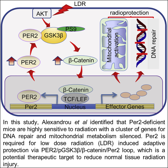
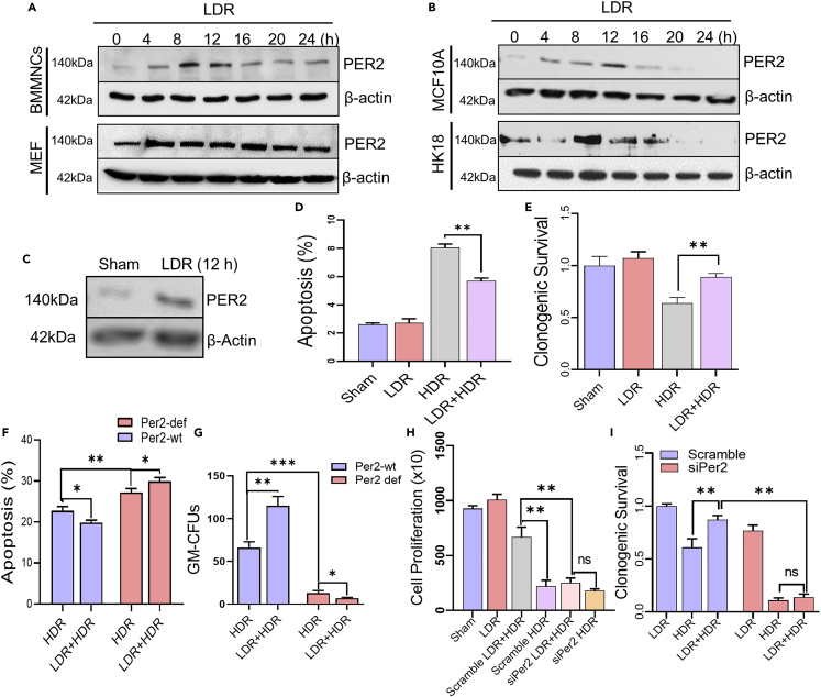
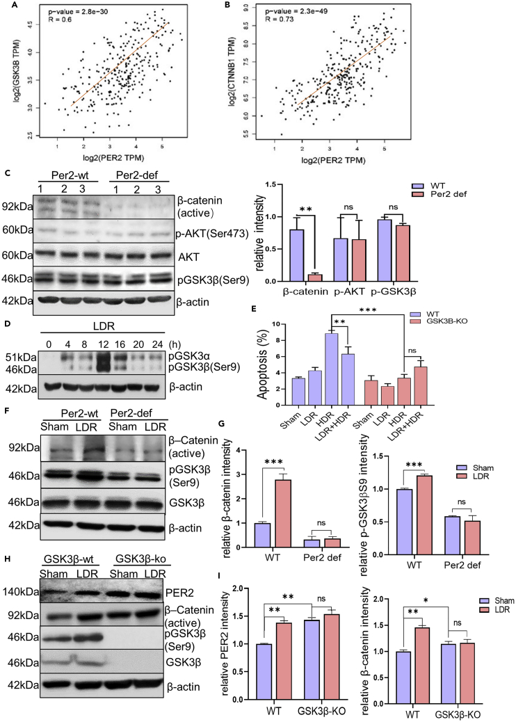
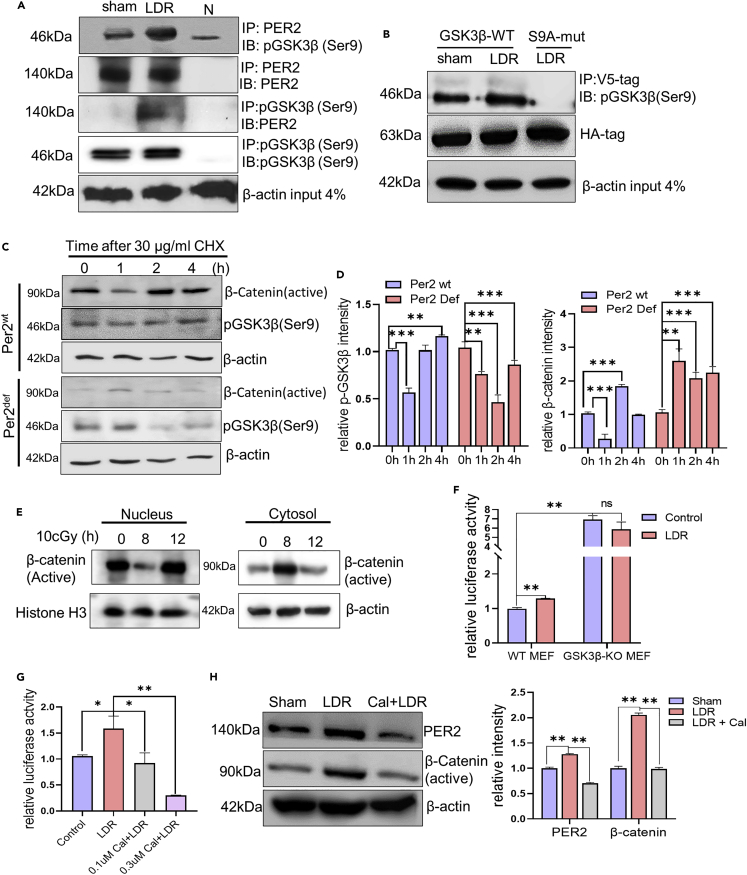
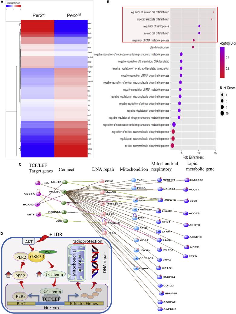
During evolution, humans are acclimatized to the stresses of natural radiation and circadian rhythmicity. Radiosensitivity of mammalian cells varies in the circadian period and adaptive radioprotection can be induced by pre-exposure to low-level radiation (LDR). It is unclear, however, if clock proteins participate in signaling LDR radioprotection. Herein, we demonstrate that radiosensitivity is increased in mice with the deficient Period 2 gene (Per2def) due to impaired DNA repair and mitochondrial function in progenitor bone marrow hematopoietic stem cells and monocytes. Per2 induction and radioprotection are also identified in LDR-treated Per2wt mouse cells and in human skin (HK18) and breast (MCF-10A) epithelial cells. LDR-boosted PER2 interacts with pGSK3β(S9) which activates β-catenin and the LEF/TCF mediated gene transcription including Per2 and genes involved in DNA repair and mitochondrial functions. This study demonstrates that PER2 plays an active role in LDR adaptive radioprotection via PER2/pGSK3β/β-catenin/Per2 loop, a potential target for protecting normal cells from radiation injury.

Keywords: Biological sciences; cell biology; molecular biology.


Conflict of interest statement

The authors declare no competing interests.

Figures

None
Graphical abstract
Figure 1
Figure 1
Per2def mice are radiosensitive with reduced DNA repair capacity (A) Kaplan-Meier survival of Per2 wild-type (Per2wt, blue line) or Per2 deficient (Per2def, red line) C57BL/6 mice following whole body exposure to different radiation doses. WBR:7-12 Gy; Data are represented as mean ± SEM, n = 12-20/group, Kaplan-Meier survival analysis Log rank test. (B) DNA repair capacity showing representative images of alkaline comet assay of Per2wt and Per2def BMMNCs at indicated times (30 min or 1 h) after 1 Gy radiation; Scale bar, 50 μm. (C) Quantitation of DNA repair capacity by tailed DNA (%) and tail moment (%). Data are represented as mean ± SEM, n = 50, ∗∗p < 0.01, ∗∗∗p < 0.001, Student’s t test. (D) Representative images of NHEJ DNA repair capacity by 53BP1 foci analysis in Per2wt and Per2def BMMNCs 2 h after 2 Gy radiation; Scale bar, 2 μm. (E) Quantitation of NHEJ DNA repair capacity by counting 53BP1 foci (left) and γH2AX foci (right) per nucleus. Data are represented as mean ± SEM, n = 40, ∗p < 0.05, ∗∗p < 0.01, ns p > 0.05, Student’s t test. (F) Representative images of HR DNA repair capacity by Rad51 foci analysis in Per2wt and Per2def BMMNCs 2 h after 2 Gy radiation; Scale bar, 2 μm. (G) Quantitation of HR DNA repair capacity by counting Rad51 foci (left) and γH2AX foci (right) per nucleus. Data are represented as mean ± SEM, n = 40, ∗p < 0.05, ∗∗p < 0.01, ns p > 0.05, Student’s t test.
Figure 2
Figure 2
DNA repair function was impaired in Per2def cells (see also Figures S1–S3) (A) Lin/Sca-1+/c-Kit+ bone marrow derived progenitor hematopoietic stem cells (BM-pHSCs) were calculated by sorting of bone marrow cells in Per2wt and Per2def mice. Data are represented as mean ± SEM, n = 3, ∗∗p < 0.01, Student’s t test. (B) Representative flow cytometry sorting images of Lin/Sca-1+/c-Kit+ BM-pHSCs by indicating gating region with anti-c-Kit and anti-Sca-l in Per2wt and Per2def BM-pHSCs. (C) A cluster of genes related to DNA damage repair by RNAseq analysis of Per2wt versus Per2def BM-pHSCs with 1.2-fold cutoff. (D) Gene ontology biological process enrichment analysis of up-regulated DNA repair genes with 1.2-fold cutoff in Per2wt versus Per2def Lin/Sca-1+/c-Kit+ BM-pHSCs. (E) Western blot of a cluster of DNA repair factors of Mre11, Brca1, Rad51, Chk1, and Chk2 in Per2wt and Per2def BMMNCs. (F) The effect of LDR on apoptosis measured with flow cytometry in Per2wt and Per2def BMMNCs 24 h after irradiation with LDR (10 cGy). Data are represented as mean ± SEM, n = 6, ∗∗∗p < 0.001, Student’s t test. (G) The effect of LDR on the proliferation capacity of Per2wt and Per2def Lin/Sca-1+/c-Kit+ BM-pHSCs was measured by GM-CFU assay. Data are represented as mean ± SEM, n = 3, ∗∗∗p < 0.01, Student’s t test.
Figure 3
Figure 3
Per2 is required for LDR-induced mitochondrial activation (see also Figure S4) (A) A cluster of genes related to mitochondrial metabolic functions by RNAseq analysis of Per2wt versus Per2def BM-HSCs with 1.2-fold cutoff. (B) Gene ontology biological process enrichment analysis of up-regulated genes related to mitochondrial metabolic functions with 1.2-fold cutoff in Per2wt versus Per2def BM-pHSCs. (C–F) Western blot of a cluster of mitochondrial metabolic factors CPT1A, CPT2, NDUFA12, and NDUFV3 in Per2wt and Per2def BMMNCs. Mitochondrial membrane potential (D), oxygen consumption (E), and ATP generation (F) were measured in Per2wt and Per2def BMMNCs 24 h after LDR. Data are represented as mean ± SEM, n = 3, ∗p < 0.05; ∗∗p < 0.01, ns p > 0.05, Student’s t test.
Figure 4
Figure 4
Per2 is required for LDR-induced adaptive radioprotection (see also Figure S5) (A and B). Per2 expression in mouse BMMNCs and MEFs (A) and in human mammary epithelial MCF-10A cells and skin keratinocytes HK18 (B) at different times after LDR. (C-E) Western blot of Per2 in primary cultured epithelial cells derived from healthy human mammary tissues 12 h following exposure to LDR. Cell apoptosis (D) and clonogenic survival (E) of MCF-10A cell exposed to Sham, LDR, HDR or LDR 8 h before HDR. Data are represented as mean ± SEM, n = 3; ∗∗p < 0.01, Student’s t test. (F) LDR-induced radioprotection was measured by apoptosis with flow cytometry in Per2wt and Per2def BMMNCs 24 h after HDR or LDR 8 h before HDR. Data are represented as mean ± SEM, n = 6, ∗p < 0.05, ∗∗p < 0.01, ANOVA two-way test was applied. (G–I) LDR-induced radioprotection was measured by GM-CFU assay in Per2wt and Per2def BMMNCs after HDR or LDR 8 h before HDR. Data are represented as mean ± SEM, n = 3, ∗p < 0.05, ∗∗p < 0.01, ∗∗∗p < 0.001, ANOVA two-way test was applied. Cell proliferation (H) and clonogenic survival (I) of MCF-10A cells transfected with siPer2 or scrambled siPer2 following exposure to Sham, LDR, HDR or LDR 8 h before HDR. Data are represented as mean ± SEM, n = 3, ∗∗p < 0.01, ns p > 0.05, ANOVA two-way test was applied.
Figure 5
Figure 5
GSK3β/β-catenin pathway is involved in Per2 mediated adaptive radioprotection (see also Figure S6) (A) Correlation of Per2 and GSK3β in human mammalian tissue analyzed with the GEPIA database. Pearson correlation analysis, R = Pearson correlation coefficient. (B) Correlation of Per2 and β-catenin in human mammalian tissue analyzed with the GEPIA database. Pearson correlation analysis, R = Pearson correlation coefficient. (C) Left, western blot of phosphorylated GSK3β (Ser9), AKT, p-AKT and active β-catenin in Per2wt and Per2def BMMNCs; right, relative expression of indicated proteins quantified with ImageJ and normalized with β-actin levels. Data are represented as mean ± SEM, n = 3, ∗∗p < 0.01, ns p > 0.05, Student’s t test. (D) Western blot of phosphorylated GSK3β (Ser9) in Per2wt/GSKßwt MEFs following exposure to LDR with estimated GSK3β phosphorylation peak at 12 h. (E) Cell apoptosis of Per2wt/GSKßwt MEFs compared to Per2wt/GSKßko MEFs after treated with LDR (10 cGy), HDR (5 Gy), or LDR 12 h before HDR. Data are represented as mean ± SEM, n = 3, ∗∗p < 0.01, ∗∗∗p < 0.001, ns p > 0.05, ANOVA two-way test was applied. (F) Western blot of pGSK3β(Ser9), GSK3β, and active β-catenin in Per2wt and Per2def mice BMMNCs 12 h after sham and LDR. (G) Relative expression of pGSK3β and active β-catenin in Per2wt and Per2def mice BMMNCs 12 h after LDR was quantified with ImageJ and normalized with β-actin levels. Data are represented as mean ± SEM, n = 3, ∗∗∗p < 0.001, ns p > 0.05, Student’s t test. (H) Western blot of pGSK3β(Ser9), GSK3β, active β-catenin, and Per2 in Per2wt/GSK3ßwt and Per2wt/GSK3ßko MEFs 12 h after sham and LDR. (I) Relative expression of PER2 and active β-catenin in Per2wt/GSK3ßwt and Per2wt/GSK3ßko MEFs 12 h after sham and LDR was quantified with ImageJ and normalized with β-actin levels. Data are represented as mean ± SEM, n = 3, ∗p < 0.05, ∗∗p < 0.01, ns p > 0.05, ANOVA two-way test was applied.
Figure 6
Figure 6
PER2 interacts with pGSK3β(Ser9) to enhance active β-catenin mediated Per2 transactivation (see also Figures S7-S9) (A) Interaction of Per2 with pGSK3β (Ser9) in Per2wt/GSKßwt MEFs detected by immunoprecipitation 12 h after LDR followed by immunoblot with anti-pGSK3β(Ser9), or reversely, immunoprecipitation of pGSK3β(Ser9) followed by immunoblot with anti-Per2 antibody (N = negative control without antibody). (B) Immunoprecipitation of co-transfected V5-Per2 with HA-pGSK3β or HA-pGSK3β S9A-mut 293T cells 12 h following LDR. (C) Degradation of pGSK3β(Ser9) and active β-catenin measured in Per2wt and Per2def BMMNCs 12 h after LDR followed by cycloheximide (30 μg/ml) for indicated times. (D) Relative expression of pGSK3β(Ser9) and active β-catenin in in Per2wt and Per2def BMMNCs 12 h after LDR followed by cycloheximide (30 μg/ml) was quantified with ImageJ and normalized with β-actin levels. Data are represented as mean ± SEM, n = 3, ∗∗p < 0.01, ∗∗∗p < 0.001, ANOVA two-way test was applied. (E) Western blot of active β-catenin in nucleus and cytosol of Per2wt/GSK3ßwt MEFs 8 h after LDR, using histone and β-actin as loading controls respectively for nuclear and cytosol proteins. (F) Luciferase reporter activity driven by mouse Per2 promoter in Per2wt/GSK3ßwt MEFs compared to Per2wt/GSKßko MEFs 12 h after LDR; Per2 luciferase transcription activity was normalized with Renilla activity. Data are represented as mean ± SEM, n = 3, ∗∗p < 0.01, ns p > 0.05, ANOVA two-way test was applied. (G) Luciferase reporter activity driven by mouse Per2 promoter in Per2wt/GSK3ßwt MEFs 12 h after LDR or LDR incubation with 0.1 and 0.3 μm β-catenin inhibitor Calphostin C (Cal, blocking β-catenin transactivation), Per2 luciferase transcription activity was normalized with Renilla activity. Data are represented as mean ± SEM, n = 3, ∗p < 0.05, ∗∗p < 0.01, ANOVA two-way test was applied. (H) Western blot of PER2 and active β-catenin in Per2wt/GSK3ßwt MEFs 12 h after LDR or LDR with Cal (0.1 μm); right, relative expression of PER2 and active β-catenin in Per2wt/GSK3ßwt MEFs 12 h after LDR or LDR with Cal (0.1 μm) was quantified with ImageJ and normalized with β-actin levels. Data are represented as mean ± SEM, n = 3, ∗∗p < 0.01, ANOVA two-way test was applied.
Figure 7
Figure 7
A Per2/pGSK3β/β-catenin/Per2 loop in Adaptive Radioprotection (see also Figure S10) (A) A cluster of β-catenin/TCF/LEF regulated prosurvival effector genes screened with 1.2-fold cutoff in RNAseq profile of Per2wt versus Per2def BMHSCs. (B) Gene ontology biological process enrichment analysis of up-regulated TCF/LEF targeted genes in Per2wt versus Per2def BMHSCs. (C) The interaction between four representatives upregulated TCF/LEF targeted genes (1.25-1.52-fold increased expression) with DNA repair genes and mitochondria metabolism genes in Per2wt versus Per2def BMpHSCs performed by VisANT, where k represents the potential KEGG pathways existing. (D) Schematic pathway of PER2/pGSK3β/β-catenin/PER2 loop in low-dose radiation-induced radioprotection. LDR-induced interaction of PER2 and pGSK3β(Ser9) leading to stabilization of pGSK3β(Ser9) enhancing active β-catenin that enters the nucleus and upregulates Per2 and a cluster of prosurvival genes involved in DNA repair and mitochondrial metabolism via β-catenin/TCF/LEF regulation. Thus, PER2/pGSK3β/β-catenin/PER2 loop may sustain the adaptive cellular homeostasis enhancing cell survival under severe genotoxic condition induced by high dose radiation.

References

    1. Adams C., Blacker E., Burke W. Night shifts: circadian biology for public health. Nature. 2017;551:33. - PubMed
    1. Pekovic-Vaughan V., Gibbs J., Yoshitane H., Yang N., Pathiranage D., Guo B., Sagami A., Taguchi K., Bechtold D., Loudon A., et al. The circadian clock regulates rhythmic activation of the NRF2/glutathione-mediated antioxidant defense pathway to modulate pulmonary fibrosis. Genes Dev. 2014;28:548–560. - PMC - PubMed
    1. Verlande A., Masri S. Circadian clocks and cancer: timekeeping governs cellular metabolism. Trends Endocrinol. Metab. 2019;30:445–458. - PMC - PubMed
    1. Le A.N., Harton J., Desai H., Powers J., Zelley K., Bradbury A.R., Nathanson K.L., Shah P.D., Doucette A., Freedman G.M., et al. Frequency of radiation-induced malignancies post-adjuvant radiotherapy for breast cancer in patients with Li-Fraumeni syndrome. Breast Cancer Res. Treat. 2020;181:181–188. - PMC - PubMed
    1. Ishihara H., Tanaka I., Yakumaru H., Chikamori M., Ishihara F., Tanaka M., Ishiwata A., Kurematsu A., Satoh A., Ueda J., et al. Circadian transitions in radiation dose-dependent augmentation of mRNA levels for DNA damage-induced genes elicited by accurate real-time RT-PCR quantification. J. Radiat. Res. 2010;51:265–275. - PubMed