Review
. 2022 Dec 1;13(6):1787-1822.
doi: 10.14336/AD.2022.0413.

SIRT6 in Aging, Metabolism, Inflammation and Cardiovascular Diseases

Affiliations
Review

SIRT6 in Aging, Metabolism, Inflammation and Cardiovascular Diseases

Zhenyang Guo et al. Aging Dis. .

Abstract

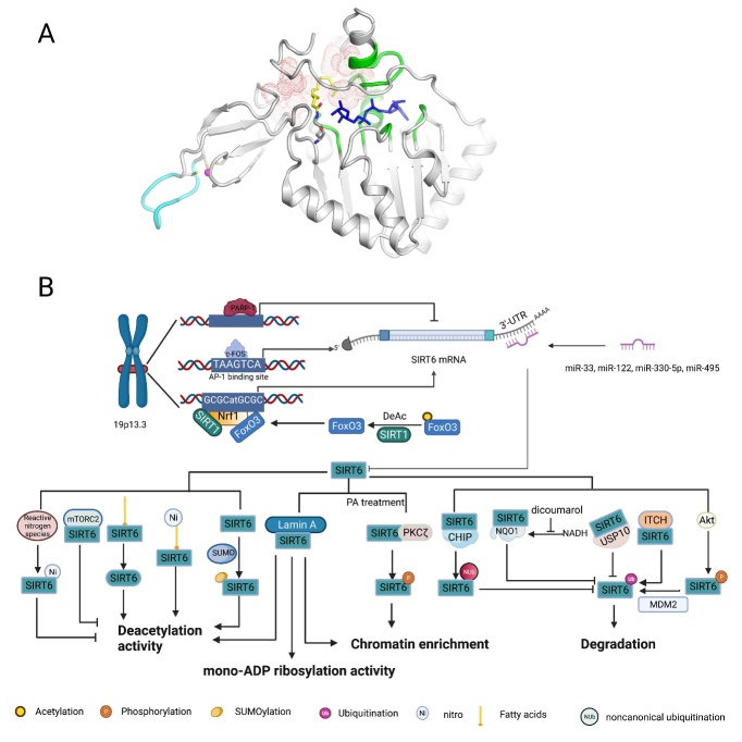
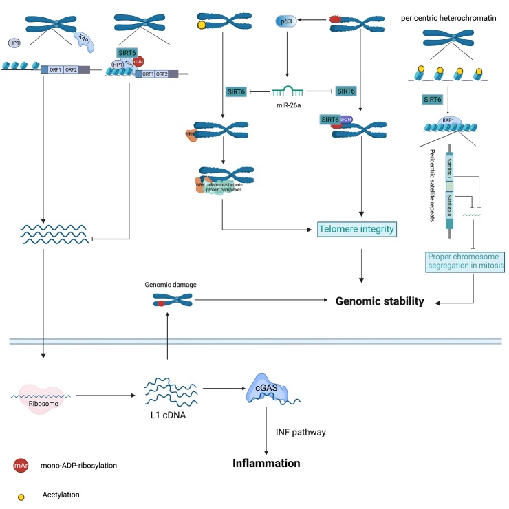
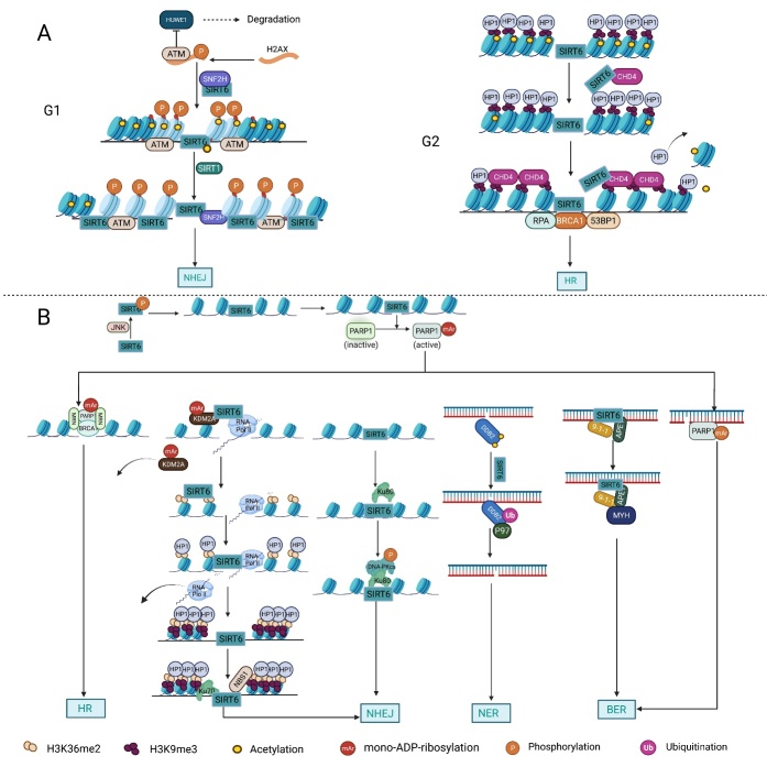
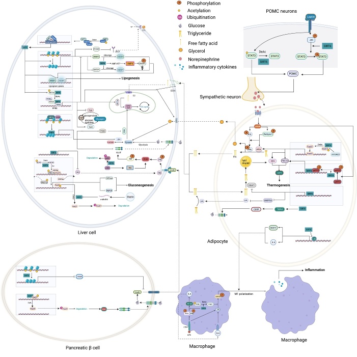
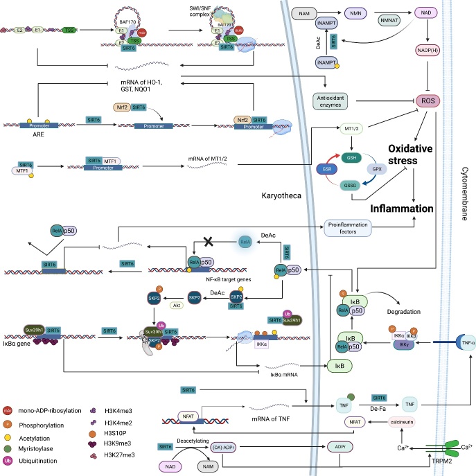
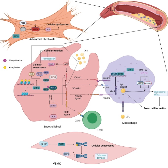
As an important NAD+-dependent enzyme, SIRT6 has received significant attention since its discovery. In view of observations that SIRT6-deficient animals exhibit genomic instability and metabolic disorders and undergo early death, SIRT6 has long been considered a protein of longevity. Recently, growing evidence has demonstrated that SIRT6 functions as a deacetylase, mono-ADP-ribosyltransferase and long fatty deacylase and participates in a variety of cellular signaling pathways from DNA damage repair in the early stage to disease progression. In this review, we elaborate on the specific substrates and molecular mechanisms of SIRT6 in various physiological and pathological processes in detail, emphasizing its links to aging (genomic damage, telomere integrity, DNA repair), metabolism (glycolysis, gluconeogenesis, insulin secretion and lipid synthesis, lipolysis, thermogenesis), inflammation and cardiovascular diseases (atherosclerosis, cardiac hypertrophy, heart failure, ischemia-reperfusion injury). In addition, the most recent advances regarding SIRT6 modulators (agonists and inhibitors) as potential therapeutic agents for SIRT6-mediated diseases are reviewed.

Keywords: SIRT6; ageing; cardiovascular diseases; inflammation; metabolism; molecular network.


Conflict of interest statement

Competing interests There is no conflict of interest involved in this review.

Figures

Figure 1.
Figure 1.
The structural features and molecular regulation of SIRT6. (A) The structural features of SIRT6: Structure of human SIRT6 in complex with H3K9-Myr (yellow) and ADP-ribose (blue) bound (PDB ID: 3ZG6). The zinc ion (pink)-binding structure is shown in the bottom left, and the unique flexible loop in the zinc-binding module is shown in azure. The stable structure of NAD+ binding with SIRT6 is colored green, and the long and wide hydrophobic-channel pocket is shown as red dots. (B) At the transcriptional level, p53 and pharmacological inhibition of PARP1 both upregulate SIRT6 expression. Similarly, c-FOS binds to the AP-1-binding site (TAAGTCA) in the SIRT6 promoter to directly promote SIRT6 expression. Under nutrient stress, SIRT1 interacts with and deacetylates FOXO3a, which is favorable for the formation of the SIRT1-FOXO3a-NRF1 (SFN) complex on the promoter of SIRT6, which upregulates SIRT6 expression. In addition, endogenous microRNAs (miRNAs), such as miR-33, miR-122, miR-330-5p and miR-495, silence translation by binding to the 3’-untranslated region (UTR) of SIRT6 mRNA. At the protein level, fatty acids (FAs) and nitrated FAs can activate SIRT6 deacetylase activity. The binding of electrophilic nitro-FAs and SIRT6 induces efficient activation (40-fold at 20 μM). In contrast, reactive nitrogen-induced nitration of tyrosine 257 (Y257) in SIRT6 causes loss of SIRT6 activity. Interaction between mTORC2 and SIRT6 suppresses SIRT6 deacetylase activity in adipose tissue. Lamin A is an endogenous activator of SIRT6, promoting SIRT6 recruitment to chromatin and activating both its deacetylase and mono-ADP ribosyltransferase activity. In addition, SUMOylation of SIRT6 specifically regulates SIRT6 deacetylation on H3K56 in vivo, and four lysine residues of SIRT6, K296, K300, K316 and K332, are thought to be SUMOylated. Under PA treatment, PKCζ binds to SIRT6 and phosphorylates SIRT6 at threonine 294 (T294) to promote SIRT6 recruitment to chromatin. Regarding stability, Akt-mediated phosphorylation of SIRT6 at serine 338 (S338) makes SIRT6 favorable for the ubiquitination via MDM2, promoting SIRT6 degradation. In contrast, SIRT6 interaction with USP10, CHIP and NQO1 blocks ubiquitin-mediated degradation of SIRT6. Among these factors, CHIP induces noncanonical ubiquitination of SIRT6 at K170, preventing canonical ubiquitination by other ubiquitin ligases. In addition, the NQO1 cofactor NADH promotes the binding of NQO1 to SIRT6, whereas DIC compromised the interaction between NQO1 and SIRT6. Ni, nitration; SUMOylation, SUMO-induced modification; Nub, noncanonical ubiquitination; Ub, ubiquitination.
Figure 2.
Figure 2.
SIRT6 inhibits genomic and telomeric instability. SIRT6 binds to the 5’-untranslated region (UTR) of LINE1 loci and mono-ADP-ribosylates KAP1, which promotes the interaction between KAP1 and HP1α, leading to LINE1 elements packaging into transcriptionally silent heterochromatin. In contrast, loss of SIRT6 in the 5’-UTR of LINE1 elevates LINE1 activity. Accumulation of cytoplasmic LINE1 cDNA instigates chromosomal rearrangements and provokes a strong type I interferon response and subsequent inflammation through the cytoplasmic DNA sensor cGAS. Impaired telomeres upregulate p53 expression, which activates miR-26, thereby decreasing SIRT6 content. During the S phase, SIRT6 deacetylates telomeric H3K9, enabling the efficient binding of the WRN protein to telomeres, leading to the recruitment of telomere/shelterin protein complexes to protect telomere integrity. In response to oxidative stress, SIRT6 recruits SNF2H to damaged telomeres to promote chromatin relaxation, which is associated with the maintenance of telomeres. In addition, SIRT6-mediated deacetylation of H3K18 within pericentric satellite sequences favors the retention of KAP1, repressing satellite transcription and maintaining proper chromosome segregation in mitosis.
Figure 3.
Figure 3.
The multitasking roles played by SIRT6 in DNA repair. (A) Chromatin accessibility: In response to DNA damage, both the deacetylation of H3K56 by SIRT6 and the interaction between SIRT6 and SNH2F are important for the recruitment of SNH2F to double-strand break (DSB) sites. The SIRT6/SNF2H complex and ATM cooperatively phosphorylate H2AX at S139 upon DSB formation to block HUWE1-induced H2AX polyubiquitination and degradation, ensuring the efficient formation of γH2AX near damaged sites. γH2AX promotes the retention of DNA repair factors, including ATM, to facilitate nonhomologous end joining (NHEJ) in the G1 phase, and in turn, ATM further phosphorylates H2AX. In addition, SIRT1-mediated deacetylation of SIRT6 at K33 enhances the interaction between SIRT6 and γH2AX. Subsequently, this interaction promotes SIRT6 retention on the chromatin flanking DSBs, enhancing the levels of deacetylation of H3K9 and H3K56 near damaged DNA sites. Similarly, SIRT6-mediated deacetylation of H3K9 and the interaction between SIRT6 and CHD4 both promote the loading of CHD4 onto H3K9me3 to competitively exclude HP1α from chromatin, leading to chromatin relaxation and the recruitment of DNA repair factors to induce efficient HR. (B) DNA repair: Under oxidative stress, SIRT6 is phosphorylated by JNK at S10, which in turn recruits PARP1 to DBSs, and SIRT6 mono-ADP-ribosylates PARP1 at K521. PARP1 mono-ADP-ribosylation is necessary for the efficient recruitment of DNA repair factors such as the MRN complex and BRCA1. SIRT6 directly binds to Ku80 and promotes the interaction between Ku80 and DNA-PKcs, which, in turn, promotes the phosphorylation of DNA-PKcs at S2056 and enhances NHEJ. In addition, SIRT6 mono-ADP-ribosylates KDM2A at R1020 within a leucine rich repeat (LRR), which facilitates displacement of KDM2A from chromatin and the subsequent increase in H3K36 dimethylation near damaged DNA sites. The accumulated H3K36me2 serves as a platform to recruit both early DNA repair components and HP1α; the former promotes NHEJ, and the latter promotes the deposition and spreading of H3K9me3 marks around DSB sites to reduce the abundance of RNA Pol II and thus ensure replication fidelity. SIRT6 deacetylates DDB2 at K35 and K77 to promote DDB2 ubiquitination, which enhances the affinity between DDB2 and ubiquitin-selective p97 segregation and subsequently releases DDB2 from damaged DNA sites. Removal of DDB2 results in relaxation of the nucleosomes around the damaged site and accumulation of downstream DNA repair factors to initiate the nucleotide excision repair (NER) cascade to repair DNA damage. The mono-ADP-ribosylation activity of SIRT6 is necessary for the functional interactions between SIRT6, Rad9-Rad1-Hus1, MYH and APE1, which promote efficient base excision repair (BER). In addition, SIRT6 promotes BER in a PARP1-dependent manner.
Figure 4.
Figure 4.
The roles played by SIRT6 in metabolic homeostasis. Glucose metabolism: By deacetylating H3K9 on HIF1α target genes, SIRT6 downregulates PDK4 expression to maintain the catalytic activity of the mitochondrial pyruvate dehydrogenase complex (PDC); LDH suppresses lactate production; and GLUT decreases glucose uptake. SIRT6 also inhibits insulin receptor and insulin receptor substrate (IRS) expression, repressing the phosphorylation of AKT at serine 473 (S473) and threonine 308 (T308) and subsequent glucose uptake. In pancreatic β-cells, SIRT6 inhibits Txnip expression to maintain cell function and survival. In addition, SIRT6-induced FoxO1 deacetylation promotes FoxO1 nuclear export and subsequent degradation, releasing the FoxO1 transcriptional repression of Pdx1 and Glut2. Increased Glut2 and Pdx1 expression promote glucose uptake by β-cells and subsequent insulin production and secretion, increasing glucose uptake and consumption by liver cells. In adipocytes, SIRT6 activates the TRPV1-CGRP-GLUT4 signaling axis to promote glucose uptake. However, in liver cells, SIRT6 downregulates p300 in a ubiquitin-proteasome system-dependent manner to reduce the expression of the estrogen receptor ERα, which restricts PI3K and AKT phosphorylation, eventually disrupting insulin signal transduction and cellular insulin sensitivity. SIRT6 deacetylates GCN5 at K549 to promote its phosphorylation and subsequent acetyltransferase activity on PGC-1α. The acetylation of PGC-1α compromises its ability to promote the expression of gluconeogenic genes, such as PCK1 and G6PC. In addition, statins increase endogenous miR-495 expression to downregulate SIRT6 expression, inhibiting PGC-1α coactivation factor FoxO1 deacetylation and subsequent ubiquitination and degradation. In a PPARα-dependent manner, SIRT6 inhibits Pygl expression to decrease glycogenolysis and promotes Gys2 expression to increase glycogen synthesis. Lipid metabolism: In liver cells, SIRT1 deacetylates FoxO3 to promote the formation of the SIRT1-FOXO3a-NRF1 (SFN) complex on the SIRT6 promoter, increasing SIRT6 protein expression levels. FoxO3 also recruits SIRT6 to the promoter of SREBP2. By deacetylating H3K9 and H3K56, SIRT6 inhibits SREBP1/2 and PCSK9 expression to suppress lipogenic gene expression and low-density lipoprotein (LDL) receptor degradation, respectively. SIRT6 inhibits the cleavage of SREBP1/2 to prevent their activation. Sirt6 also increases the AMP/ATP ratio to promote AMPK-mediated phosphorylation of SREBP1, which inhibits its cleavage. SIRT6 deacetylates NCOA2 at K780 to activate PPARα transcriptional activity. Activated PPARα binds to the retinoic acid receptor RXRα to form a heterodimer that regulates different metabolic pathways. In a PPARα-dependent manner, SIRT6 inhibits SREBP1/2 and their target gene expression to suppress cholesterol biogenesis; stimulates the expression of the FA transporter cluster of differentiation 36 (CD36), acetyl carnitine (C2) and the β-oxidation activator Cpt1α to promote fatty acid utilization; and promotes glycerol transporter Aqp3 expression, leading to increased glycerol uptake. In POMC-expressing neurons, SIRT6 maintains the leptin-induced phosphorylation of STAT3, which promotes POMC production, while SIRT6 can reduce POMC production by deacetylating STAT3. POMC promotes sympathetic activity in adipose tissues, increasing norepinephrine release to increase the cAMP level in adipocytes. An increased cAMP level promotes phosphorylation of hormone-sensitive triglyceride lipase (HSL), Perilipin-1 and ATF2. On the one hand, phosphorylated Perilipin-1 activates ATGL, and on the other hand, it transfers activated HSL from the cytoplasm to the lipid droplet surface, thereby promoting lipolysis. The breakdown of triglycerides yields free fatty acids (FFAs) and glycerol, which are then transported to liver cells through the long-chain FA transporter CD36 and the glycerol transporter aquaporin 3 (AQP3). In adipocytes, SIRT6 promotes deacetylation of FoxO1 to increase ATGL expression. In addition, SIRT6 suppresses DGAT1 expression to inhibit the synthesis of triglycerides. SIRT6 also suppresses the expression of ANGPTL4 to upregulate lipoprotein lipase (LPL) production, thereby enhancing the clearance of serum triglycerides. In brown adipocytes, SIRT6-mediated deacetylation of FoxO1 promotes interferon regulatory factor 4 (IRF4) and PGC1α expression to promote UCP1 expression, thereby enhancing thermogenesis. In addition, SIRT6 promotes p-ATF2 binding to the promoter of PGC1α, upregulating PGC1αexpression. However, SIRT6 suppresses the expression of c-JUN target genes (MCP-1 and IL-6) to inhibit inflammation in adipose tissue. In macrophages, SIRT6 not only inhibits the expression of the NF-KB target gene IL-6 but also prevents STAT3 phosphorylation by deacetylating PKM2 at K433, thereby disrupting the activation of the NF-κB-IL-6-STAT3 axis and preventing macrophage polarization and migration toward adipose tissue.
Figure 5.
Figure 5.
The roles played by SIRT6 in the regulation of oxidative stress and inflammation. (A) Oxidative stress: SIRT6 deacetylates H3K56 at the promoter of Nrf2 target genes and is a bridge that recruits the NRF2-RNAP II transcription complex, thereby upregulating antioxidant enzyme expression. Furthermore, in response to oxidative stress, SIRT6 mono-ADP-ribosylates BAF170 at K312 to promote the recruitment of the SWI/SNF complex to the promoter region of HO-1, facilitating chromatin loop formation at the HO-1 locus to reduce the total physical volume between the enhancer and transcription start site. On the promoter of Mt1/2, SIRT6 physically interacts with and deacetylates Mtf1, coactivating the transcription of Mt1 and Mt2 to boost cell defenses against reactive oxygen species (ROS). Moreover, increased Mt1 and Mt2 levels promote the GSH-GSSH system to counteract oxidative stress. SIRT6 deacetylates NAMPT at K53 to promote its activity, increasing cellular NAD+ and NADPH levels, which confers cell resistance against oxidative stress damage. Increased NAD+ levels also upregulate SIRT6 deacetylase activity. (B) Inflammation: Through deacetylating H3K9 on the promoter of NF-κB target genes, SIRT6 prevents the binding of RelA to chromatin, repressing proinflammatory factor expression. In addition, SIRT6 directly interacts with and deacetylates RelA at K310, which restricts its DNA binding activity, terminating NF-κB signaling. Under TNF-α stimulation, IκBα is phosphorylated, subsequently ubiquitinated and eventually degraded, which leads to the translocation of NF-κB (RelA/p50) to the nucleus, where it binds to target gene promoters and regulates their expression. Moreover, TNF-α-induced activation of NF-κB activates SIRT6, which in turn deacetylates the E3 ubiquitin ligase SKP2 at K73 and K77, resulting in subsequent phosphorylation of SKP2. These modifications enhance the stability and nuclear content of SKP2, contributing to SKP2-mediated monoubiquitination of Suv39h1 and subsequent Suv39h1 release from the promoter of IκBα. Then, the H3K9 demethylation rate is increased, and H3S10 is phosphorylated by IKKα on the IκBα promoter, eventually promoting the transcription of IκBα. The product of SIRT6-mediated deacetylation OAADPr and its derivative ADPr activate TRPM2 to promote Ca2+ influx, thereby promoting TNF-α and IL-8 expression through the calcineurin-nuclear factor of activated T cells (NFAT) pathway. SIRT6 promotes TNF-α secretion by removing the fatty acyl groups from K19/20. SIRT6 upregulates TNF-α mRNA translation efficiency.
Figure 6.
Figure 6.
The roles played by SIRT6 in vascular atherosclerosis. In endothelial cells (ECs), SIRT6 downregulates the expression of proinflammatory cytokines, including VCAM-1, ICAM-1, NKG2D ligand and OX40 ligand, by deacetylating H3K9 on their respective promoters, reducing the number of inflammatory cells to inhibit vascular inflammation. SIRT6 also inhibits the cholesterol crystal-induced high expression of ICAM-1 and VCAM-1 by activating Nrf2. In addition, by deacetylating H3K9, SIRT6 suppresses Nkx3.2 expression to promote GATA5, which induces zonula occluden-1 (ZO-1) and NO activity, thereby decreasing cellular permeability and maintaining endothelial cell function. SIRT6 upregulates FOXM1 expression to prevent cellular senescence. Under hypoxic stress, SIRT6 deubiquitinates HIF-1α at K37 and K532 to protect it from decomposition, thus upregulating VEGF expression, which increases angiogenesis. In addition, SIRT6 inhibits catalase activity by deacetylating H3K56 on its promoter, which inhibits reactive oxygen species (ROS) clearance, further aggravating injury and hemorrhage of the neovasculature. In macrophages, SIRT6 downregulates Msr1 expression by suppressing c-Myc transcriptional activity and reducing oxidized low-density lipoprotein (ox-LDL) uptake and foam cell formation. SIRT6 also upregulates ABCA1 and ABCG1 expression by inhibiting miR-33 production and promoting cholesterol efflux. In vascular smoot muscle cells (VSMCs), SIRT6 maintains telomere stability by deacetylating H3K9 to prevent 53BP1 binding, thereby delaying cellular senescence and attenuating atherosclerosis development. In adventitial fibroblasts, SIRT6-ELA-ACE2 signaling can upregulate AMPK activity to reduce the ROS levels, thus protecting against oxidative stress and apoptosis.
Figure 7.
Figure 7.
The roles played by SIRT6 in myocardial diseases. At c-Jun target gene promoters, SIRT6 interacts with c-Jun and deacetylates H3K9 to inhibit the expression of IGF signaling-related genes downstream of C-Jun, such as FoxO1, IGF2, IGF2R and Akt, attenuating cardiac hypertrophy and heart failure. SIRT6 also inhibits the phosphorylation of Akt to suppress its activation. Phosphorylated Akt promotes p300 phosphorylation and subsequent expression of NF-κB target genes, such as BNP and ANF, leading to the development of myocardial hypertrophy. In addition, phosphorylated Akt facilitates phosphorylation of FoxO3 and its subsequent nuclear export to inhibit autophagic gene expression, including Atg8, Atg12 and Gabarapl1, promoting pathological growth of cardiomyocytes. EGCG has been shown to upregulate SIRT6 activity by enhancing NMNAT activity and subsequently increasing NAD+ levels. In addition, the novel PARP-1 inhibitor AG-690 inhibits PARP-1 activity to maintain NAD+ intracellular levels, thereby upregulating SIRT6 activity. SIRT6 inhibits the expression and transcriptional activity STAT3 to hinder the expression of its target genes BNP and ANF. In addition, SIRT6 both inhibits NFATc4 expression and deacetylates NFATc4 to promote its nuclear export and subsequent degradation, suppressing the expression of its downstream genes BNP and ANF. On the promoters of mTOR signaling genes, SIRT6 interacts with Sp1 to repress the expression of related proteins, including mTOR, Rheb and p70S6K, thereby inhibiting both the synthesis of abnormal proteins and development of myocardial hypertrophy. In the promoter of Bcl-2, SIRT6 initially occupies this region via its property of high nucleosome-binding affinity and recruits TIP60. GATA4 recognizes the GATA sequence and subsequently interacts with SIRT6 via its C-terminal Zn-finger to form the SIRT6-TIP60-GATA4 complex. In this complex, SIRT6 deacetylase activity is repressed by GATA4, while TIP60 enhances GATA4 transcriptional activity and the acetylation level of local histones, ensuring the transcription of Blc-2. CircITCH sponges miR-330-5p to upregulate SIRT6 expression, attenuating reactive oxygen species (ROS) formation, DNA damage and cardiotoxicity. By deacetylating H3K9 and H3K56, SIRT6 inhibits the expression of TGF-β signaling-related proteins, such as SMAD3, TGF-β1 and TGF-β2. In addition, SIRT6 deacetylates SMAD3 at K333 and K378 to repress its transcriptional activity. SIRT6 deacetylates FoxO1 to promote its degradation, and SIRT6-mediated deacetylation of H3K9 within the PDK4 promoter also suppresses FoxO1 binding to this region, thereby reducing PDK4 expression and the subsequent accumulation of pyruvate and maintaining cardiac function. SIRT6 also inhibits myostatin expression to prevent the development of heart failure and its complications. After cardiac ischemia/reperfusion (I/R), SIRT6 upregulates AMPK activity and downregulates NF-κB signaling pathway activation, reducing cellular ROS levels and attenuating myocardial apoptosis. In addition, SIRT6 inhibits FoxO3 phosphorylation in an AMPK-dependent manner, and SIRT6 promotes FoxO3 nuclear translocation to induce FoxO3 target gene expression and reduce ROS production.

Similar articles

Cited by

References

    1. Imai S, Armstrong CM, Kaeberlein M, Guarente L (2000). Transcriptional silencing and longevity protein Sir2 is an NAD-dependent histone deacetylase. Nature, 403:795-800. - PubMed
    1. Seto E, Yoshida M (2014). Erasers of histone acetylation: the histone deacetylase enzymes. Cold Spring Harb Perspect Biol, 6:a018713. - PMC - PubMed
    1. Finkel T, Deng CX, Mostoslavsky R (2009). Recent progress in the biology and physiology of sirtuins. Nature, 460:587-591. - PMC - PubMed
    1. Schutkowski M, Fischer F, Roessler C, Steegborn C (2014). New assays and approaches for discovery and design of Sirtuin modulators. Expert Opin Drug Discov, 9:183-199. - PubMed
    1. Simeoni F, Tasselli L, Tanaka S, Villanova L, Hayashi M, Kubota K, et al.. (2013). Proteomic analysis of the SIRT6 interactome: novel links to genome maintenance and cellular stress signaling. 3:3085. - PMC - PubMed

LinkOut - more resources