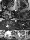
Post treatment imaging in patients with local advanced cervical carcinoma
- PMID: 36465360
- PMCID: PMC9710522
- DOI: 10.3389/fonc.2022.1003930
Post treatment imaging in patients with local advanced cervical carcinoma
Abstract
Cervical cancer (CC) is the fourth leading cause of death in women worldwide and despite the introduction of screening programs about 30% of patients presents advanced disease at diagnosis and 30-50% of them relapse in the first 5-years after treatment. According to FIGO staging system 2018, stage IB3-IVA are classified as locally advanced cervical cancer (LACC); its correct therapeutic choice remains still controversial and includes neoadjuvant chemo-radiotherapy, external beam radiotherapy, brachytherapy, hysterectomy or a combination of these modalities. In this review we focus on the most appropriated therapeutic options for LACC and imaging protocols used for its correct follow-up. We explore the imaging findings after radiotherapy and surgery and discuss the role of imaging in evaluating the response rate to treatment, selecting patients for salvage surgery and evaluating recurrence of disease. We also introduce and evaluate the advances of the emerging imaging techniques mainly represented by spectroscopy, PET-MRI, and radiomics which have improved diagnostic accuracy and are approaching to future direction.
Keywords: MRI; cervical cancer; gynecologic malignancies; gynecology; oncology.
Copyright © 2022 Ciulla, Celli, Aiello, Gigli, Ninkova, Miceli, Ercolani, Dolciami, Ricci, Palaia, Catalano and Manganaro.
Conflict of interest statement
The authors declare that the research was conducted in the absence of any commercial or financial relationships that could be construed as a potential conflict of interest.
Figures


Similar articles
-
Optimal treatment in locally advanced cervical cancer.Expert Rev Anticancer Ther. 2021 Jun;21(6):657-671. doi: 10.1080/14737140.2021.1879646. Epub 2021 Mar 11. Expert Rev Anticancer Ther. 2021. PMID: 33472018 Review.
-
MRI-based radiomics: promise for locally advanced cervical cancer treated with a tailored integrated therapeutic approach.Tumori. 2022 Aug;108(4):376-385. doi: 10.1177/03008916211014274. Epub 2021 Jul 8. Tumori. 2022. PMID: 34235995
-
Patterns of recurrence and prognosis in locally advanced FIGO stage IB2 to IIB cervical cancer: Retrospective multicentre study from the FRANCOGYN group.Eur J Surg Oncol. 2019 Apr;45(4):659-665. doi: 10.1016/j.ejso.2018.11.014. Epub 2018 Dec 30. Eur J Surg Oncol. 2019. PMID: 30685326
-
The management of locally advanced cervical cancer.Curr Opin Oncol. 2018 Sep;30(5):323-329. doi: 10.1097/CCO.0000000000000471. Curr Opin Oncol. 2018. PMID: 29994902 Review.
-
Five-fraction HDR brachytherapy in locally advanced cervical cancer: A monocentric experience.Cancer Radiother. 2021 Jul;25(5):463-468. doi: 10.1016/j.canrad.2021.03.006. Epub 2021 May 19. Cancer Radiother. 2021. PMID: 34023215
Cited by
-
Daily AI-Based Treatment Adaptation under Weekly Offline MR Guidance in Chemoradiotherapy for Cervical Cancer 1: The AIM-C1 Trial.J Clin Med. 2024 Feb 7;13(4):957. doi: 10.3390/jcm13040957. J Clin Med. 2024. PMID: 38398270 Free PMC article.
-
Dramatic Radiographic Response of Pelvis-Filling Locally Advanced Cervical Cancer Treated With Radiation and Chemotherapy.Cureus. 2024 Jun 2;16(6):e61544. doi: 10.7759/cureus.61544. eCollection 2024 Jun. Cureus. 2024. PMID: 38962615 Free PMC article.
-
An Update on the Role of MRI in Treatment Stratification of Patients with Cervical Cancer.Cancers (Basel). 2023 Oct 23;15(20):5105. doi: 10.3390/cancers15205105. Cancers (Basel). 2023. PMID: 37894476 Free PMC article. Review.
-
Comparing Methods to Determine Complete Response to Chemoradiation in Patients with Locally Advanced Cervical Cancer.Cancers (Basel). 2023 Dec 31;16(1):198. doi: 10.3390/cancers16010198. Cancers (Basel). 2023. PMID: 38201625 Free PMC article.
References
Publication types
LinkOut - more resources
Full Text Sources

