

Evolving imaging methods of prostate cancer and the emergence of magnetic resonance imaging guided ablation techniques
- PMID: 36465377
- PMCID: PMC9714456
- DOI: 10.3389/fonc.2022.1043688
Evolving imaging methods of prostate cancer and the emergence of magnetic resonance imaging guided ablation techniques
Abstract
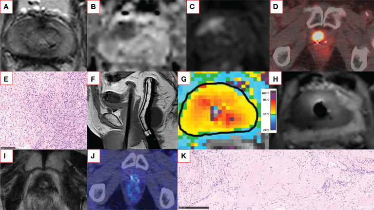
Established therapies for prostate cancer (PCa), surgery and radiotherapy, treat the entire gland regardless of the location of the cancerous lesion within the prostate. Although effective, these methods include a significant risk of worsening genitourinary outcomes. Targeted image-guided cancer therapy has gained acceptance through improved PCa detection, localization, and characterization by magnetic resonance imaging (MRI). Minimally-invasive ablative techniques aim to achieve comparable oncological outcomes to radical treatment while preserving genitourinary function. Transurethral ultrasound ablation (TULSA) and next-generation transrectal high-intensity focused ultrasound (HIFU) utilize MRI guidance to thermally ablate prostate tissue under real-time MRI monitoring and active temperature feedback control. Previous trials performed by our group and others, including a large multicenter study in men with localized favorable-risk disease, have demonstrated that TULSA provides effective prostate ablation with a favorable safety profile and low impact on quality of life. Recently, MRI-guided HIFU focal therapy was also shown as a safe and effective treatment of intermediate-risk PCa. Here we review the current literature on ablative techniques in the treatment of localized PCa with a focus on TULSA and HIFU methods.
Keywords: HIFU; MRI; Tulsa; ablation therapy; high-intensity focused ultrasound; magnetic resonance imaging; prostate cancer; transurethral ultrasound ablation.
Copyright © 2022 Anttinen, Blanco Sequeiros, Boström and Taimen.
Conflict of interest statement
MA reports grants from Profound Medical Inc, Finnish Urological Research Foundation, and Finnish Urological Association, and personal fees from Astellas, Bayer, Orion, and Janssen-Cilag, all outside the submitted work. PB reports personal fees from Profound Medical Inc and Janssen-Cilag Company outside the submitted work. PT reports personal fees from Roche, AstraZeneca, and MSD and non-financial support from MSD, all outside the submitted work. The remaining author declares that the research was conducted in the absence of any commercial or financial relationships that could be construed as a potential conflict of interest.
Figures

Similar articles
-
Magnetic resonance imaging-guided ultrasound ablation for prostate cancer - A contemporary review of performance.Front Oncol. 2023 Jan 4;12:1069518. doi: 10.3389/fonc.2022.1069518. eCollection 2022. Front Oncol. 2023. PMID: 36686753 Free PMC article. Review.
-
Magnetic Resonance Imaging-Guided Transurethral Ultrasound Ablation of Prostate Tissue in Patients with Localized Prostate Cancer: A Prospective Phase 1 Clinical Trial.Eur Urol. 2016 Sep;70(3):447-55. doi: 10.1016/j.eururo.2015.12.029. Epub 2016 Jan 6. Eur Urol. 2016. PMID: 26777228 Clinical Trial.
-
Magnetic Resonance Imaging-Guided Transurethral Ultrasound Ablation of Prostate Cancer: A Systematic Review.J Endourol. 2022 Jun;36(6):841-854. doi: 10.1089/end.2021.0866. Epub 2022 Mar 7. J Endourol. 2022. PMID: 35029127 Free PMC article.
-
Contrast enhanced ultrasound (CEUS) with MRI image fusion for monitoring focal therapy of prostate cancer with high intensity focused ultrasound (HIFU)1.Clin Hemorheol Microcirc. 2018;69(1-2):93-100. doi: 10.3233/CH-189123. Clin Hemorheol Microcirc. 2018. PMID: 29660918
-
New kids on the block: MRI guided transrectal focused US, TULSA, focal laser ablation, histotripsy - a comprehensive review.Prostate Cancer Prostatic Dis. 2025 Mar 27. doi: 10.1038/s41391-025-00956-x. Online ahead of print. Prostate Cancer Prostatic Dis. 2025. PMID: 40140552 Review.
Cited by
-
MR-Guided Transurethral Ultrasound Ablation (TULSA)-An Emerging Minimally Invasive Treatment Option for Localised Prostate Cancer.Cardiovasc Intervent Radiol. 2024 Jun;47(6):689-701. doi: 10.1007/s00270-024-03696-y. Epub 2024 Mar 15. Cardiovasc Intervent Radiol. 2024. PMID: 38491163 Review.
-
Evolution of non-perfused volume after transurethral ultrasound ablation of prostate: A retrospective 12-month analysis.Eur J Radiol Open. 2023 Jul 6;11:100506. doi: 10.1016/j.ejro.2023.100506. eCollection 2023 Dec. Eur J Radiol Open. 2023. PMID: 37456928 Free PMC article.
-
Salvage Magnetic Resonance Imaging-guided Transurethral Ultrasound Ablation for Localized Radiorecurrent Prostate Cancer.Eur Urol Open Sci. 2024 Dec 5;71:69-77. doi: 10.1016/j.euros.2024.11.001. eCollection 2025 Jan. Eur Urol Open Sci. 2024. PMID: 39703741 Free PMC article.
References
-
- Neal DE, Metcalfe C, Donovan JL, Lane JA, Davis M, Young GJ, et al. . Ten-year mortality, disease progression, and treatment-related side effects in men with localised prostate cancer from the ProtecT randomised controlled trial according to treatment received. Eur Urol (2020) 77(3):320–30. doi: 10.1016/j.eururo.2019.10.030 - DOI - PubMed
Publication types
LinkOut - more resources
Full Text Sources
Medical