

Slow flow induces endothelial dysfunction by regulating thioredoxin-interacting protein-mediated oxidative metabolism and vascular inflammation
- PMID: 36465470
- PMCID: PMC9708747
- DOI: 10.3389/fcvm.2022.1064375
Slow flow induces endothelial dysfunction by regulating thioredoxin-interacting protein-mediated oxidative metabolism and vascular inflammation
Abstract
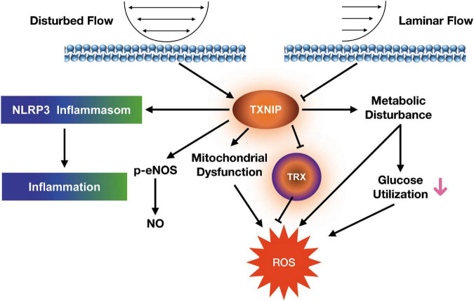
Endothelial cells are highly sensitive to hemodynamic shear stresses, which act in the blood flow's direction on the blood vessel's luminal surface. Thus, endothelial cells on that surface are exposed to various physiological and pathological stimuli, such as disturbed flow-induced shear stress, which may exert effects on adaptive vascular diameter or structural wall remodeling. Here we showed that plasma thioredoxin-interactive protein (TXNIP) and malondialdehyde levels were significantly increased in patients with slow coronary flow. In addition, human endothelial cells exposed to disturbed flow exhibited increased levels of TXNIP in vitro. On the other hand, deletion of human endothelial TXNIP increased capillary formation, nitric oxide production and mitochondrial function, as well as lessened oxidative stress response and endothelial cell inflammation. Additional beneficial impacts from TXNIP deletion were also seen in a glucose utilization study, as reflected by augmented glucose uptake, lactate secretion and extracellular acidification rate. Taken together, our results suggested that TXNIP is a key component involved in mediating shear stress-induced inflammation, energy homeostasis, and glucose utilization, and that TXNIP may serve as a potentially novel endothelial dysfunction regulator.
Keywords: disturbed flow; endothelial dysfunction; mitochondrial dysfunction; oxidative metabolism; thioredoxin-interacting protein (TXNIP).
Copyright © 2022 Wang, Liu, Liu, Sun, Chen, Liao, Zeng, Zhang, Dong, Xia and Yuan.
Conflict of interest statement
The authors declare that the research was conducted in the absence of any commercial or financial relationships that could be construed as a potential conflict of interest.
Figures


Similar articles
-
Thioredoxin-interacting protein is a biomechanical regulator of Src activity: key role in endothelial cell stress fiber formation.Circ Res. 2014 Mar 28;114(7):1125-32. doi: 10.1161/CIRCRESAHA.114.301315. Epub 2014 Feb 10. Circ Res. 2014. PMID: 24515523 Free PMC article.
-
Role of thioredoxin-interacting protein in mediating endothelial dysfunction in hypertension.Genes Dis. 2020 Aug 31;9(3):753-765. doi: 10.1016/j.gendis.2020.08.008. eCollection 2022 May. Genes Dis. 2020. PMID: 35782967 Free PMC article.
-
Fluid shear stress inhibits vascular inflammation by decreasing thioredoxin-interacting protein in endothelial cells.J Clin Invest. 2005 Mar;115(3):733-8. doi: 10.1172/JCI23001. J Clin Invest. 2005. PMID: 15696199 Free PMC article.
-
Thioredoxin-Interacting Protein (TXNIP) with Focus on Brain and Neurodegenerative Diseases.Int J Mol Sci. 2020 Dec 8;21(24):9357. doi: 10.3390/ijms21249357. Int J Mol Sci. 2020. PMID: 33302545 Free PMC article. Review.
-
Thioredoxin-Interacting Protein (TXNIP) in Cerebrovascular and Neurodegenerative Diseases: Regulation and Implication.Mol Neurobiol. 2018 Oct;55(10):7900-7920. doi: 10.1007/s12035-018-0917-z. Epub 2018 Feb 27. Mol Neurobiol. 2018. PMID: 29488135 Free PMC article. Review.
Cited by
-
Near-Wall Slow Flow Contributes to Wall Enhancement of Middle Cerebral Artery Bifurcation Aneurysms on Vessel Wall MRI.Diagnostics (Basel). 2024 Dec 3;14(23):2722. doi: 10.3390/diagnostics14232722. Diagnostics (Basel). 2024. PMID: 39682630 Free PMC article.
-
Glutamine metabolism is systemically different between primary and induced pluripotent stem cell-derived brain microvascular endothelial cells.J Cereb Blood Flow Metab. 2025 Jun;45(6):1082-1099. doi: 10.1177/0271678X241310729. Epub 2025 Jan 7. J Cereb Blood Flow Metab. 2025. PMID: 39763385 Free PMC article.
-
Inhibition of the Foxo3/Txnip Axis Alleviates Ventilator-Induced Diaphragmatic Dysfunction by Downregulating MuRF1.Appl Biochem Biotechnol. 2025 Aug;197(8):4949-4968. doi: 10.1007/s12010-025-05261-w. Epub 2025 May 16. Appl Biochem Biotechnol. 2025. PMID: 40377847
References
-
- Griffith TM. Endothelial control of vascular tone by nitric oxide and gap junctions: a haemodynamic perspective. Biorheology. (2002) 39:307–18. - PubMed
LinkOut - more resources
Full Text Sources