. 2023 Jan 20;26(1):105696.
doi: 10.1016/j.isci.2022.105696. Epub 2022 Nov 30.

Conformational stability of SARS-CoV-2 glycoprotein spike variants

Affiliations

Conformational stability of SARS-CoV-2 glycoprotein spike variants

Hiam R S Arruda et al. iScience. .

Abstract

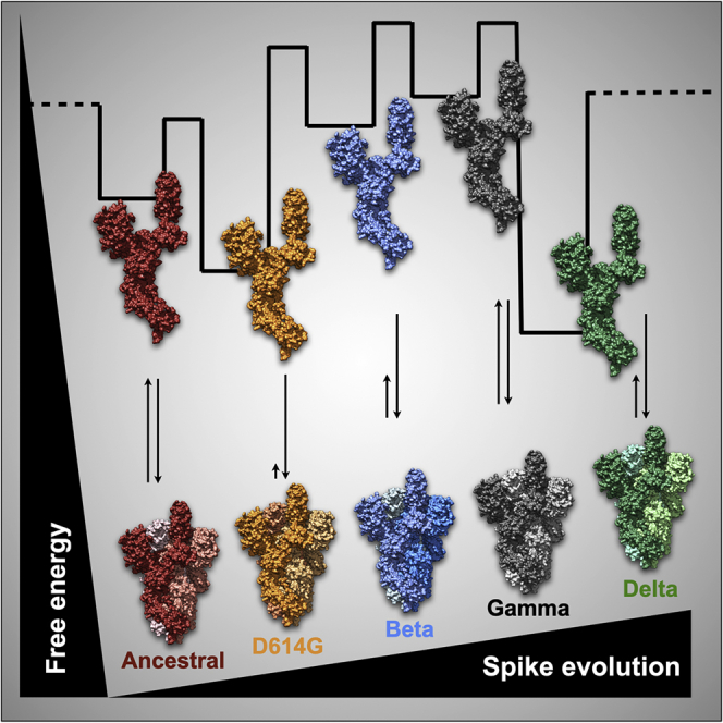
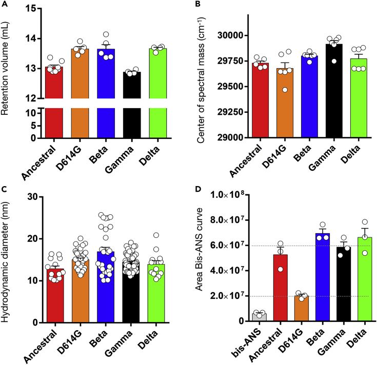
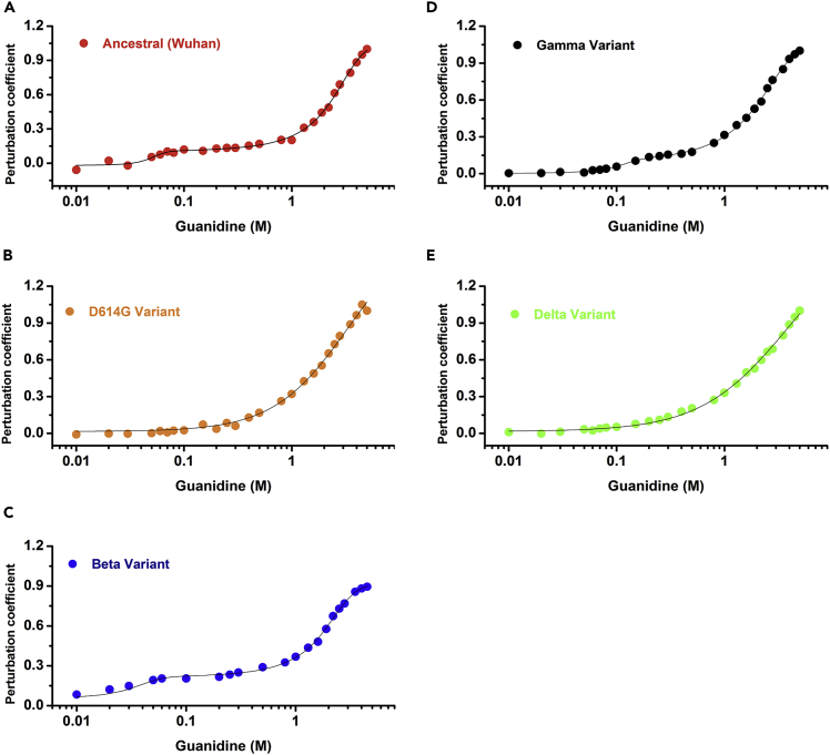
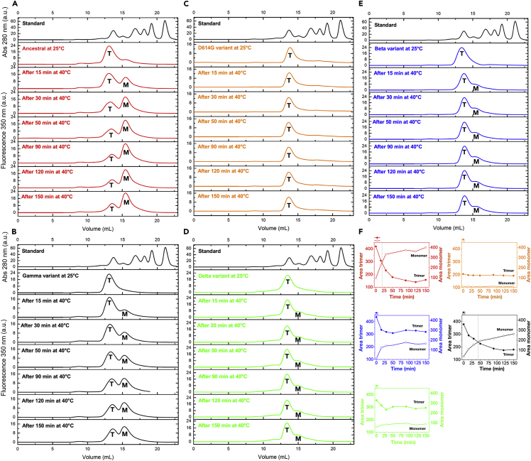
The severe acute respiratory syndrome spread worldwide, causing a pandemic. SARS-CoV-2 mutations have arisen in the spike, a glycoprotein at the viral envelope and an antigenic candidate for vaccines against COVID-19. Here, we present comparative data of the glycosylated full-length ancestral and D614G spike together with three other transmissible strains classified by the World Health Organization as variants of concern: beta, gamma, and delta. By showing that D614G has less hydrophobic surface exposure and trimer persistence, we place D614G with features that support a model of temporary fitness advantage for virus spillover. Furthermore, during the SARS-CoV-2 adaptation, the spike accumulates alterations leading to less structural stability for some variants. The decreased trimer stability of the ancestral and gamma and the presence of D614G uncoupled conformations mean higher ACE-2 affinities compared to the beta and delta strains. Mapping the energetics and flexibility of variants is necessary to improve vaccine development.

Keywords: Biochemistry; Protein structure aspects; Structural biology; Virology.


Conflict of interest statement

The authors declare no competing interests.

Figures

None
Graphical abstract
Figure 1
Figure 1
Spike glycoprotein and the variants of concern (A) Atomic model (PDB: 6VSB) showing the ribbon representation of the spike protomer obtained from cryo-EM maps. Schematics is color-coded for the N-terminal domain (NTD, blue); receptor-binding domain (RBD, green); sub-domain 1 and 2 (SD1/SD2, tan); fusion peptide (FP, turquoise); central helix (CH, orange); heptad repeat (HR1, gold yellow); S2 subunit (dark red); connector domain (CD, purple). The NTD and RBD form the S1 subunit. (B) Atomic model highlighting two spike protomers and the D614 interprotomer interaction. Zoomed images show D614-T859 H-bond interaction (PDB: 6VSB) at 2.7 Å distance and the D614-K854 salt bridge at 3.6 Å distance (PDB: 6ZGE). (C–E) Atomic model (PDB: 6VSB) showing altered sites for (C) beta variant in blue, (D) gamma variant in black, (E) delta variant in green. Altered sites are shown as spheres. Labels in red refer to residues not resolved on the cryo-EM map.
Figure 2
Figure 2
Spike protein glycosylation profile (A and B) Images of SDS-PAGE gels showing (A) two representative batches (#1 and #2) of purified spikes and (B) spikes in the absence (−) or in the presence (+) of PNGase F. Positions of the glycosylated and deglycosylated (“null”) spikes are shown in the gel. (C–F) Line plots showing the relative fluorescence as a function of time from hydrophilic chromatography (HILIC) of (C) ancestral vs. D614G, (D) ancestral vs. beta, (E) ancestral vs. gamma, and (F) ancestral vs. delta. Results are flipped to facilitate visualization. Black asterisks show abundant glycan peaks. Thick horizontal bars show major differences relative to the ancestral spike. See also Figures S1, S2, and S3.
Figure 3
Figure 3
Structural features of SARS-CoV-2 spikes (A–D) Scatterplots showing (A) the retention volumes of studied spikes obtained from analytical size-exclusion chromatography. Data show the avg. ± SEM of independent protein batch preparations (n = number of data points for each series); (B) the center of spectral mass of studied spikes obtained from the emission fluorescence spectrum upon excitation at 280 nm. Data show the avg. ± SEM of at least three independent protein batches; (C) the hydrodynamic diameter of studied spikes determined by dynamic light scattering. Data are the avg. ± SD of several technical replicates from the same protein batch; (D) Area under the Bis-ANS curve of studied spikes obtained from the emission fluorescence spectrum upon excitation at 360 nm. Data show the avg. ± SEM of independent protein batches (n = 3). The gray bar shows bis-ANS fluorescence noise in the presence of buffer. See also Figures S5, S6, and S7.
Figure 4
Figure 4
Structural stability of the ancestral spike (A) Dot plots showing the perturbation coefficient as a function of guanidine increments for the ancestral spike in the presence of tris-based buffer (tagged protein, see STAR methods for details). Abbreviations on panel A stand for FT, folded trimer; HL, hinge-like; FM, folded monomer; UM, unfolded monomer. Data are represented as avg. ± SEM of (n = 3, independent protein batches). Check Figure S8Bb. (B) Line plots showing SEC runs for the ancestral spike in the presence of the tris-based buffering (thick red line) and in the presence of 0.3 M (dashed red line) and 1 M of guanidine (thin red line). Insert shows the y-scaled chromatogram at 1 M of guanidine to visualize the peaks corresponding to the trimer (T) and the monomer (M). Standard is colored black. (C) DLS number-weighted distribution as a function of hydrodynamic diameter in the absence (ancestral) and presence of 0.3 and 1 M of guanidine. (D) Schematics showing spike top views to illustrate the interpretation of the data. At 0.3 M of guanidine, the data supports RBD hinge-like motions. At this stage of knowledge, we cannot rule out whether all three RBDs are facing up. At 1 M of guanidine, the data indicate trimer-to-monomer dissociation. At concentrations higher than 3.5 M guanidine, monomer unfolding is achieved. See also Figure S8.
Figure 5
Figure 5
Structural stability of spike variants Dot plots showing the perturbation coefficient as a function of guanidine increments for the (A) ancestral, (B) D614G, (C) beta, (D) gamma, and (E) delta variants. Curves were carried out in phosphate buffer and tagless proteins (see STAR methods for details).
Figure 6
Figure 6
Trimer-to-monomer spike dissociation (A–E) Collection of line plots showing the fluorescence signal at 350 nm as a function of retention volume for (A) the ancestral strain in red, (B) gamma in black, (C) D614G in orange, (D) delta in green, and (E) beta in blue at 25°C and after several times at 40°C. T and M stand for trimer and monomer, respectively. The absorbance at 280 nm of the calibration standard is shown at the top of each experiment. (F) Double-Y plots show the integration area corresponding to the trimers (filled dots) and the monomers (empty dots). The color code is the same as for panels (A–E). Crossover points (T50%) are highlighted by vertical dashed lines for the ancestral strain (red) and gamma variant (black).

References

    1. Mittal A., Manjunath K., Ranjan R.K., Kaushik S., Kumar S., Verma V. COVID-19 pandemic: insights into structure, function, and hACE2 receptor recognition by SARS-CoV-2. PLoS Pathog. 2020;16 doi: 10.1371/journal.ppat.1008762. - DOI - PMC - PubMed
    1. Huang Y., Yang C., Xu X.F., Xu W., Liu S.W. Structural and functional properties of SARS-CoV-2 spike protein: potential antivirus drug development for COVID-19. Acta Pharmacol. Sin. 2020;41:1141–1149. doi: 10.1038/s41401-020-0485-4. - DOI - PMC - PubMed
    1. Heikkinen T., Järvinen A. The common cold. Lancet. 2003;361:51–59. doi: 10.1016/S0140-6736(03)12162-9. - DOI - PMC - PubMed
    1. Zhu N., Zhang D., Wang W., Li X., Yang B., Song J., Zhao X., Huang B., Shi W., Lu R., et al. A novel coronavirus from patients with pneumonia in China, 2019. N. Engl. J. Med. 2020;382:727–733. doi: 10.1056/NEJMoa2001017. - DOI - PMC - PubMed
    1. Ou X., Liu Y., Lei X., Li P., Mi D., Ren L., Guo L., Guo R., Chen T., Hu J., et al. Characterization of spike glycoprotein of SARS-CoV-2 on virus entry and its immune cross-reactivity with SARS-CoV. Nat. Commun. 2020;11:1620. doi: 10.1038/s41467-020-15562-9. - DOI - PMC - PubMed

LinkOut - more resources