Review
. 2022 Nov 25:28:e20220053.
doi: 10.1590/1678-9199-JVATITD-2022-0053. eCollection 2022.

A scoping study of pulmonary paracoccidioidomycosis: severity classification based on radiographic and tomographic evaluation

Affiliations
Review

A scoping study of pulmonary paracoccidioidomycosis: severity classification based on radiographic and tomographic evaluation

Sergio Marrone Ribeiro et al. J Venom Anim Toxins Incl Trop Dis. .

Abstract

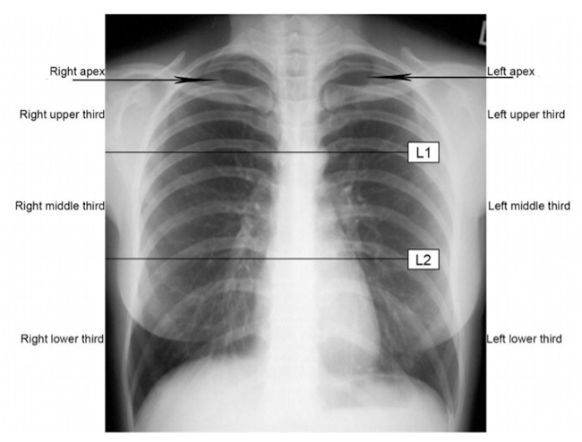
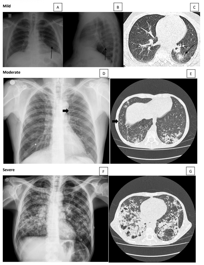
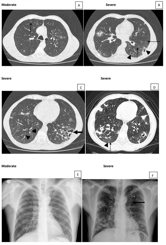
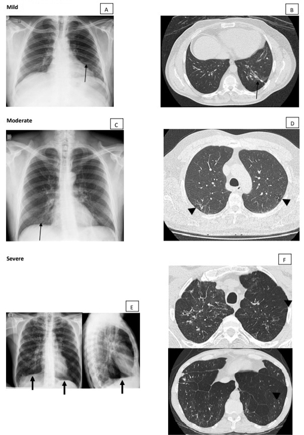
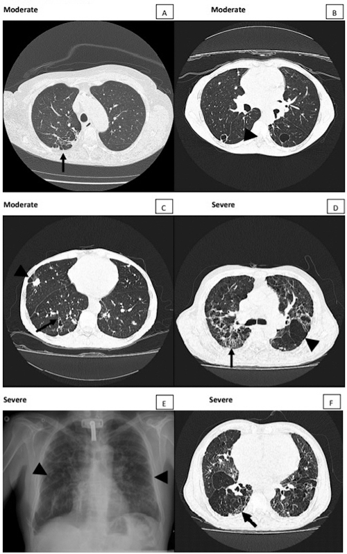
The lungs have great importance in patients with paracoccidioidomycosis since they are the portal of entry for the infecting fungi, the site of quiescent foci, and one of the most frequently affected organs. Although they have been the subject of many studies with different approaches, the severity classification of the pulmonary involvement, using imaging procedures, has not been carried out yet. This study aimed to classify the active and the residual pulmonary damage using radiographic and tomographic evaluations, according to the area involved and types of lesions.

Keywords: Paracoccidioides sp; Paracoccidioidomycosis; Radiographic evaluation; Severity classification.


Conflict of interest statement

Competing interests: The authors declare that they have no competing interests.

Figures

Figure 1.
Figure 1.. Areas of the chest radiograph.
Figure 2.
Figure 2.. (A, B, C) Mild active lesion. Chest radiograph and computed tomography (CT) scan of the same patient showing a solitary pulmonary nodule. Moderate active lesion: (D) chest radiograph showing intersticial lesions with bronchial wall thickening (arrow) and (E) CT scan showing consolidation, ground-glass opacities, and intersticial lesions with tree-in-bud pattern (arrow). (F, G) Severe active lesion. Mixed lesions involving > 1/3 of the total pulmonary parenchyma with cavitary lesions.
Figure 3.
Figure 3.. Moderate and severe active lesions. (A) Moderate lesion showing > 3 ground-glass nodules (arrow). (B) Besides ground-glass nodules (arrows), cavitary lesions with and without septations (arrowheads) in greater amount, characterizing severe classification. (C) Bronchiectasis inside consolidation opacity (arrow) and bronchial wall thickening (arrowheads). (D) Focal lesions, ground-glass and intersticial opacities with interlobular septal thickening (arrowhead) and small centrilobular nodules (arrow). (E) Chest radiograph showing only interstitial opacities, a finding in which CT helps to better characterize and classify the lesions. (F) Bronchial wall thickening (tram-track sign) better observed in this patient with severe lesion (arrow).
Figure 4.
Figure 4.. (A, B) Mild residual lesion. Chest radiograph and CT scan of the same patient showing parenchymal band associated with traction bronchiectasis on CT (arrows). Moderate residual lesion: (C) chest radiograph showing intersticial lesions in subhilar regions (arrow) and (D) CT scan showing focal paracicatricial emphysema adjacent to irregular septal thickening (arrowhead). Severe residual lesion: (E) chest radiograph showing interstitial fibrotic lesions and signs of pulmonary hyperinflation, such as low set and flattened diaphragm (arrow). (F) These emphysema signs are better characterized by CT scan (arrowheads).
Figure 5.
Figure 5.. Moderate and severe residual lesions. (A) Fibrotic lesions with bronchial wall thickening, traction bronchiectasis, paracicatricial emphysema, and pleural adherences simulating tuberculosis sequalae. (B) Moderate residual lesion showing thin wall cavity-gas-filled spaces. (C) Paracicatricial emphysema beside nodule (arrowhead) and bronchial wall thickening (arrow) in a moderate residual lesion. Severe residual lesions showing fibrotic lesions with honeycomb lung (arrows in D and F) and emphysema signs (arrowheads in D and E).

Similar articles

Cited by

References

    1. Mendes RP. Paracoccidioidomycosis surveillance and control. J Venom Anim Toxins incl Trop Dis. 2010;16(2):194–197. doi: 10.1590/S1678-91992010000200003. - DOI
    1. Mendes RP, Cavalcante RS, Marques SA, Marques MEA, Venturini J, Sylvestre TF, Paniago AMM, Pereira AC, Silva JF, Fabro AT, Bosco SMG, Bagagli E, Hahn RC, Levorato AD. Paracoccidioidomycosis: current perspectives from Brazil. Open Microbiol J. 2017;11:224–282. - PMC - PubMed
    1. Londero AT, Ramos CD. Paracoccidioidomycosis: a clinical and mycologic study of forty-one cases observed in Santa Maria, RS, Brazil. Am J Med. 1972;52(6):771–775. - PubMed
    1. Londero AT, Ramos CD, Lopes JOS. Progressive pulmonary paracoccidioidomycosis a study of 34 cases observed in Rio Grande do Sul (Brazil) Mycopathologia. 1978;63(1):53–56. doi: 10.1007/BF00473160. - DOI - PubMed
    1. Londero AT, Severo LC. The gamut of progressive pulmonary paracoccidioidomycosis. Mycopathologia. 1981;75(2):65–74. doi: 10.1007/BF00505780. - DOI - PubMed

LinkOut - more resources