Inter and intra-observer agreement of arterial wall contrast-enhanced ultrasonography in giant cell arteritis
- PMID: 36465936
- PMCID: PMC9715579
- DOI: 10.3389/fmed.2022.1042366
Inter and intra-observer agreement of arterial wall contrast-enhanced ultrasonography in giant cell arteritis
Abstract
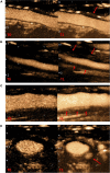
Objective: The aim of this study was to analyze inter- and intra-observer agreement for contrast-enhanced ultrasonography (CEUS) for monitoring disease activity in Giant Cell Arteritis (GCA) in the wall of axillary arteries, and common carotid arteries.
Methods: Giant cell arteritis patients have CEUS of axillary arteries and common carotid. These images were rated by seven vascular medicine physicians from four hospitals who were experienced in duplex ultrasonography of GCA patients. Two weeks later, observers again rated the same images. GCA patients were recruited in from December 2019 to February 2021. An analysis of the contrast of the ultrasound images with a gradation in three classes (grade 0, 1, and 2) was performed. Grade 0 corresponds to no contrast, grade 1 to moderate wall contrast and grade 2 to intense contrast. A new analysis in 2 classes: positive or negative wall contrast; was then performed on new series of images.
Results: Sixty arterial segments were evaluated in 30 patients. For the three-class scale, intra-rater agreement was substantial: κ 0.70; inter-rater agreement was fair: κ from 0.22 to 0.27. Thirty-four videos had a wall thickness of less than 2 mm and 26 videos had a wall thickness greater than 2 mm. For walls with a thickness lower than 2 mm: intra-rater agreement was substantial: κ 0.69; inter-rater agreement was fair: κ 0.35. For walls with a thickness of 2 mm or more: intra-rater agreement was substantial: κ 0.53; inter-rater agreement was fair: κ 0.25. For analysis of parietal contrast uptake in two classes: inter-rater agreement was fair to moderate: κ from 0.35 to 0.41; and for walls with a thickness of 2 mm or more: inter-rater agreement was fair to substantial κ from 0.22 to 0.63.
Conclusion: The visual analysis of contrast uptake in the wall of the axillary and common carotid arteries showed good intra-rater agreement in GCA patients. The inter-rater agreement was low, especially when contrast was analyzed in three classes. The inter-rater agreement for the analysis in two classes was also low. The inter-rater agreement was higher in two-class analysis for walls of 2 mm thickness or more.
Keywords: agreement; contrast-enhanced ultrasonography (CEUS); giant cell arteritis; giant cell arteritis–large-vessel; large-vessel vasculitis (LVV).
Copyright © 2022 Espitia, Robin, Hersant, Roncato, Théry, Vibet, Gautier, Raimbeau and Lapébie.
Conflict of interest statement
The authors declare that the research was conducted in the absence of any commercial or financial relationships that could be construed as a potential conflict of interest.
Figures

References
-
- Prieto-González S, Arguis P, García-Martínez A, Espígol-Frigolé G, Tavera-Bahillo I, Butjosa M, et al. Large vessel involvement in biopsy-proven giant cell arteritis: prospective study in 40 newly diagnosed patients using CT angiography. Ann Rheum Dis. (2012) 71:1170–6. 10.1136/annrheumdis-2011-200865 - DOI - PubMed
-
- Blockmans D. The use of (18F) fluoro-deoxyglucose positron emission tomography in the assessment of large vessel vasculitis. Clin Exp Rheumatol. (2003) 21:S15–22. - PubMed
LinkOut - more resources
Full Text Sources

