

Vibration-controlled Transient Elastography in NAFLD: Review Study
- PMID: 36466098
- PMCID: PMC9681576
- DOI: 10.5005/jp-journals-10018-1365
Vibration-controlled Transient Elastography in NAFLD: Review Study
Abstract
Aim: In this study, we aimed to provide information about transient elastography, a noninvasive method that shows liver steatosis and fibrosis, and to review diagnostic accuracy studies in the literature.
Background: Nonalcoholic fatty liver disease (NAFLD) is the most common cause of chronic liver diseases. It has a wide clinical spectrum, ranging from asymptomatic steatosis to cirrhosis with complications that can lead to mortality. Although its frequency varies geographically, it is believed that one out of every four people in the world has NAFLD. Recently, the number of studies about the noninvasive diagnosis of NAFLD and liver fibrosis is increasing. Vibration-controlled transient elastography (VCTE) is a method used for about two decades and provides important information in determining steatosis and fibrosis in the liver.
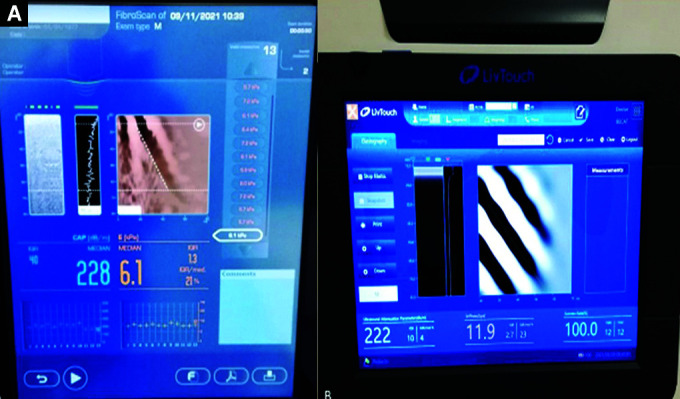
Review results: Area under curve (AUC) levels for ≥S1 are between 0.8 and 0.95 in studies showing the accuracy of the CAP score in detecting steatosis. Sensitivity is between 68 and 87% and specificity is 74 and 91%. AUC levels for steatosis ≥S2 range from 0.73 to 0.88. Sensitivity is between 77 and 85% and specificity is 59 and 81%. For detecting ≥S3, AUC levels were 0.69 to 0.94 and the sensitivity and specificity were 71 to 88%, and 58 to 89%, respectively. In studies, evaluating the effectiveness of elastography in determining the level of fibrosis in patients with NAFLD: AUC was between 0.79 and 0.87, sensitivity was 62 and 94%, and specificity was 61 and 100% for F ≥2. Area under curve was 0.76 to 0.98, sensitivity was 65 to 100% and specificity was 75 to 97% for ≥F3. Area under curve was ranged from 0.91 to 0.99 and sensitivity was 78 to 100% and specificity was 76 to 98% for ≥F4. The studies about the comparison of FibroScan and novel transient elastography device (FibroTouch) reported that results are correlated (r = 0.5-0.6) and the AUC of FibroTouch to detect fibrosis is nearly 0.8.
Conclusion: AUROC in studies are mostly above 0.80 in detecting steatosis and detecting the presence of fibrosis in patients diagnosed with NAFLD indicates the reliability of the data obtained. Transient elastography is suggested by the international guidelines for diagnosing NAFLD, especially the decision of biopsy. FibroTouch was found correlated with FibroScan but further studies are necessary to indicate that FibroTouch can be used instead of FibroScan.
How to cite this article: Ozercan AM, Ozkan H. Vibration-controlled Transient Elastography in NAFLD: Review Study. Euroasian J Hepato-Gastroenterol 2022;12(Suppl 1):S41-S45.
Keywords: Fibrosis; Nonalcoholic fatty liver disease; Steatosis; Transient elastography.
Copyright © 2022; Jaypee Brothers Medical Publishers (P) Ltd.
Conflict of interest statement
Source of support: Nil Conflict of interest: None
Figures
References
Publication types
LinkOut - more resources
Full Text Sources
Miscellaneous