. 2022;17(4):263-280.
doi: 10.1080/17469899.2022.2100764. Epub 2022 Sep 6.

Hyalocytes in proliferative vitreo-retinal diseases

Affiliations

Hyalocytes in proliferative vitreo-retinal diseases

Charlotte H Jones et al. Expert Rev Ophthalmol. 2022.

Abstract

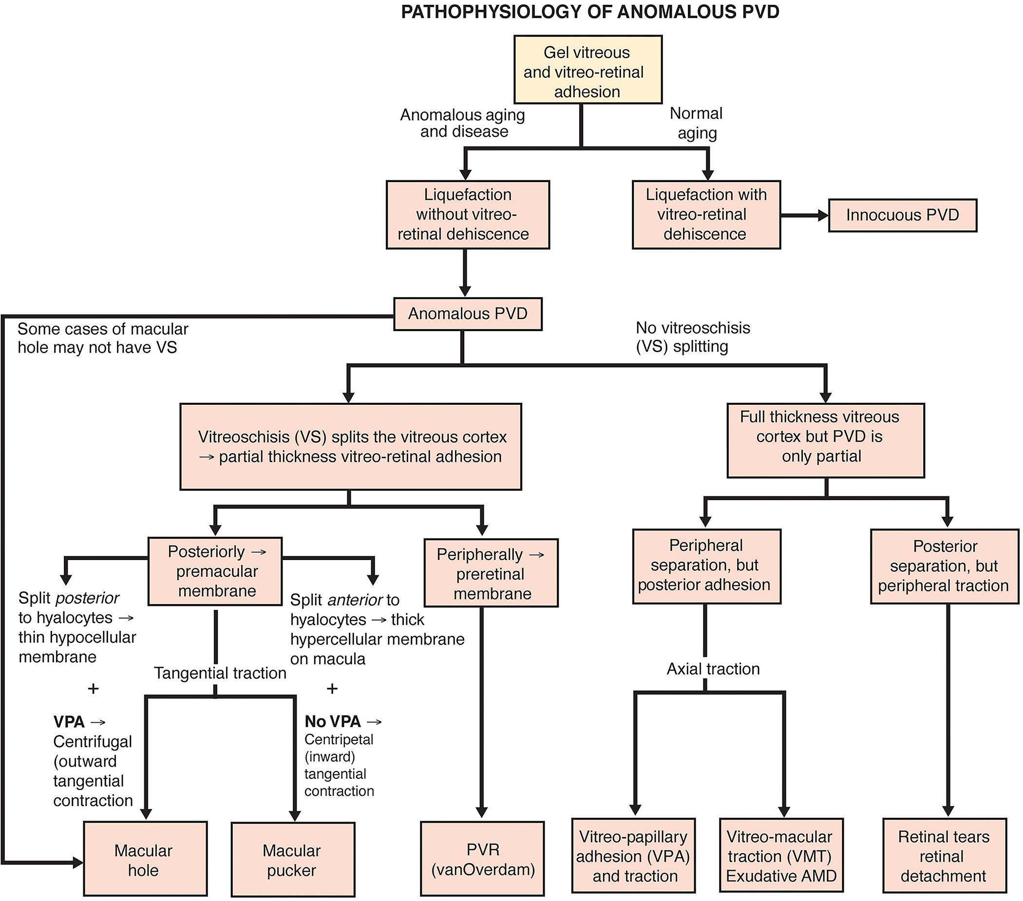
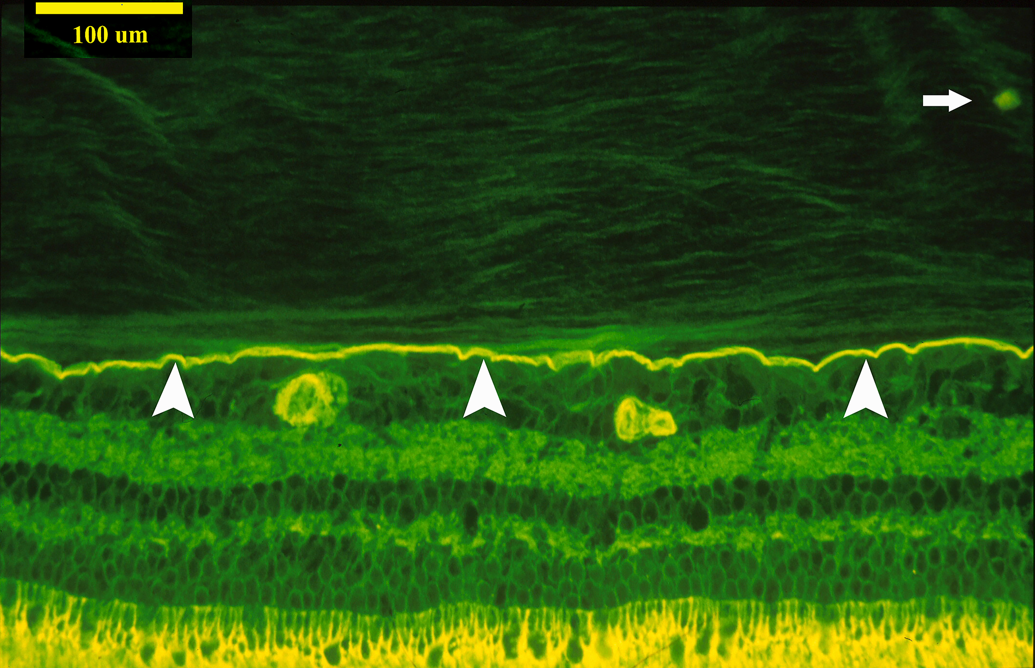
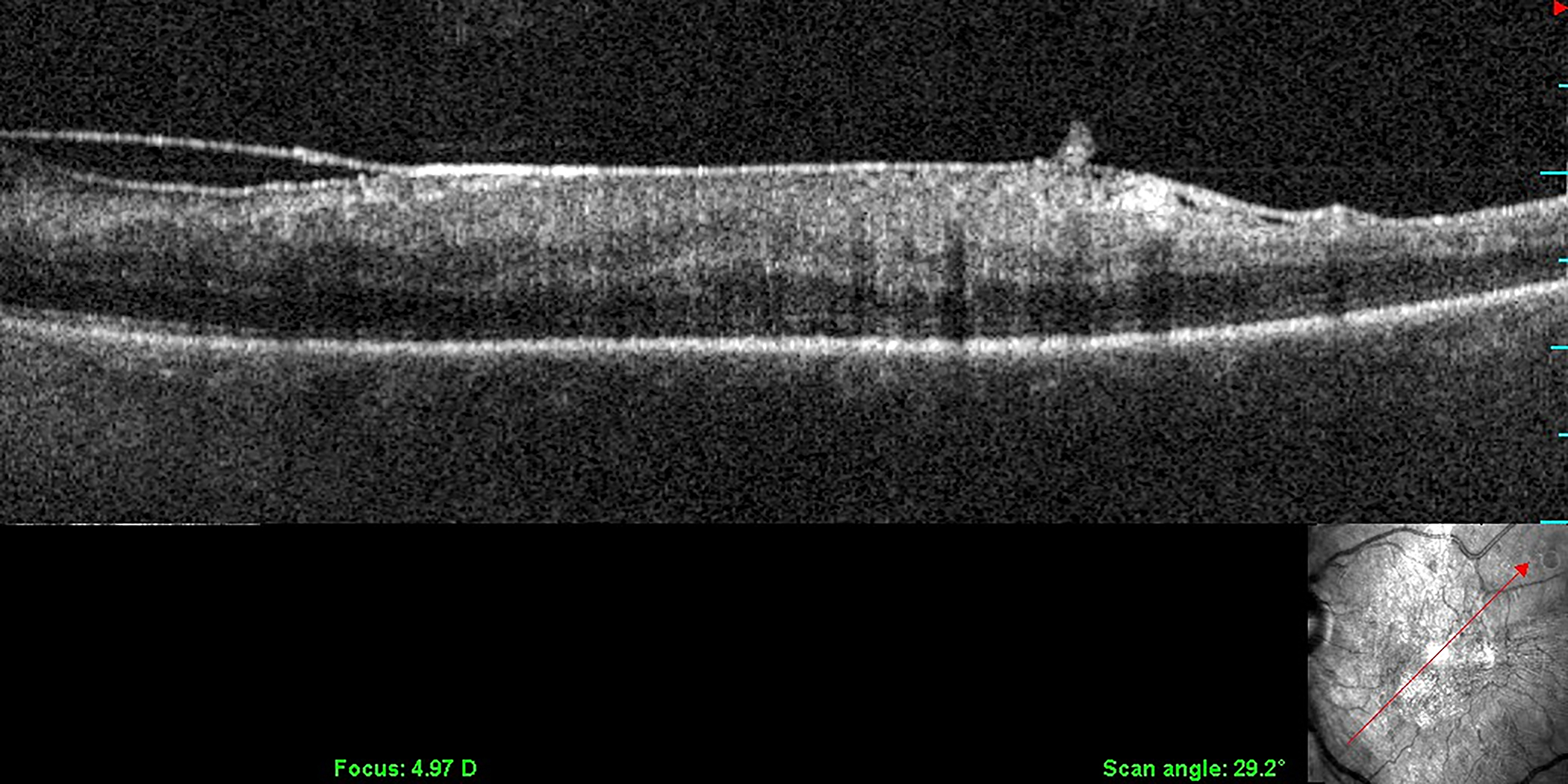
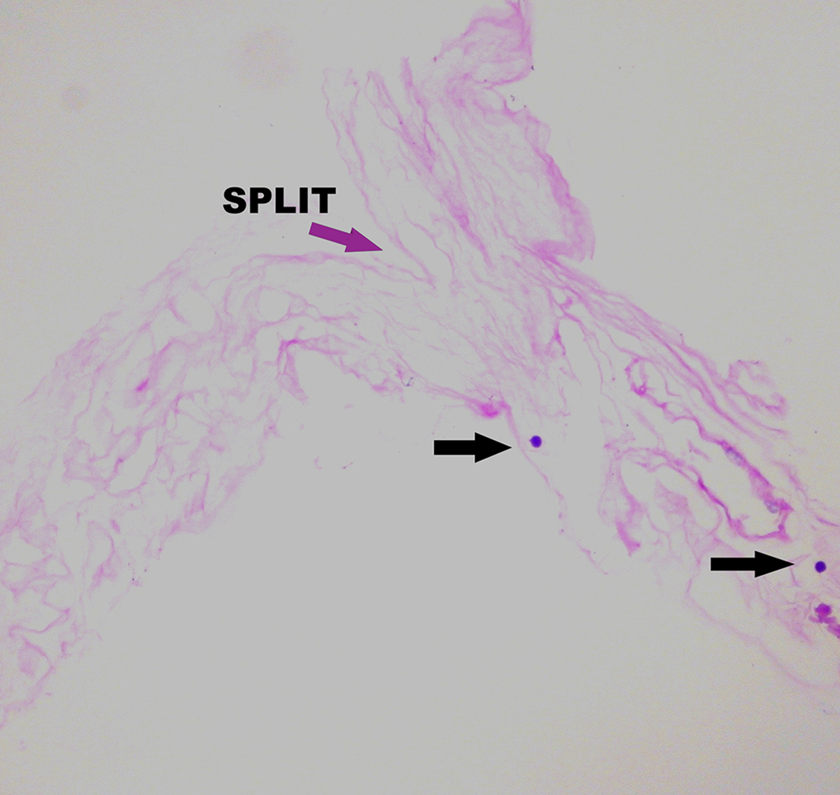
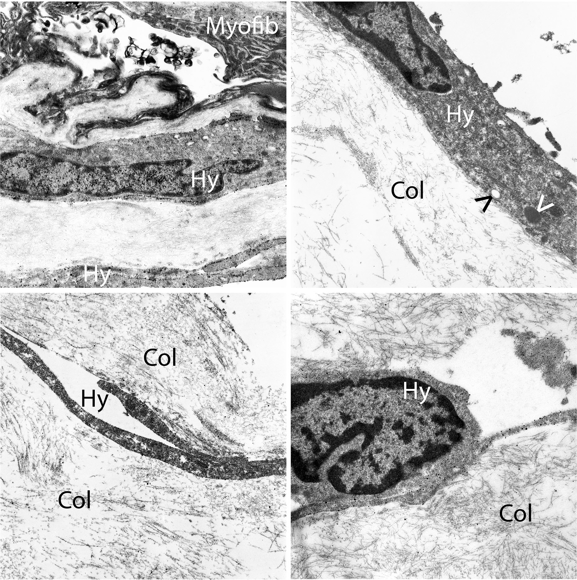
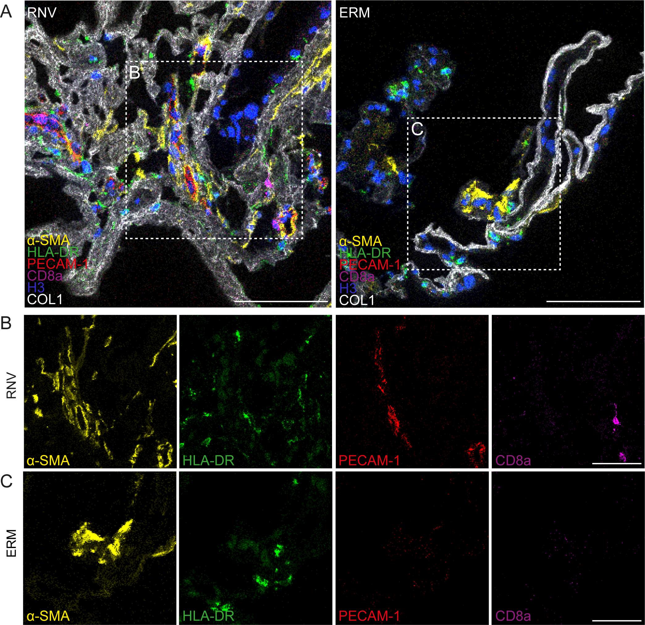
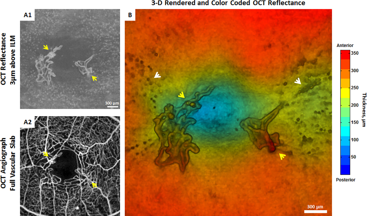
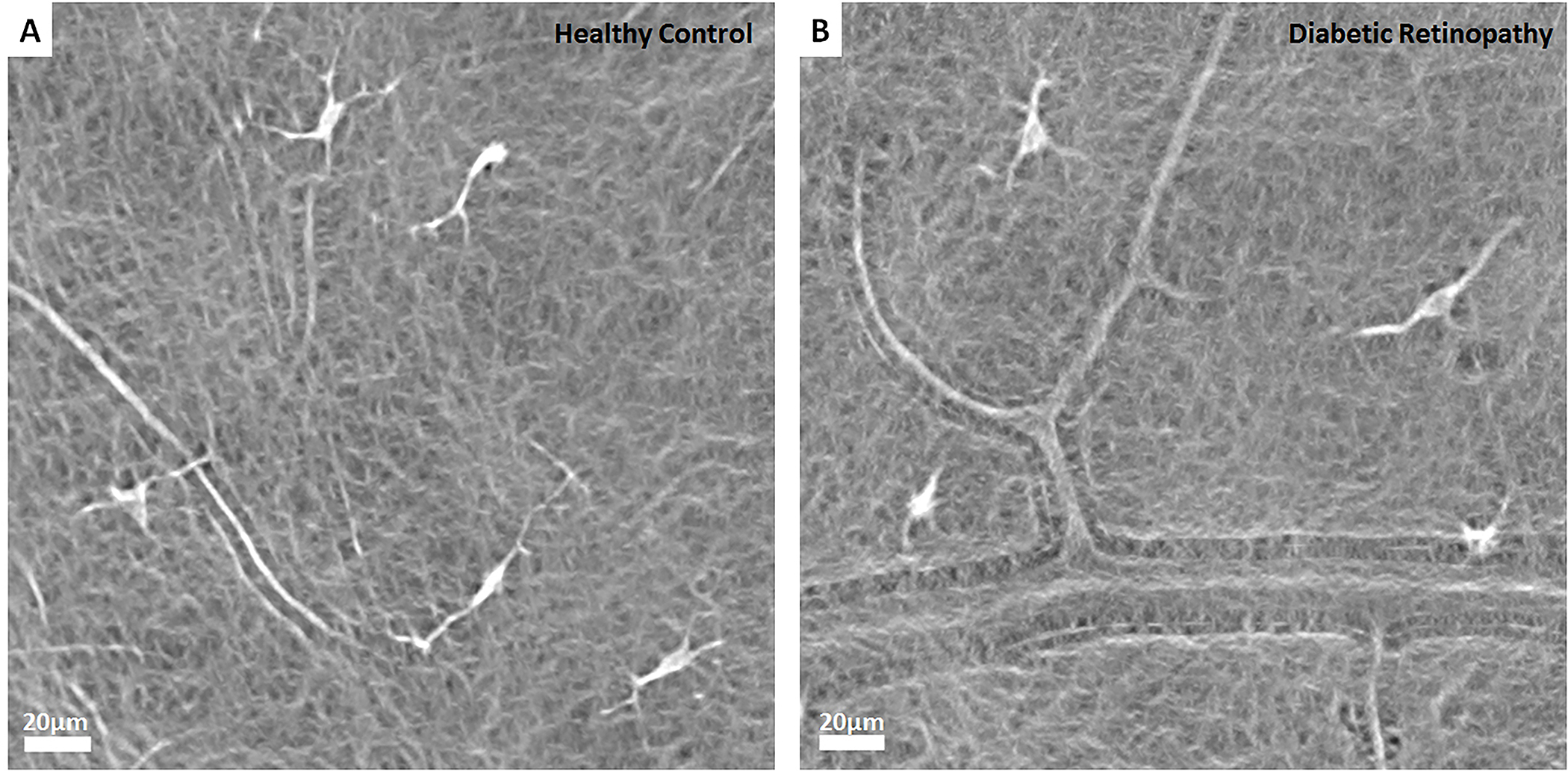
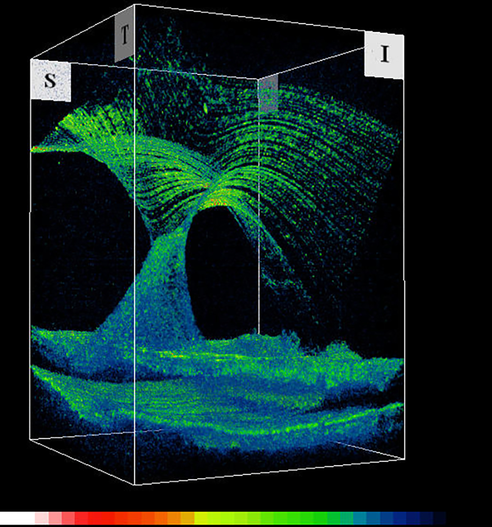
Introduction: Hyalocytes are sentinel macrophages residing within the posterior vitreous cortex anterior to the retinal inner limiting membrane (ILM). Following anomalous PVD and vitreoschisis, hyalocytes contribute to paucicellular (vitreo-macular traction syndrome, macular holes) and hypercellular (macular pucker, proliferative vitreo-retinopathy, proliferative diabetic vitreo-retinopathy) diseases.

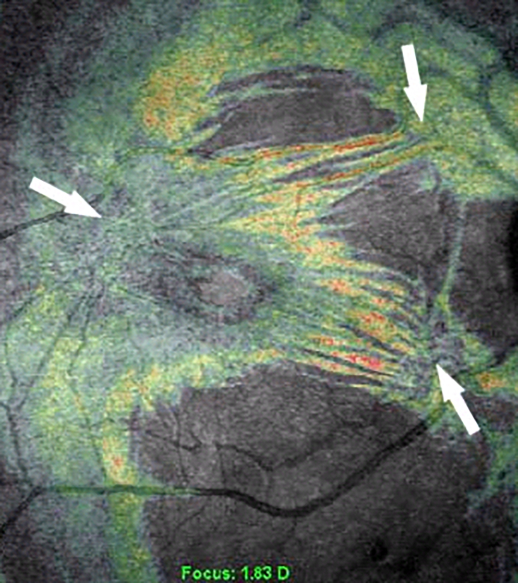
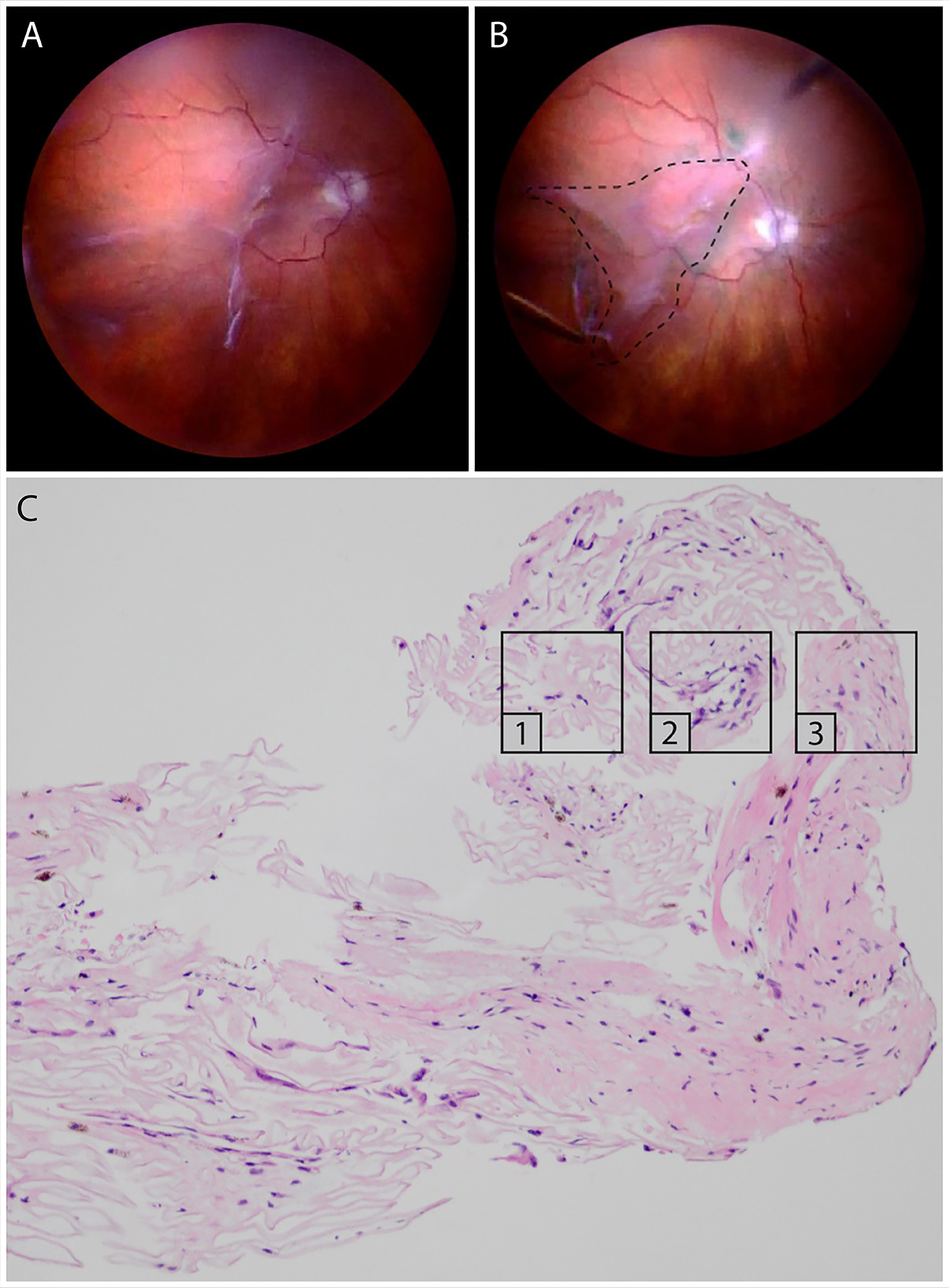
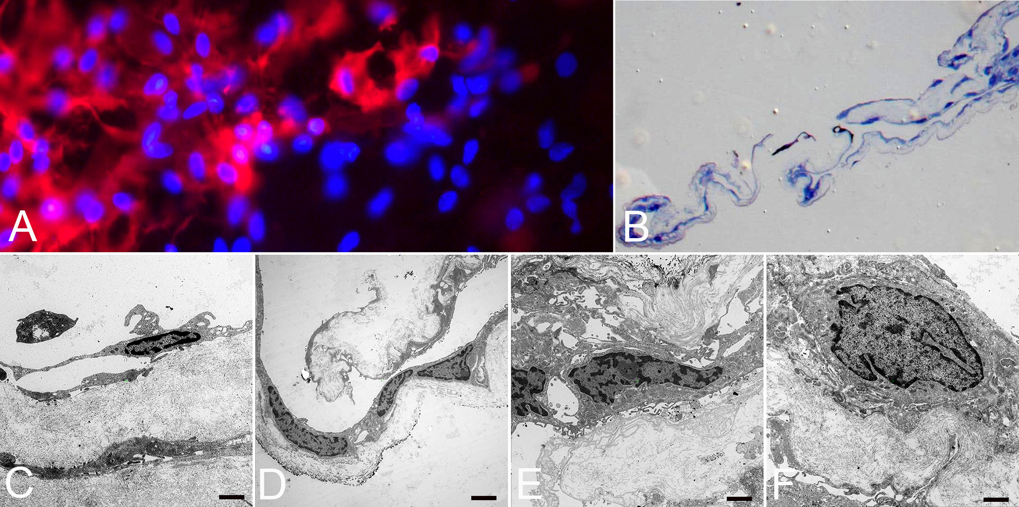
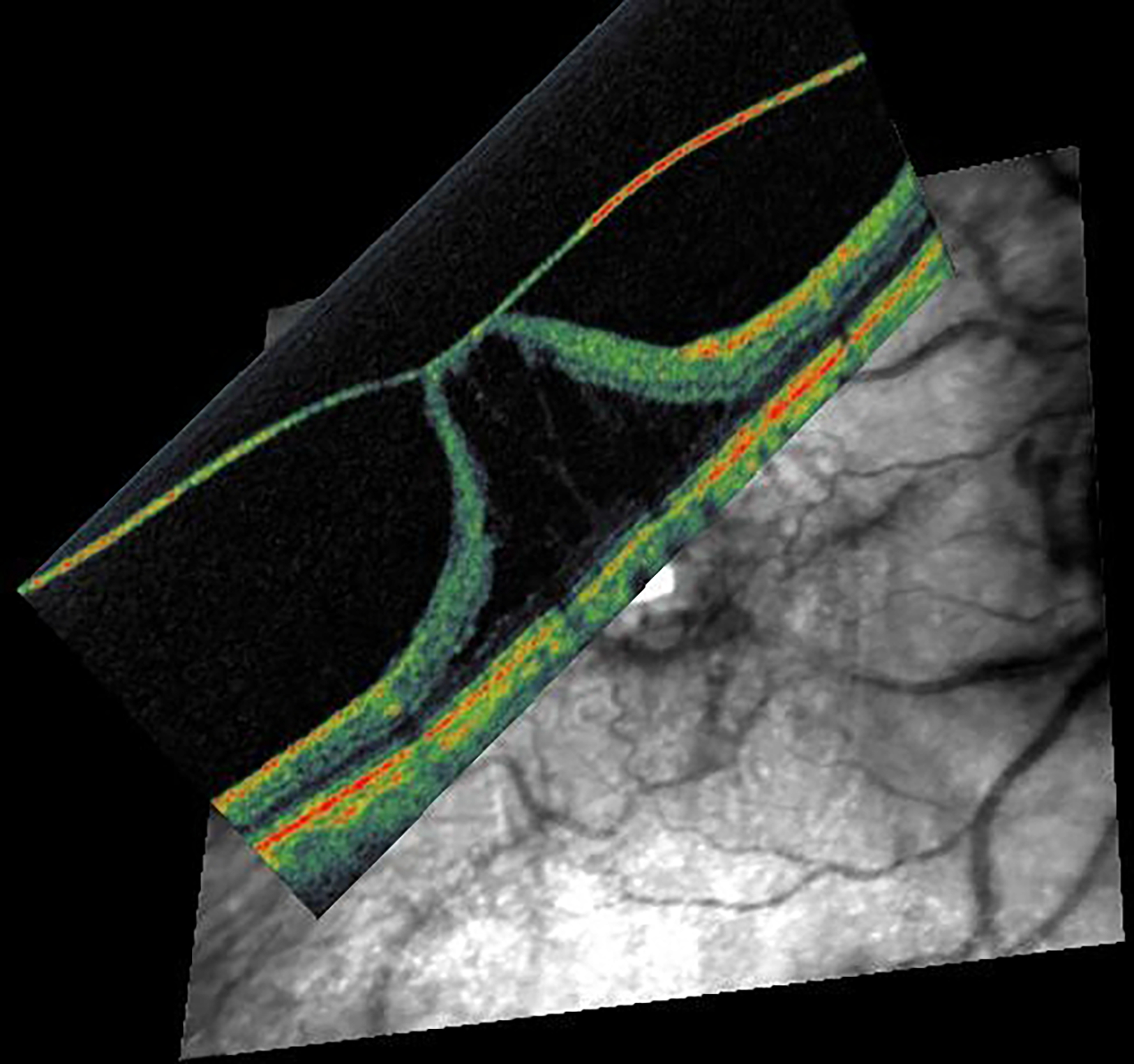
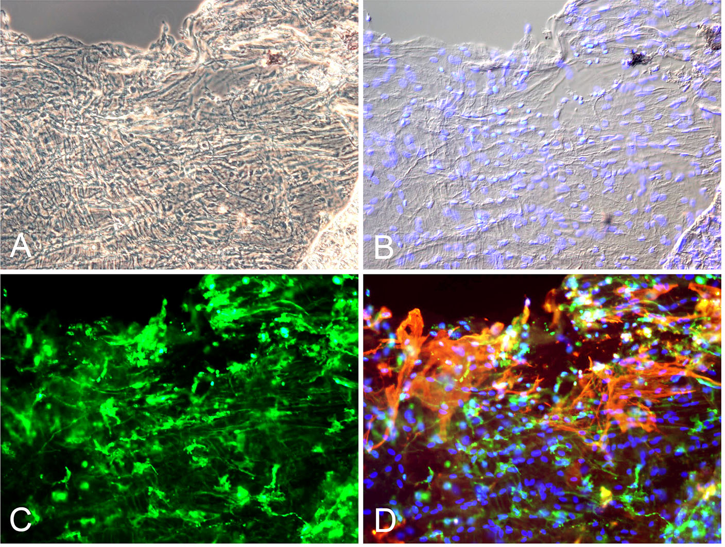
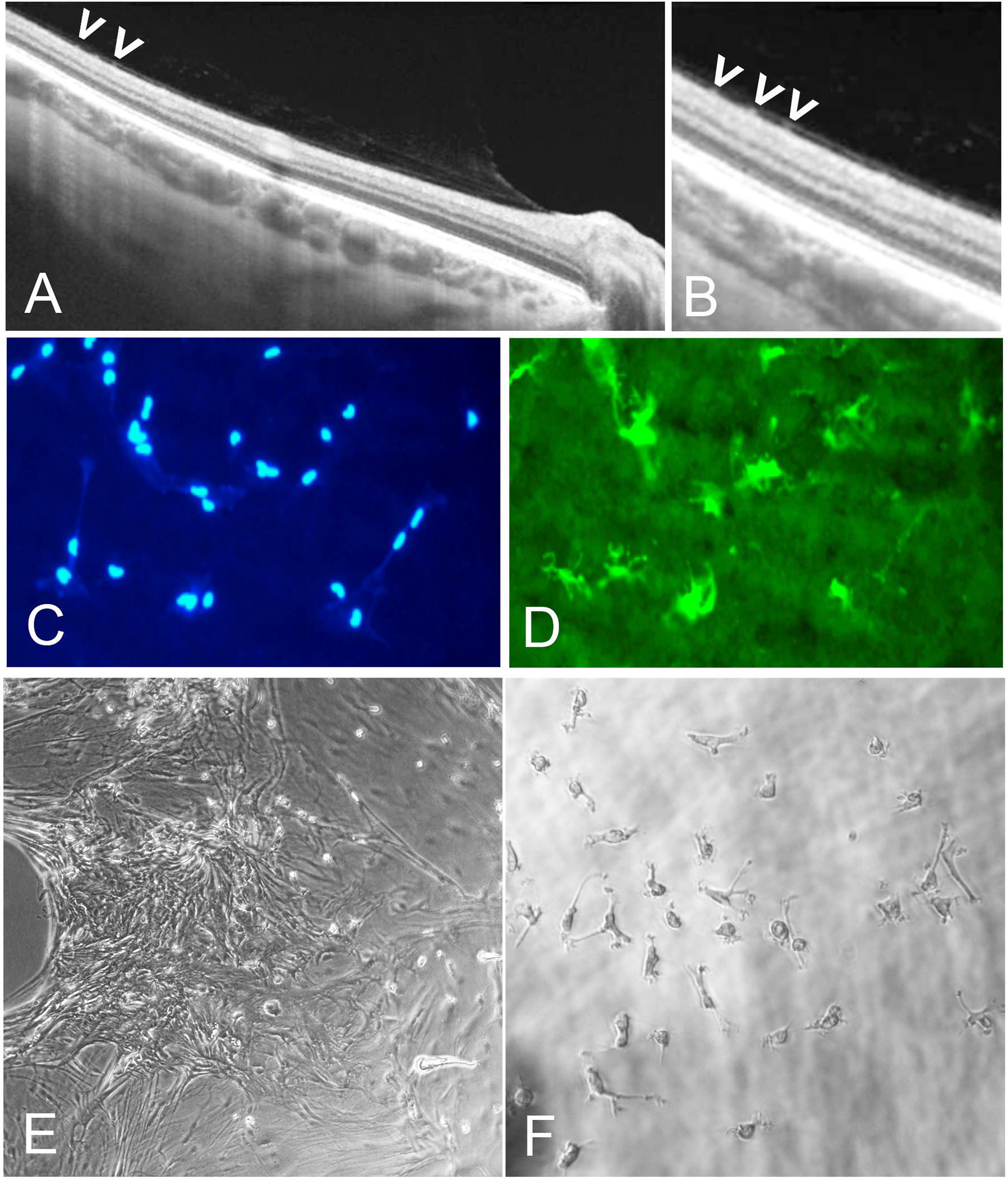
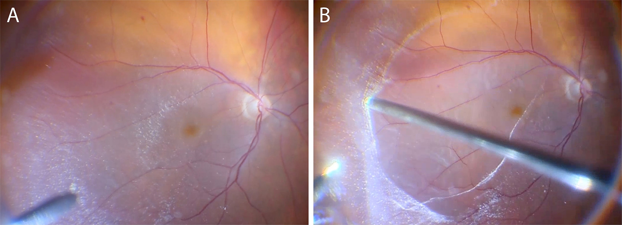
Areas covered: Studies of human tissues employing dark-field, phase, and electron microscopy; immunohistochemistry; and in vivo imaging of human hyalocytes.

Expert opinion: Hyalocytes are important in early pathophysiology, stimulating cell migration and proliferation, as well as subsequent membrane contraction and vitreo-retinal traction. Targeting hyalocytes early could mitigate advanced disease. Ultimately, eliminating the role of vitreous and hyalocytes may prevent proliferative vitreo-retinal diseases entirely.

Keywords: Vitreous; anomalous PVD; hyalocytes; macular pucker; proliferative diabetic vitreo-retinopathy; proliferative vitreo-retinopathy; vitreoschisis.


Figures

Figure 1:
Figure 1:. Anomalous PVD
When gel liquefaction and weakening of vitreo-retinal adhesion occur concurrently, the vitreous body separates away from the retina without sequelae. If the gel liquefies without concurrent dehiscence at the vitreo-retinal interface, there can be various untoward consequences, depending upon where vitreous is most adherent. If separation of vitreous from retina is full-thickness but topographically incomplete, there can be different forms of partial PVD (right side of diagram). Posterior separation with persistent peripheral vitreo-retinal attachment can induce retinal breaks and detachments. Peripheral vitreo-retinal separation with persistent full-thickness attachment of vitreous to the retina posteriorly can induce traction upon the macula (VMT), known as the vitreo-macular traction syndrome. Persistent full-thickness adherence to the macula is associated with (and may promote) exudative age-related macular degeneration (AMD). Persistent attachment to the optic disc can induce vitreo-papillopathies and also contribute to neovascularization and vitreous hemorrhage in ischemic retinopathies, as well as some cases of full-thickness macular hole. If, during PVD, the posterior vitreous cortex splits (vitreoschisis), there can be different effects depending upon the level (plane) of the split. Vitreoschisis more anteriorly leaves a relatively thick, cellular membrane adherent to the macula with embedded hyalocytes which recruit circulatiung monocytes and retinal glial cells. If there is also separation from the optic disc (found in 90% of macular pucker cases), inward (centripetal) contraction of this premacular membrane induces macular pucker. In the periphery, vitreoschisis in eyes with retinal detachment can result in vitreous cortex remnants (VCRs, as named by van Overdam[56,57]) with hyalocytes that promote proliferative vitreo-retinopathy (PVR) and recurrent retinal detachment[111]. If the vitreoschisis split occurs more posteriorly, the remaining premacular membrane is relatively thin and hypocellular. Persistent vitreopapillary adhesion (VPA, present in 87.5% or more of cases of macular holes) influences the vector of force in the tangential plane, resulting in outward (centrifugal) tangential traction (especially nasally), opening a central dehiscnece which is recognized clinically as a macular hole. Reproduced with permission from [10], © 2014 Springer Science+Business Media New York.
Figure 2:
Figure 2:. Vitreoschisis.
Left: The lamellar structure of the mammalian posterior vitreous cortex is demonstrated in an adult monkey eye stained with fluorescein-conjugated ABA lectin stain. The retina (below) and vitreous (above) are separated by the ILM (white arrowheads), above which is the posterior vitreous cortex whose lamellar structure is clearly evident. These potential cleavage planes can split apart during anomalous PVD or during vitrectomy surgery with membrane peeling, in each instance leaving a layer of vitreous attached to the macula. A hyalocyte (white arrow) is embedded in the posterior vitreous cortex (bar = 100 μM) Image courtesy of G Hageman. Middle: Spectral domain OCT/SLO imaging of macular pucker demonstrating splitting of the posterior vitreous cortex (left side of scan), known as “vitreoschisis”. Reproduced with permission from [13], © 2011 BMJ Publishing Group Ltd. Right: The eye shown in the middle panel underwent vitrectomy surgery with membrane peeling and the specimen was examined histologically. The area designated as “split’ is the site where the posterior vitreous cortex splits into the vitreoschisis cavity seen on OCT (middle panel). The arrows indicate hyalocytes embedded in the posterior vitreous cortex (Periodic Acid Schiff stain, magnification = 225x). Image courtesy of N Rao, Doheny Eye Institute.
Figure 3:
Figure 3:. Transmission electron microscopy of hyalocytes in macular pucker.
Multilayers of vitreous collagen (col) with hyalocytes (Hy) and myofibroblasts (My) are evident in this fibrocellular membrane. Hyalocytes possess an oval nucleus with marginal chromatin, vacuoles, dense granules, and thin cytoplasmic protrusions. Myofibroblasts are distinguished by cytoplasmic aggregates of actin microfilaments forming stress bundles. Image courtesy of author R Schumann.
Figure 4:
Figure 4:. Coronal plane (en face) imaging of macular pucker.
Combined OCT-SLO imaging with superimposition of coronal plane OCT (color) onto SLO (grayscale) images demonstrate macular pucker with multiple centers of retinal contraction (arrows): bi-centric (left), compared to three centers of retinal contraction (right). Eyes with 3 or 4 centers of retinal contraction were found to have a higher prevalence of intraretinal cysts and a thicker macula than eyes with 1 or 2 centers of retinal contraction. Reproduced with permission from [26], © 2008 The Ophthalmic Communications Society, Inc.
Figure 5:
Figure 5:. Proliferative vitreo-retinopathy (PVR).
Intraoperative imaging before (A) and after (B) peeling of a PVR membrane, extending from the superior arcade to the inferotemporal periphery. The dashed line indicates the area from where the membrane was excised. (C) Light microscopy of the PVR membrane stained with hematoxylin and eosin. Different continuous membrane areas can be distinguished, representing different stages of PVR: paucicellular, lamellar collagen-rich areas with hyalocytes, suggestive for VCR (1); areas with increased cellular infiltration by RPE and glial cells (2); more fibrotic areas with low cellularity and myofibroblasts (3). Images courtesy of author K van Overdam.
Figure 6:
Figure 6:. Histopathology of proliferative diabetic vitreo-retinopathy (PDVR).
Immunofluorescence microscopy (A), light (B) and transmission electron microscopy (C-F) of a fibrocellular, premacular membrane surgically removed from an eye with PDVR. A) Flat-mounted membrane with anti-CD45 (red) positive staining merged with cell nuclei staining (blue) indicating the presence of hyalocytes (original magnification x400). B) Semithin section of membrane demonstrated folded fibrocellular composition with thick collagen strands (original magnification x100). C) Ultrastructural analysis revealed hyalocytes with elongated cell bodies and thin cell processes in abundance of native vitreous collagen (original magnification x7000, bar = 1000 nm). D) Hyalocytes and myofibroblasts situated on layers of vitreous collagen strands (original magnification x3000, bar = 2000 nm). E) Myofibroblasts surrounded by newly formed collagen (original magnification x7000, bar = 1000 nm). F) Fibroblast with large cell nucleus and newly formed collagen (original magnification x7000, bar = 1000 nm). Images courtesy of author R Schumann.
Figure 7:
Figure 7:. Imaging mass cytometry of preretinal diabetic retinal neovascularization membranes compared to premacular membranes in macular pucker (non-diabetic).
Imaging mass cytometry of human retinal neovascularization (“RNV”) and macular pucker (“ERM”) tissue samples from humans. Multiplexed stainings for α-SMA (α-smooth muscle actin, yellow), HLA-DR (human leukocyte antigen – DR isotype, green), PECAM-1 (platelet endothelial cell adhesion molecule, red), CD8a (cluster of differentiation 8a, magenta), Histone H3 (blue) and COL1 (collagen type I, white) are presented. Higher magnification of the sections within the dashed white squares are shown in the panels in B and C, respectively. Scale bars correspond to 100 μm (A) and 50 μm (B and C). Reproduced from [89], licensed under CC-BY4.0 (http://creativecommons.org/licenses/by/4.0/)
Figure 8:
Figure 8:. Imaging human hyalocytes in vivo.
In vivo imaging of hyalocytes in a 61-year-old patient with proliferative diabetic vitreo-retinopathy using clinical OCT. A1) 3μm OCT reflectance slab located above the ILM centered at the fovea. Clustering of hyalocytes near the sites of neovascularization is observed. A2) Corresponding OCTA image shows the foveal full vascular layer. B) 3-D rendered and color-coded OCT reflectance image. Yellow arrows indicate neovascularization near the margin of the foveal avascular zone. White arrows indicate clustering of hyalocytes near neovascularization. (See also Movie 1 for a 3-D rendered and color-coded OCT reflectance movie). Images courtesy of authors TYP Chui and RB Rosen.
Figure 9:
Figure 9:. Human hyalocytes in vivo.
Comparison of hyalocytes imaged using non-confocal quadrant detection Adaptive Optics Scanning Light Ophthalmoscopy in a 32-year-old healthy control (A) and a 26-year-old patient with proliferative diabetic vitreo-retinopathy (B). Hyalocytes in the healthy control appear more ramified with multiple fine and long processes. In contrast, hyalocytes in the diabetic patient look relatively less ramified (top two cells) and more amoeboid shaped (bottom two cells). Images courtesy of authors TYP Chui and RB Rosen.
Figure 10:
Figure 10:. Vitreo-macular traction syndrome.
Anomalous PVD with persistent full-thickness attachment to the fovea can induce significant axial traction. There is relatively less cellular involvement in this process compared to macular pucker and PVR. Left: 3D-OCT. Image courtesy of author Michael Engelbert. Right: Combined OCT (color) – SLO (scanning laser ophthalmoscopy, in greyscale). Reproduced with permission from [23], © 2014 Springer Science+Business Media New York.
Figure 11:
Figure 11:. Immunofluorescence microscopy of ILM surgical specimen removed in vitreo-macular traction syndrome.
(A, B) Interference microscopy shows a complex cellular membrane with numerous cell nuclei (blue). (C, D) Positive immunostaining for anti-CD64 (green) and alpha-smooth muscle actin (red) indicates presence of hyalocytes and large myofibroblasts with intracytoplasmatic stress bundles. (Magnification x200). Images courtesy of author R Schumann.
Figure 12:
Figure 12:. Cells of the vitreo-macular interface.
(A) Spectral-domain OCT of vitreoschisis with dots (presumed cells) on retinal surface. (B) Higher magnification of image A indicates premacular cells (arrowheads). (C, D) Fluorescence microscopy with cell nuclei staining (blue) of IBA1-positive (green) premacular cells of flat mounted inner limiting membrane (ILM) specimen removed during macular surgery for macular hole. (E, F) Interference microscopy of ILM with premacular cells in tissue culture demonstrates presence of small dot-like premacular cells that represent macrophage-like phenotype and behavior, consistent with their identity as hyalocytes. Images courtesy of author R Schumann.
Figure 13:
Figure 13:. Chromodissection of preretinal membranes in rhegmatogenous retinal detachment.
Intraoperative imaging during vitrectomy for primary rhegmatogenous retinal detachment with presumed pre-operative PVD. After core vitrectomy and repeated, targeted use of triamcinolone acetonide for vitreous and VCR visualization, VCR membranes were detected (A) and removed (B) from the macula and peripheral retinal surface. Images courtesy of author K van Overdam, and P van Etten.

References

    1. Wieghofer P, Engelbert M, Chui TY et al. Hyalocyte origin, structure, and imaging. Exp. Rev. Opthalmol. [In Press] (2022). - PMC - PubMed
    1. Boneva S, Wolf J, Wieghofer P et al. Hyalocyte functions and immunology. Exp. Rev. Opthalmol. [In Press] (2022).
    1. Sebag J. Vitreous & Vitreo-Retinal Interface. In: Schachat AP, editor. Ryan’s Retina. 6th ed. Elsevier; 2018. p. 544–581.
    1. Sebag J. Vitreous and Vision Degrading Myodesopsia. Progress in Retinal and Eye Research. 2020;79:100847. - PubMed
    1. Chew L, Sebag J. Vitreous. Adler’s Physiology of the Eye. 12th ed. Philadelphia: Elsevier; 2022. p. (in press).