. 2022 Nov 16:13:1005321.
doi: 10.3389/fimmu.2022.1005321. eCollection 2022.

An intranasal vaccine targeting the receptor binding domain of SARS-CoV-2 elicits a protective immune response

Affiliations

An intranasal vaccine targeting the receptor binding domain of SARS-CoV-2 elicits a protective immune response

Li Chen et al. Front Immunol. .

Abstract

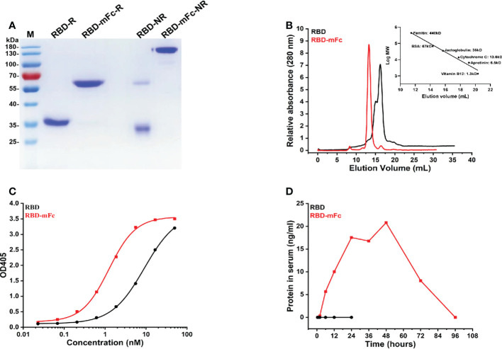
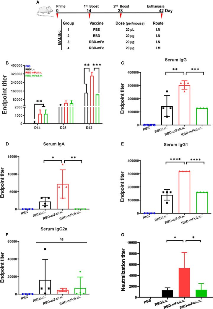
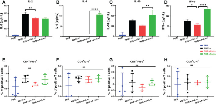
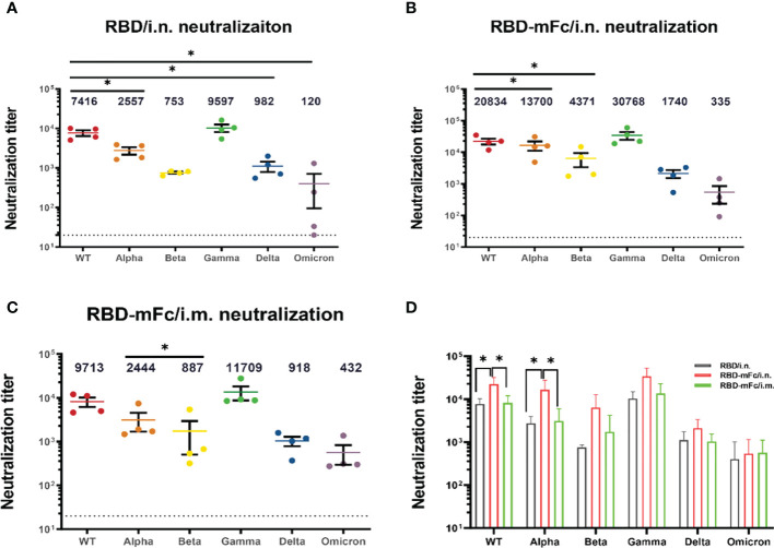
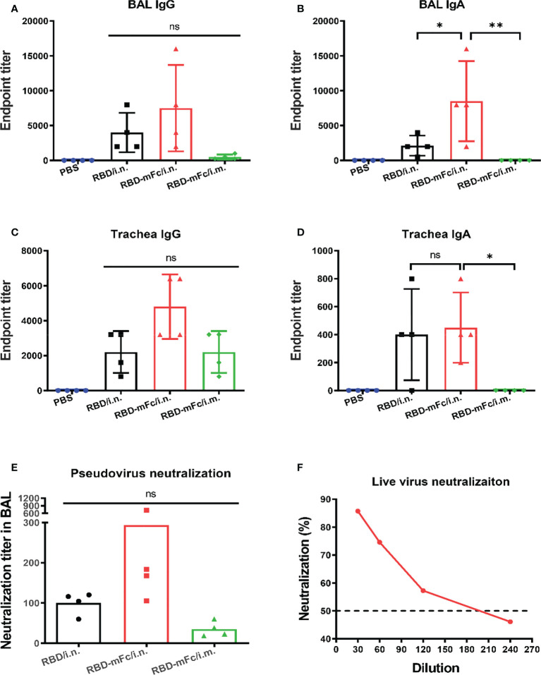
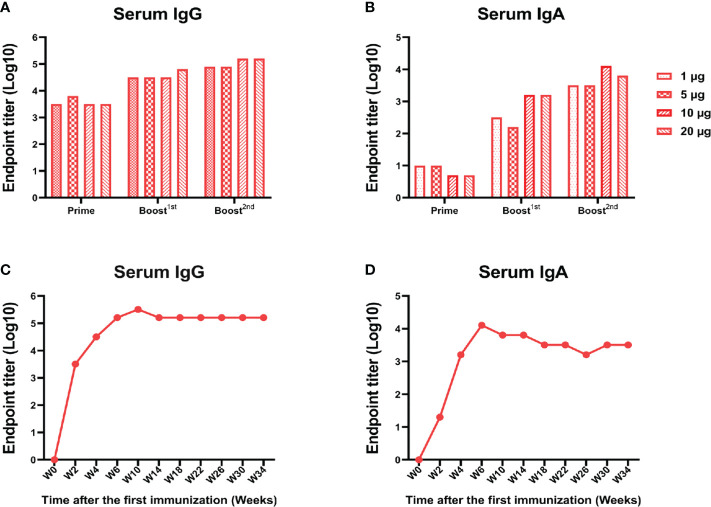
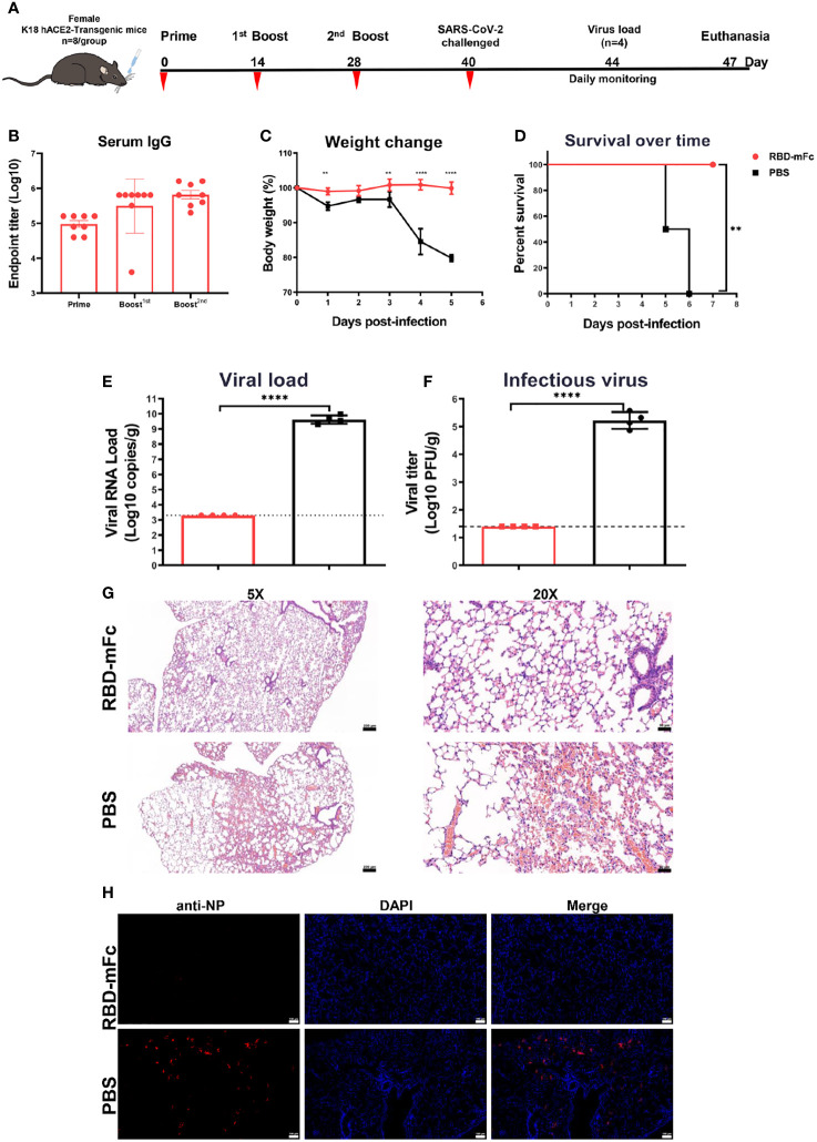
Severe acute respiratory syndrome coronavirus 2 (SARS-CoV-2), the pathogen responsible for COVID-19, has caused an ongoing worldwide pandemic. Due to the rapid emergence of variants of concern (VOCs), novel vaccines and vaccination strategies are urgently needed. We developed an intranasal vaccine consisting of the SARS-CoV-2 receptor binding domain (RBD) fused to the antibody Fc fragment (RBD-Fc). RBD-Fc could induce strong humoral immune responses via intranasal vaccination. Notably, this immunogen could efficiently induce IgG and IgA and establish mucosal immunity in the respiratory tract. The induced antibodies could efficiently neutralize wild-type SARS-CoV-2 and currently identified SARS-CoV-2 VOCs, including the Omicron variant. In a mouse model, intranasal immunization could provide complete protection against a lethal SARS-CoV-2 challenge. Unfortunately, the limitation of our study is the small number of animals used in the immune response analysis. Our results suggest that recombinant RBD-Fc delivered via intranasal vaccination has considerable potential as a mucosal vaccine that may reduce the risk of SARS-CoV-2 infection.

Keywords: RBD-Fc; SARS-CoV-2; intranasal vaccine; mucosal immunity; mucosal vaccine.


Conflict of interest statement

The authors declare that the research was conducted in the absence of any commercial or financial relationships that could be construed as a potential conflict of interest.

Figures

Figure 1
Figure 1
RBD-mFc fusion protein is dimeric, functional and long-lasting in serum. (A) Analysis of RBD-mFc and RBD protein (approximately 4 μg each) under reducing and nonreducing conditions through SDS–PAGE (4-12% gradient gel) which indicated that RBD-mFc existed as a disulfide-linked dimer while RBD is a mixture of monomer and dimer. M, marker; R, reduced form; NR, nonreduced form. (B) Analysis of RBD-mFc and RBD proteins by size exclusion chromatography on Superdex 200 10/300 (GE). X-axis: elution volume (mL), Y-axis: A280 nm (a.u.) The right inset shows the standard curve. Two peaks were observed in the eluted RBD, while only one peak was separated from the eluted RBD-mFc, indicating that RBD-mFc was uniform. (C) Binding of RBD-mFc and RBD to hACE2-Fc, as measured by ELISA. The EC50 values of hACE2-Fc binding to coated RBD-mFc and RBD were estimated at 1.2 nM and 9.1 nM, respectively. Data are average values of two replicates. (D) Plasma half-life of RBD-mFc and RBD via intranasal administration. Purified RBD-mFc and RBD proteins (50 μg) were intranasally inoculated into BALB/c mice (n=3). Serum was collected at the times indicated on the abscissa. The protein concentration in pooled blood circulation were measured by ELISA. Substantial RBD-mFc accumulation and persistence in sera were observed while RBD was barely detectable.
Figure 2
Figure 2
A strong immune response was elicited by RBD-mFc intranasal immunization. (A) Schematic of the BALB/c immunization strategy. Four mice from each group were immunized with different vaccines by different routes at the times indicated (days). Serum was collected every two weeks and assessed for specific antibody response to RBD. (B) Overall immune response of the four immunized groups. The data show the reciprocal endpoint dilution titers, with each data point representing the mean of four animals. Mice immunized with RBD-mFc protein developed a more rapid and efficient response after prime and boost vaccination compared to RBD. Serum antibody responses were analyzed 14 days after the 2nd boost immunization. RBD-specific IgG (C) and IgA (D) were assessed by ELISA. Intranasal immunization with RBD-mFc induced a stronger immune effect than immunization in the other two groups. Analysis of IgG subclasses of the RBD-specific antibody response (E, F). (G) The neutralizing antibody response to SARS-CoV-2 was determined by PRNT and represented as the reciprocal half-maximal inhibition concentration (IC50). All RBD protein-immunized sera could efficiently inhibit authentic SARS-CoV-2 infection, whereas sera from the PBS control showed no neutralization activity The data in (B–G) represent the mean ± SD. Significant differences were determined by one-way ANOVA with Tukey’s multiple comparisons test. ns, not significant; *p < 0.05; **p < 0.01; ***p < 0.001; ****p < 0.0001.
Figure 3
Figure 3
Cellular response induced by RBD-mFc intranasal immunization. Vaccinated BALB/c mice were euthanized at 2 weeks post-2nd boost immunization. The splenocytes harvested from immunized mice (n=4) were stimulated with RBD protein for 42 hours. Cultured supernatants were harvested and pooled. Cytokine production was measured using an ELISA kit. RBD-immunized mice generated a certain amount of IL-2 (A), IL-10 (C) and IFN-γ (D). IL-4 (B) was detected only in RBD-mFc intramuscularly immunized mice. Data represent the mean of three parallel wells in one experiment. Splenocytes were incubated with SARS-CoV-2 RBD protein. The responding CD4+ (E, F) and CD8+ T (G, H) cells were identified by intracellular staining for effector cytokines. The gating strategies are shown in Supplementary Figure S1 . Data are represented as the mean ± SD. Significant differences were determined by one-way ANOVA with Tukey’s multiple comparisons test. ns, not significant; *p < 0.05; **p < 0.01; ****p < 0.0001.
Figure 4
Figure 4
Broader neutralizing antibodies against SARS-CoV-2 variants were induced by RBD-mFc intranasal immunization. The cross-reactive neutralization of immune sera against wild-type and variants of SARS-CoV-2 was analyzed by pseudovirus neutralization. (A–C) Neutralization titers for wild-type and VOC pseudoviruses generated by immune sera from the (A) RBD/i.n., (B) RBD-mFc/i.n. and (C) RBD-mFc/i.m. groups 14 days after the 2nd boost immunization. The majority of immune sera showed broad-spectrum neutralization capacity. All groups n=4. The dotted lines represent the limit of detection (1:20 dilution). Geometric mean titers calculated by GraphPad Prism are shown above each column. (D) Comparison of neutralization titers against individual viruses between immune sera from the RBD/i.n., RBD-mFc/i.n. and RBD-mFc/i.m. groups. Data are presented as the mean ± SD; nonparametric ANOVA with Dunn’s multiple comparison test was used to test for significant differences in (A). Significant differences in (B–D) were determined by one-way ANOVA with Tukey’s multiple comparisons test. *p < 0.05.
Figure 5
Figure 5
Robust mucosal immunity in the respiratory system was induced by RBD-mFc via intranasal immunization. The fully immunized mice were euthanized 14 days after the 2nd boost immunization, and samples from the respiratory tract, including bronchoalveolar lavage fluid (BAL) and trachea wash, were collected. (A–D) The antibody response in these samples were determined by evaluating RBD-specific IgG and IgA. In contrast to intramuscular vaccination, intranasal immunization could additionally induce mucosal immunity. (E) The neutralization titer in BAL fluid were determined by a SARS-CoV-2 pseudovirus neutralization assay. (F) Neutralization of authentic SARS-CoV-2 virus by pooled BAL from RBD-mFc/i.n. group. The data in (A–D) represent the mean ± SD. Significant differences were determined by one-way ANOVA with Tukey’s multiple comparisons test. ns, not significant; *p < 0.05; **p < 0.01.
Figure 6
Figure 6
A low dose of RBD-mFc induced a lasting and strong immune response.BALB/c mice (n=4) were intranasally immunized with different doses of RBD-mFc. Serum was collected and pooled every two weeks after the prime immunization. RBD-specific IgG (A) and IgA (B) were assessed by ELISA. Increasing antibody responses were induced by increasing the immune cycle in a dose-dependent manner. To test for antibody persistence after immunization, a group of 10 μg immunized mice was chosen to assess RBD-specific IgG (C) and IgA (D) duration. Antibodies in serum underwent dynamic change and plateaued until the end of detection.
Figure 7
Figure 7
RBD-mFc protects mice from lethal challenge with SARS-CoV-2. (A) Immunization and challenge schedules for hACE2 transgenic mice. Female hACE2 transgenic mice (n=8) were intranasally immunized with 10 μg RBD-mFc protein on days 0, 14 and 28. Equal volumes of adjuvant mixed with PBS were used as controls. Blood samples were collected at the times indicated. All vaccinated mice were challenged with 20 μL of 1×10⁴ TCID50/mouse wild-type SARS-CoV-2. Half of the mice were sacrificed at day 4 postinfection to assess the viral load; the remaining mice were monitored until 7 days postinfection. (B) Overall RBD-specific antibody response after the prime immunization. A potent antibody response was successfully induced in all mice. (C) Body weight change of mice for 5 days. (D) Survival curve. Mice in the PBS group experienced a shaped weight change at 4 dpi and gradually started to die. In the RBD-mFc group, the mice were well maintained. SARS-CoV-2 RNA copies detected by RT‒qPCR (E) and titers of infectious viral particles assessed by plaque assay (F) at 4 dpi in homogenized lung. A large amount of virus was detected in the control group, while all detected samples from the RBD-mFc group were under the detection limit. HE staining (G) and IFA against N protein (H) were evaluated in lungs at 4 dpi. No observed in the RBD-mFc group compared with the PBS group. The scale bar in (G, H) represents 100 μm. Data are represented as the mean ± SD. Significant differences in (C) were determined by two-way ANOVA with Sidak’s multiple comparisons test. Data in (D) were analyzed by the log-rank (Mantel‒Cox) test. Significant differences in (E, F) were determined by a two-tailed unpaired t test. **p < 0.01; ****p < 0.0001.

Similar articles

Cited by

References

    1. Miao X, Luo Y, Huang X, Lee SMY, Yuan Z, Tang Y, et al. . A novel biparatopic hybrid antibody-ACE2 fusion that blocks SARS-CoV-2 infection: Implications for therapy. MAbs (2020) 12(1):1804241. doi: 10.1080/19420862.2020.1804241 - DOI - PMC - PubMed
    1. Zhu N, Zhang D, Wang W, Li X, Yang B, Song J, et al. . A noncompeting pair of human neutralizing antibodies block COVID-19 virus binding to its receptor ACE2. science (2020) 382(8):727–33. doi: 10.1056/NEJMoa2001017 - DOI - PMC - PubMed
    1. Pinto D, Park YJ, Beltramello M, Walls AC, Tortorici MA, Bianchi S, et al. . Cross-neutralization of SARS-CoV-2 by a human monoclonal SARS-CoV antibody. Nature (2020) 583(7815):290–5. doi: 10.1038/s41586-020-2349-y - DOI - PubMed
    1. Ejemel M, Li Q, Hou S, Schiller ZA, Tree JA, Wallace A, et al. . A cross-reactive human IgA monoclonal antibody blocks SARS-CoV-2 spike-ACE2 interaction. Nat Commun (2020) 11(1):4198. doi: 10.1038/s41467-020-18058-8 - DOI - PMC - PubMed
    1. Walls AC, Park YJ, Tortorici MA, Wall A, McGuire AT, Veesler D. Structure, function, and antigenicity of the SARS-CoV-2 spike glycoprotein. Cell (2020) 181(2):281–292 e6. doi: 10.1016/j.cell.2020.02.058 - DOI - PMC - PubMed

Publication types

Supplementary concepts