. 2022 Nov 17:13:1039120.
doi: 10.3389/fimmu.2022.1039120. eCollection 2022.

Age-dependent NK cell dysfunctions in severe COVID-19 patients

Affiliations

Age-dependent NK cell dysfunctions in severe COVID-19 patients

Cinzia Fionda et al. Front Immunol. .

Abstract

Natural Killer (NK) cells are key innate effectors of antiviral immune response, and their activity changes in ageing and severe acute respiratory syndrome coronavirus 2 (SARS-CoV-2) infection. Here, we investigated the age-related changes of NK cell phenotype and function during SARS-CoV-2 infection, by comparing adult and elderly patients both requiring mechanical ventilation. Adult patients had a reduced number of total NK cells, while elderly showed a peculiar skewing of NK cell subsets towards the CD56lowCD16high and CD56neg phenotypes, expressing activation markers and check-point inhibitory receptors. Although NK cell degranulation ability is significantly compromised in both cohorts, IFN-γ production is impaired only in adult patients in a TGF-β-dependent manner. This inhibitory effect was associated with a shorter hospitalization time of adult patients suggesting a role for TGF-β in preventing an excessive NK cell activation and systemic inflammation. Our data highlight an age-dependent role of NK cells in shaping SARS-CoV-2 infection toward a pathophysiological evolution.

Keywords: COVID-19; NK cell subsets; Natural Killer cells; T-BET; TGF-β; ageing; inflammation.


Conflict of interest statement

The authors declare that the research was conducted in the absence of any commercial or financial relationships that could be construed as a potential conflict of interest.

Figures

Figure 1
Figure 1
Overview of study design. (A) Experimental design and workflow of the study. (B) Schematic representation of the major demographic (age and sex) and clinical data [PaO2/FiO2, intensive care unit (ICU), hospital days, white blood count (WBC) and outcome] of patients enrolled in the study.
Figure 2
Figure 2
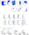
Changes in adult and elderly innate lymphocytes and NK cells during SARS-CoV-2 infection. (A) Gating strategy used to identify conventional NK cells (NK), CD56negEOMES+/T-BET+ cells and ILC2. NK and EOMES-/T-BET- cells were gated on CD56pos and CD56neg NK cell populations respectively within Lin- (CD3, CD4, CD5, CD14, CD19) CD45+CD7+ cells. ILC2 were gated on EOMES-/T-BET- as GATA3+. Representative overlays displaying CD127, CD117 and CD294 (as ILC2 markers). (B) Representative overlays displaying CD127, CD117 and CD294 (as ILC2 markers) as well as CD16, CD57, KIRs and NKG2A (as NK cell markers) expression on the indicated cell populations in a healthy donor. (C, D) Histograms represent the median ± SD of the frequency and the total cell count/μl of the indicated cell populations in adult (n = 19) and elderly (n = 8) patients (Pt) compared to age-matched volunteers (HD; adult = 16, elderly = 9). * p< 0.05, ** p< 0.01, *** p< 0.001, **** p< 0.0001. One way ANOVA Kruskal-Wallies test multiple comparisons).
Figure 3
Figure 3
Alterations in circulating CD56pos cells in adult and elderly COVID-19 patients at hospital admission. Gating strategies used to identify total and relative frequency of CD56high and CD56low cell populations and their subsets are shown on a representative adult c COVID-19 patient and age-matched controls (A, C, E). CD56high and CD56low cell populations were gated on Lin- CD45+CD7+ CD56+ and distinguished based on CD16 expression. Frequency and total cell count/μl of total CD56high and CD56low cell (B) as well as of specific subsets CD56highCD16+ and CD56highCD16- (D), and CD56lowCD16+ and CD56lowCD16low/- (F) in COVID-19 patients and healthy donors are shown. Histograms show mean ± SD. * p< 0.05, ** p< 0.01, *** p< 0.001, **** p< 0.0001. Two-way analysis of variance (ANOVA) Tukey’s multiple comparisons test and One way ANOVA Kruskal-Wallies test multiple comparisons.
Figure 4
Figure 4
Alterations in circulating CD56neg cells in adult and elderly COVID-19 patients at hospital admission. Gating strategies used to identify EOMES- T-BET- and EOMES+ T-BET+ subsets within CD56neg cells (A) and ILC2 among EOMES- T-BET- cells (D) and their relative frequency are shown on a representative adult and elderly COVID-19 patient and age-matched controls. Lin- CD45+CD7+CD56- cells were analyzed for the expression of transcription factors EOMES and T-BET. ILC2 were identified within EOMES-/T-BET- CD56- cells as EOMES-T-BET- GATA3+. Percentage and absolute counts of CD56- EOMES-/T-BET- and EOMES+ T-BET+ (B, C) and ILC2 (E, F) are reported. Histograms show mean ± SD. * p< 0.05, ** p< 0.01, *** p< 0.001, **** p< 0.0001. Two-way analysis of variance (ANOVA) Tukey’s multiple comparisons test and One way ANOVA Kruskal-Wallies test multiple comparisons.
Figure 5
Figure 5
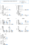
Longitudinal analysis of NK/ILC populations in adult COVID-19 patients. Histograms represent the mean ± SD of the frequency and the absolute count of CD7+ (A), CD56pos and CD56neg (B), CD56high (C) and CD56low (D) and ILC2 (E) in adult patients (n = 7) at 1week post-hospitalization. Cell populations were gated as described above. * p< 0.05, ** p< 0.01, *** p< 0.001, **** p< 0.0001. Two-way analysis of variance (ANOVA) Tukey’s multiple comparisons test and One way ANOVA Kruskal-Wallies test multiple comparisons.
Figure 6
Figure 6
Longitudinal analysis of NK/ILC populations in elderly COVID-19 patients. Histograms represent the mean ± SD of the frequency and the absolute count of CD7+ (A), CD56pos and CD56neg (B), CD56high (C) CD56low (D) and ILC2 (E) in elderly patients at the indicated period. Cell populations were gated as described above. Elderly patients: hospital admission n = 8; week 1 n = 8; week 2 n = 6; week 3 n = 3. * p< 0.05, ** p< 0.01, *** p< 0.001. Two-way analysis of variance (ANOVA) Tukey’s multiple comparisons test and One way ANOVA Kruskal-Wallies test multiple comparisons.
Figure 7
Figure 7
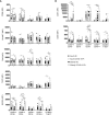
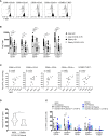
An impairment of degranulation ability characterizes both adult and elderly NK cell subsets. The percentage of K562 target cell-induced CD107a positive NK cell subsets from adult and elderly COVID-19 patients and healthy controls is shown early after hospital admission (A) and at different times after hospitalization in adult (B) and elderly (C) patients. Representative overlays and mean fluorescence intensity (MFI) of Perforin (D), Granzyme B (GrzB) (E) and FasL (F) expression by distinct NK cell subsets are reported for COVID-19 patients and age-matched controls. Histograms represent the mean ± SD. * p< 0.05, ** p< 0.01, *** p< 0.001, **** p< 0.0001. Two-way analysis of variance (ANOVA) Tukey’s multiple comparisons test and One way ANOVA Kruskal-Wallies test multiple comparisons.
Figure 8
Figure 8
Receptor prolife of adult and elderly NK cell subsets. Expression levels of inhibitory (A) and activation/maturation (B) markers on NK cell subsets in adult and elderly COVID-19 patients alongside healthy controls early after hospital admission. MFI median values ± SD for each marker are shown. * p< 0.05, ** p< 0.01, *** p< 0.001, **** p< 0.0001. Two-way analysis of variance (ANOVA) Tukey’s multiple comparisons test and One way ANOVA Kruskal-Wallies test multiple comparisons.
Figure 9
Figure 9
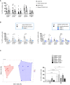
Differential ability of producing IFN-γ by adult and elderly COVID-19 NK cell subsets. The frequency of IFN-γ positive cells, upon IL-12 plus IL-18 stimulation, is shown for each NK cell subset from adult and elderly COVID-19 patients compared with healthy controls early after hospital admission (A) and at different times after hospitalization in adult and elderly patients (B). Histograms represent the mean ± SD. K-means clustering of our patient cohort identified two clusters based on IFN-γ production by NK cell subsets and the hospitalization time ( Supplementary Table S3 ) (left panel) (C). These two clusters differentially present the indicated population for IFN-γ production (right panel). *p< 0.05, **p< 0.01, *** p< 0.001, **** p< 0.0001 . Two-way analysis of variance (ANOVA) and Sidak’s multipletest.
Figure 10
Figure 10
TGF-β regulates IFN-γ production in adult COVID-19 NK cell subsets. Representative overlays displaying T-BET expression of distinct NK cell subsets from adult and elderly COVID-19 patients and age-matched controls (A). Summary of data showing the median values ± SD of T-BET MFI expression on each subset (B). Spearman’s rank correlation of T-BET expression (MFI) and the oxygenation index PaO2/FiO2 ratio at hospital admission in adult patients (C). Plasma levels of TGF-β of elderly and adult COVID-19 patients at hospital admission (adult = 12, elderly = 7). * p< 0.05 unpaired t-student test analysis is indicated (D). PBMC from healthy donors were cultured in medium containing adult healthy or COVID-19 plasma or elderly healthy or COVID-19 plasma for 48h in the absence or in the presence of α-TGF-β blocking antibody. The percentage of IFN-γ positive cells after IL-12 plus IL-18 stimulation was assessed for each NK cell subset. Histograms represent the mean ± SD (E). * p< 0.05, ** p< 0.01, *** p< 0.001, **** p< 0.0001. Two-way analysis of variance (ANOVA) Tukey’s multiple comparisons test and One way ANOVA Kruskal-Wallies test multiple comparisons.

References

    1. Wu F, Zhao S, Yu B, Chen YM, Wang W, Song ZG, et al. . A new coronavirus associated with human respiratory disease in China. Nature (2020) 579:265–9. doi: 10.1038/s41586-020-2008-3 - DOI - PMC - PubMed
    1. Zhu N, Zhang D, Wang W, Li X, Yang B, Song J, et al. . A Novel Coronavirus from Patients with Pneumonia in China, 2019. New Engl J Med (2020) 382(8):727–33. doi: 10.1056/nejmoa2001017 - DOI - PMC - PubMed
    1. Huang C, Wang Y, Li X, Ren L, Zhao J, Hu Y, et al. . Clinical features of patients infected with 2019 novel coronavirus in Wuhan, China. Lancet (2020) 395(10223):497–506. doi: 10.1016/S0140-6736(20)30183-5 - DOI - PMC - PubMed
    1. Zhou F, Yu T, Du R, Fan G, Liu Y, Liu Z, et al. . Clinical course and risk factors for mortality of adult inpatients with COVID-19 in Wuhan, China: a retrospective cohort study. Lancet (2020) 395(10229):1054–62. doi: 10.1016/S0140-6736(20)30566-3 - DOI - PMC - PubMed
    1. Williamson EJ, Walker AJ, Bhaskaran K, Bacon S, Bates C, Morton CE, et al. . Factors associated with COVID-19-related death using OpenSAFELY. Nature (2020) 584(7821):430–6. doi: 10.1038/s41586-020-2521-4 - DOI - PMC - PubMed

Publication types

Substances