

The effect of organ-specific tumor microenvironments on response patterns to immunotherapy
- PMID: 36466910
- PMCID: PMC9713699
- DOI: 10.3389/fimmu.2022.1030147
The effect of organ-specific tumor microenvironments on response patterns to immunotherapy
Abstract
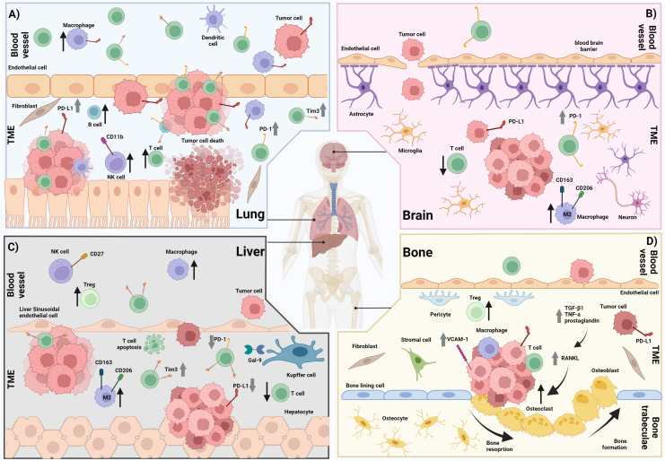
Immunotherapy, particularly immune checkpoint inhibitors, have become widely used in various settings across many different cancer types in recent years. Whilst patients are often treated on the basis of the primary cancer type and clinical stage, recent studies have highlighted disparity in response to immune checkpoint inhibitors at different sites of metastasis, and their impact on overall response and survival. Studies exploring the tumor immune microenvironment at different organ sites have provided insights into the immune-related mechanisms behind organ-specific patterns of response to immunotherapy. In this review, we aimed to highlight the key learnings from clinical studies across various cancers including melanoma, lung cancer, renal cell carcinoma, colorectal cancer, breast cancer and others, assessing the association of site of metastasis and response to immune checkpoint inhibitors. We also summarize the key clinical and pre-clinical findings from studies exploring the immune microenvironment of specific sites of metastasis. Ultimately, further characterization of the tumor immune microenvironment at different metastatic sites, and understanding the biological drivers of these differences, may identify organ-specific mechanisms of resistance, which will lead to more personalized treatment approaches for patients with innate or acquired resistance to immunotherapy.
Keywords: immunotherapy; metastasis; organ-specific immune microenvironment; organ-specific immune response; tumor microenvironment.
Copyright © 2022 Conway, Braden, Wilmott, Scolyer, Long and Pires da Silva.
Conflict of interest statement
GL is consultant advisor for Agenus, Amgen, Array Biopharma, Boehringer Ingelheim, Bristol Myers Squibb, Evaxion, Hexal AG Sandoz Company, Highlight Therapeutics S.L., Innovent Biologics USA, Merck Sharpe & Dohme, Novartis, OncoSec, PHMR Ltd, Pierre Fabre, Provectus, Qbiotics, Regeneron. RS has received fees for professional services from F. Hoffmann-La Roche Ltd, Evaxion, Provectus Biopharmaceuticals Australia, Qbiotics, Novartis, Merck Sharp & Dohme, NeraCare, AMGEN Inc., Bristol-Myers Squibb, Myriad Genetics and GlaxoSmithKline. IP had travel support by BMS and MSD, and speaker fee by Roche, Bristol-Myers Squibb and Merck Sharpe & Dohme. The remaining authors declare that the research was conducted in the absence of any commercial or financial relationships that could be construed as a potential Conflict of interest. The handling editor MA declared a past co-authorship with the author GL.
Figures
Similar articles
-
Recent advances in tumor microenvironment-targeted nanomedicine delivery approaches to overcome limitations of immune checkpoint blockade-based immunotherapy.J Control Release. 2021 Apr 10;332:109-126. doi: 10.1016/j.jconrel.2021.02.002. Epub 2021 Feb 8. J Control Release. 2021. PMID: 33571549 Review.
-
Acquired resistance for immune checkpoint inhibitors in cancer immunotherapy: challenges and prospects.Aging (Albany NY). 2022 Jan 17;14(2):1048-1064. doi: 10.18632/aging.203833. Epub 2022 Jan 17. Aging (Albany NY). 2022. PMID: 35037899 Free PMC article. Review.
-
T-cell immunoglobulin and ITIM domain, as a potential immune checkpoint target for immunotherapy of colorectal cancer.IUBMB Life. 2021 May;73(5):726-738. doi: 10.1002/iub.2461. Epub 2021 Mar 30. IUBMB Life. 2021. PMID: 33686787 Review.
-
New Strategies and Combinations to Improve Outcomes in Immunotherapy in Metastatic Non-Small-Cell Lung Cancer.Curr Oncol. 2021 Dec 23;29(1):38-55. doi: 10.3390/curroncol29010004. Curr Oncol. 2021. PMID: 35049678 Free PMC article. Review.
-
Synergistic effects of radiotherapy and targeted immunotherapy in improving tumor treatment efficacy: a review.Clin Transl Oncol. 2022 Dec;24(12):2255-2271. doi: 10.1007/s12094-022-02888-7. Epub 2022 Aug 1. Clin Transl Oncol. 2022. PMID: 35913663 Review.
Cited by
-
The Comparative Effect of Morphine on Proliferation of Cancer Cell Lines Originating from Different Organs: An In Vitro Study.Pharmaceuticals (Basel). 2024 Dec 9;17(12):1656. doi: 10.3390/ph17121656. Pharmaceuticals (Basel). 2024. PMID: 39770497 Free PMC article.
-
Organ-Specific Immune Setpoints Underlie Divergent Immune Profiles across Metastatic Sites in Breast Cancer.Cancer Immunol Res. 2024 Nov 4;12(11):1559-1573. doi: 10.1158/2326-6066.CIR-23-0718. Cancer Immunol Res. 2024. PMID: 39051632 Free PMC article.
-
Complete blood counts as potential risk factors of early dissemination to liver and lungs in resected colorectal cancer: a retrospective cohort study.Int J Colorectal Dis. 2025 Jan 21;40(1):21. doi: 10.1007/s00384-024-04802-9. Int J Colorectal Dis. 2025. PMID: 39836241 Free PMC article.
-
[Bioinformatic analysis of CCND2 expression in papillary thyroid carcinoma and its impact on immune infiltration].Nan Fang Yi Ke Da Xue Xue Bao. 2024 May 20;44(5):981-988. doi: 10.12122/j.issn.1673-4254.2024.05.21. Nan Fang Yi Ke Da Xue Xue Bao. 2024. PMID: 38862457 Free PMC article. Chinese.
-
Epithelial-Mesenchymal Transition in Non-Small Cell Lung Cancer Management: Opportunities and Challenges.Biomolecules. 2024 Nov 28;14(12):1523. doi: 10.3390/biom14121523. Biomolecules. 2024. PMID: 39766230 Free PMC article. Review.
References
Publication types
MeSH terms
Substances
LinkOut - more resources
Full Text Sources
Medical