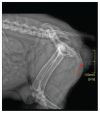
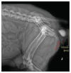
Treatment of a cystocele in a female dog 3 days after whelping
- PMID: 36467387
- PMCID: PMC9648471
Treatment of a cystocele in a female dog 3 days after whelping
Abstract
A case of a cystocele is reported in a 3-year-old intact female Doberman pinscher. The urinary bladder and vaginal body were reduced within the abdominal cavity and secured by cystopexy and cervicopexy allowing the uterus and ovaries to be spared. This is the first report describing the surgery for a cystocele in a young female dog 3 days after whelping, with sparing of the reproductive tract. Key clinical message: This is the first report to describe a cystocele in a young intact female dog after whelping with sparing of the female reproductive tract.
Traitement d’une cystocèle chez une chienne 3 jours après la mise bas. Un cas de cystocèle est rapporté chez une femelle Doberman pinscher intacte de 3 ans. La vessie et le corps vaginal ont été réduits dans la cavité abdominale et sécurisés par cystopexie et cervicopexie permettant d’épargner l’utérus et les ovaires. Il s’agit du premier rapport décrivant la chirurgie d’une cystocèle chez une jeune chienne trois jours après la mise bas, avec préservation de l’appareil reproducteur.Message clinique clé :Il s’agit du premier rapport décrivant une cystocèle chez une jeune chienne intacte après mise bas avec préservation de l’appareil reproducteur femelle.(Traduit par Dr Serge Messier).
Copyright and/or publishing rights held by the Canadian Veterinary Medical Association.
Figures


References
-
- Manothaiudom K, Johnston SD. Clinical approach to vaginal/vestibular masses in the bitch. Vet Clin North Am Small Anim Pract. 1991;21:509–521. - PubMed
-
- Johnston SD, Kustritz MVR, Olson PNS, editors. Canine and Feline Theriogenology. 1st ed. Philadelphia, Pennsylvania: WB Saunders; 2001. pp. 225–273.
-
- McNamara PS, Harvey JJ, Dykes N. Chronic vaginocervical prolapse with visceral incarceration in a dog. J Am Anim Hosp Assoc. 1997;33:533–536. - PubMed
-
- Konig GJ, Handler J, Arbeiter K. Rare case of a vaginal prolapse during the last third of pregnancy in a Golden Retriever bitch. Kleintierpraxis. 2004;49:299–305.
-
- Rani RU, Kathiresan D, Sivaseelan S. Vaginal fold prolapse in a pregnant bitch and its surgical management. Indian Vet J. 2004;81:1390–1391.
Publication types
MeSH terms
LinkOut - more resources
Full Text Sources
