Isolation of BVDV-1a, 1m, and 1v strains from diarrheal calf in china and identification of its genome sequence and cattle virulence
- PMID: 36467650
- PMCID: PMC9709263
- DOI: 10.3389/fvets.2022.1008107
Isolation of BVDV-1a, 1m, and 1v strains from diarrheal calf in china and identification of its genome sequence and cattle virulence
Abstract
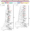
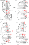
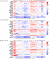
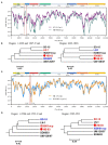
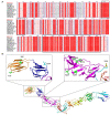
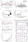
Bovine viral diarrhea virus (BVDV) is an important livestock viral pathogen responsible for causing significant economic losses. The emerging and novel BVDV isolates are clinically and biologically important, as there are highly antigenic diverse and pathogenic differences among BVDV genotypes. However, no study has yet compared the virulence of predominant genotype isolates (BVDV-1a, 1b, and 1m) in China and the emerging genotype isolate BVDV-1v. The serological relationship among these genotypes has not yet been described. In this study, we isolated three BVDV isolates from calves with severe diarrhea, characterized as BVDV-1a, 1m, and novel 1v, based on multiple genomic regions [including 5-untranslated region (5'-UTR), Npro, and E2] and the phylogenetic analysis of nearly complete genomes. For the novel genotype, genetic variation analysis of the E2 protein of the BVDV-1v HB-03 strain indicates multiple amino acid mutation sites, including potential host cell-binding sites and neutralizing epitopes. Recombination analysis of the BVDV-1v HB-03 strain hinted at the possible occurrence of cross-genotypes (among 1m, 1o, and 1q) and cross-geographical region transmission events. To compare the pathogenic characters and virulence among these BVDV-1 genotypes, newborn calves uninfected with common pathogens were infected intranasally with BVDV isolates. The calves infected with the three genotype isolates show different symptom severities (diarrhea, fever, slowing weight gain, virus shedding, leukopenia, viremia, and immune-related tissue damage). In addition, these infected calves also showed bovine respiratory disease complexes (BRDCs), such as nasal discharge, coughing, abnormal breathing, and lung damage. Based on assessing different parameters, BVDV-1m HB-01 is identified as a highly virulent strain, and BVDV-1a HN-03 and BVDV-1v HB-03 are both identified as moderately virulent strains. Furthermore, the cross-neutralization test demonstrated the antigenic diversity among these Chinese genotypes (1a, 1m, and 1v). Our findings illustrated the genetic evolution characteristics of the emerging genotype and the pathogenic mechanism and antigenic diversity of different genotype strains, These findings also provided an excellent vaccine candidate strain and a suitable BVDV challenge strain for the comprehensive prevention and control of BVDV.
Keywords: BVDV-1v; bovine viral virus diarrhea (BVDV); calf diarrhea; genetic variation; novel genotype; pathogenicity.
Copyright © 2022 Zhu, Wang, Zhang, Zhu, Li, Wang, Xue, Qi, Peng, Chen, Hu, Chen, Chen, Chen and Guo.
Conflict of interest statement
Author QP was employed by the company Wuhan Keqian Biology Co. Ltd., Wuhan, China. The remaining authors declare that the research was conducted in the absence of any commercial or financial relationships that could be construed as a potential conflict of interest.
Figures



Similar articles
-
Immunogenicity of an inactivated Chinese bovine viral diarrhea virus 1a (BVDV 1a) vaccine cross protects from BVDV 1b infection in young calves.Vet Immunol Immunopathol. 2014 Aug 15;160(3-4):288-92. doi: 10.1016/j.vetimm.2014.04.007. Epub 2014 Apr 29. Vet Immunol Immunopathol. 2014. PMID: 24880701
-
A bovine viral diarrhea virus type 1a strain in China: isolation, identification, and experimental infection in calves.Virol J. 2014 Jan 20;11:8. doi: 10.1186/1743-422X-11-8. Virol J. 2014. PMID: 24444389 Free PMC article.
-
Prevalence and genetic diversity of bovine viral diarrhea virus in dairy herds of China.Vet Microbiol. 2020 Mar;242:108565. doi: 10.1016/j.vetmic.2019.108565. Epub 2019 Dec 27. Vet Microbiol. 2020. PMID: 32122580
-
Variability and Global Distribution of Subgenotypes of Bovine Viral Diarrhea Virus.Viruses. 2017 May 26;9(6):128. doi: 10.3390/v9060128. Viruses. 2017. PMID: 28587150 Free PMC article. Review.
-
[Bovine diarrhea virus: an update].Rev Argent Microbiol. 1997 Jan-Mar;29(1):47-61. Rev Argent Microbiol. 1997. PMID: 9229725 Review. Spanish.
Cited by
-
Transcriptomic Analysis of Metformin's Effect on Bovine Viral Diarrhea Virus Infection.Vet Sci. 2024 Aug 15;11(8):376. doi: 10.3390/vetsci11080376. Vet Sci. 2024. PMID: 39195830 Free PMC article.
-
Role of nucleotide pair frequency and synonymous codon usage in the evolution of bovine viral diarrhea virus.Arch Virol. 2025 Feb 27;170(3):64. doi: 10.1007/s00705-025-06250-4. Arch Virol. 2025. PMID: 40011265
-
Diagnosis of bovine viral diarrhea virus: an overview of currently available methods.Front Microbiol. 2024 Apr 5;15:1370050. doi: 10.3389/fmicb.2024.1370050. eCollection 2024. Front Microbiol. 2024. PMID: 38646626 Free PMC article. Review.
-
A Systematic Study of Bovine Viral Diarrhoea Virus Co-Infection with Other Pathogens.Viruses. 2025 May 14;17(5):700. doi: 10.3390/v17050700. Viruses. 2025. PMID: 40431711 Free PMC article. Review.
-
Genetic analyses of the structural protein E2 bovine viral diarrhea virus isolated from dairy cattle in Yogyakarta, Indonesia.Vet World. 2024 Jul;17(7):1562-1574. doi: 10.14202/vetworld.2024.1562-1574. Epub 2024 Jul 21. Vet World. 2024. PMID: 39185050 Free PMC article.
References
-
- Pedrera M, Gomez-Villamandos JC, Molina V, Risalde MA, Rodriguez-Sanchez B, Sanchez-Cordon PJ. Quantification and determination of spread mechanisms of bovine viral diarrhoea virus in blood and tissues from colostrum-deprived calves during an experimental acute infection induced by a non-cytopathic genotype 1 strain. Transbound Emerg Dis. (2012) 59:377–84. 10.1111/j.1865-1682.2011.01281.x - DOI - PubMed
-
- Hanon JB, Van der Stede Y, Antonissen A, Mullender C, Tignon M, van den Berg T, et al. . Distinction between persistent and transient infection in a bovine viral diarrhoea (Bvd) control programme: appropriate interpretation of real-time Rt-Pcr and antigen-elisa test results. Transbound Emerg Dis. (2014) 61:156–62. 10.1111/tbed.12011 - DOI - PubMed
LinkOut - more resources
Full Text Sources

