

Aggressive, early resistant and relapsed mantle cell lymphoma distinct extrinsic microenvironment highlighted by transcriptome analysis
- PMID: 36467789
- PMCID: PMC9713019
- DOI: 10.1002/jha2.549
Aggressive, early resistant and relapsed mantle cell lymphoma distinct extrinsic microenvironment highlighted by transcriptome analysis
Abstract
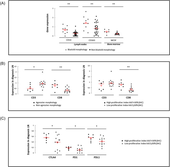
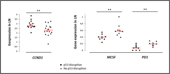
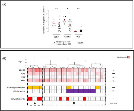
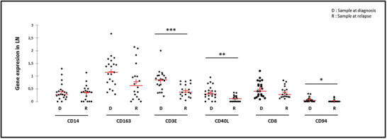
Immunotherapy strategies relying on innate or adaptive immune components are increasingly used in onco-haematology. However, little is known about the infiltrated lymph nodes (LN) or bone marrow (BM) landscape of mantle cell lymphoma (MCL). The original transcriptomic approach of reverse transcriptase multiplex ligation-dependent probe amplification (RT-MLPA) was applied here to explore the expression of 24 genes of interest in MCL at diagnosis (21 LN and 15 BM) or relapse (18 LN). This allowed us to identify that at baseline, samples from MCL patients with an aggressive morphology (i.e. blastoid or pleomorphic) or a high proliferative profile, displayed significantly higher monocyte/macrophage-associated transcripts (CD14 and CD163) in LN and BM. Regarding T-cells, aggressive MCL forms had significantly lower amounts of LN CD3E transcripts, yet an increased expression of cytotoxic markers in LN (CD8) and BM (CD94). A very high-risk group with early treatment resistance displayed, at diagnosis, high proliferation (KI67) and high macrophages and cytotoxic transcript levels. Post-immunochemotherapy relapsed samples revealed lower levels of T- and natural killer-cells markers, while monocyte/macrophage markers remained similar to diagnosis. This study suggests that rapid analysis of MCL microenvironment transcriptome signatures by RT-MLPA could allow for an early distinction of patient subgroups candidates for adapted treatment strategies.
Keywords: macrophages; mantle cell lymphoma; microenvironment; relapse; resistance.
© 2022 The Authors. eJHaem published by British Society for Haematology and John Wiley & Sons Ltd.
Conflict of interest statement
The authors declare they have no conflicts of interest.
Figures
References
-
- Le Gouill S, Thieblemont C, Oberic L, Moreau A, Bouabdallah K, Dartigeas C, et al. Rituximab after autologous stem‐cell transplantation in mantle‐cell lymphoma. N Engl J Med. 2017;377(13):1250–60. - PubMed
-
- Hoster E, Rosenwald A, Berger FO, Bernd H‐W, Hartmann S, Loddenkemper C, et al. Prognostic value of Ki‐67 index, cytology, and growth pattern in mantle‐cell lymphoma: results from randomized trials of the European Mantle Cell Lymphoma Network. J Clin Oncol. 2016;34(12):1386–94. - PubMed
-
- Le Bris Y, Magrangeas F, Moreau A, Chiron D, Theisen O, Pichon O, et al. Whole genome copy number analysis in search of new prognostic biomarkers in first‐line treatment of mantle cell lymphoma. A study by the LYSA group. Hematol Oncol. 2020;38(4):446–55. - PubMed
-
- Delfau‐Larue MH, Klapper W, Berger F, Jardin F, Briere J, Salles G, et al. High‐dose cytarabine does not overcome the adverse prognostic value of CDKN2A and TP53 deletions in mantle cell lymphoma. Blood 2015;126(5):604–11. - PubMed
LinkOut - more resources
Full Text Sources
Research Materials