. 2023 May;36(5):e4889.
doi: 10.1002/nbm.4889. Epub 2022 Dec 26.

Red cell exchange transfusions increase cerebral capillary transit times and may alter oxygen extraction in sickle cell disease

Affiliations

Red cell exchange transfusions increase cerebral capillary transit times and may alter oxygen extraction in sickle cell disease

Tonner DeBeer et al. NMR Biomed. 2023 May.

Abstract

Persons with sickle cell disease (SCD) suffer from chronic hemolytic anemia, reduced blood oxygen content, and lifelong risk of silent and overt stroke. Major conventional stroke risk factors are absent in most individuals with SCD, yet nearly 50% have evidence of brain infarcts by the age of 30 years, indicating alternative etiologies for ischemia. We investigated whether radiological evidence of accelerated blood water transit through capillaries, visible on arterial spin labeling (ASL) magnetic resonance imaging, reduces following transfusion-induced increases in hemoglobin and relates to oxygen extraction fraction (OEF). Neurological evaluation along with anatomical and hemodynamic imaging with cerebral blood flow (CBF)-weighted pseudocontinuous ASL and OEF imaging with T2 -relaxation-under-spin-tagging were applied in sequence before and after blood transfusion therapy (n = 32) and in a comparator cohort of nontransfused SCD participants on hydroxyurea therapy scanned at two time points to assess stability without interim intervention (n = 13). OEF was calculated separately using models derived from human hemoglobin-F, hemoglobin-A, and hemoglobin-S. Gray matter CBF and dural sinus signal, indicative of rapid blood transit, were evaluated at each time point and compared with OEF using paired statistical tests (significance: two-sided p < 0.05). No significant change in sinus signal was observed in nontransfused participants (p = 0.650), but a reduction was observed in transfused participants (p = 0.034), consistent with slower red cell transit following transfusion. The dural sinus signal intensity was inversely associated with OEF pretransfusion (p = 0.011), but not posttransfusion. Study findings suggest that transfusion-induced increases in total hemoglobin may lengthen blood transit times through cerebral capillaries and alter cerebral OEF in SCD.

Keywords: capillary; cerebral blood flow; oxygen extraction fraction; shunting; sickle cell disease; stroke.


Figures

Figure 1.
Figure 1.
Potential relevance of capillary transit time on arterial, venous, and tissue oxygen delivery in sickle cell disease. Warm colors depict high oxygen saturation and cool colors show low oxygen saturation. (A) In healthy brain parenchyma, capillary transit times of 0.9–1.5s lead to an oxygen extraction fraction of 30–40%. (B) In the proposed situation of compensated anemia, smooth muscle surrounding arterioles will relax to facilitate vasodilation and increase cerebral blood flow; when these changes are sufficient to maintain cerebral oxygen delivery despite anemia, normal amounts of oxygen are delivered to tissue. (C) In non-compensated anemia, the cerebral hyperemic response is typically larger and leads to a rapid transit of red cells and plasma through capillaries (white arrows). This rapid transit may result in insufficient time for oxygen to be extracted by tissue, leading to reduced oxygen delivery to brain tissue and increased oxygen concentrations in venules. In this model, the presence of arterio-venous shunting is generally associated with higher cerebral blood flow, which is demonstrated in vivo in Figure 2A. SaO2=arterial oxygen saturation. SvO2=venous oxygen saturation.
Figure 2.
Figure 2.
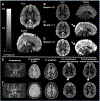
Quantification of dural sinus signal in pseudocontinuous arterial spin labeling (pCASL) data using both a (A) continuous measure of sinus signal and (B-D) categorical scoring system. (A) Location of the standardized region of interest (ROI) selected with a cross-sectional circular area of approximately 35–45 mm2. Orthogonal depictions of cerebral blood flow (CBF)-weighted pCASL maps are shown as references for shunting scores of (B) 0, (C) 1 and (D) 2, assigned according to strength and continuity of venous signal. Note that the conspicuity of the dural sinus hyperintensity generally scales with the extent of cortical hyperemia. (E) Participant with sickle cell disease (SCD) and silent cerebral infarcts on regular blood transfusion therapy with no history of vasculopathy or overt stroke. CBF-weighted pCASL maps are shown prior to and following a blood transfusion. Baseline hemoglobin (Hb) was 9.1 g/dl, which elevated to 10.2 g/dl following transfusion. White arrows indicate dural sinus hyperintensity in the superior sagittal sinus, which reduces in conspicuity post-transfusion. (F) Participant with silent cerebral infarcts and SCA stable on hydroxyurea with no blood transfusion intervention and no history of vasculopathy or overt stroke. CBF-weighted pCASL maps are shown at time 1 and time 2. This participant’s baseline hemoglobin was 11.6 g/dl, which increased slightly to 12.2 g/dl at time 2. Dural sinus hyperintensities are less conspicuous compared to the transfusion participant and are largely stable across time points. Group results are summarized quantitatively in Figure 4.
Figure 3.
Figure 3.
Arterial spin labeling images after (A) and before (B) co-registration to the 2 mm standard atlas demonstrate that dural sinus hyperintensities are conspicuous at both resolutions. It is possible to disambiguate the sinus hyperintensities from extra-axial noise and motion by ensuring that the signal traces through slice, which can be appreciated in the orthogonal planes shown. Yellow arrows depict the sagittal sinus and magenta arrows depict the straight sinus, when visible.
Figure 4.
Figure 4.
(A) Gray matter cerebral blood flow and (B) flow signal in the sagittal sinus at two time points without interval intervention or before versus after transfusion. In sickle cell disease participants not receiving interval transfusion, gray matter cerebral blood flow and sagittal sinus flow signal are not significantly different between two time points. In participants receiving interval transfusion, gray matter cerebral blood flow (p=0.031) and flow signal in the sagittal sinus (p=0.034) both reduce. Note that the flow signal is shown in calibrated units of ml/100g/min to allow for direct comparison with cerebral blood flow, however, this measurement depicts the intravascular signal and not the rate of blood delivery to tissue. * p < 0.05.
Figure 5.
Figure 5.
Before transfusion, there is an inverse relationship (p=0.011) between flow signal in the sagittal sinus and oxygen extraction fraction (OEF), whereas after transfusion (gray) the relationship becomes non-significant. Findings are shown for the human HbAA calibration model (see Methods and Discussion). Note that the flow signal is shown in calibrated units of ml/100g/min to allow for direct comparison with cerebral blood flow, however, this measurement depicts the intravascular signal and not the rate of blood delivery to tissue.

References

    1. Kassim AA, Pruthi S, Day M, Rodeghier M, Gindville MC, Brodsky MA, DeBaun MR, Jordan LC. Silent cerebral infarcts and cerebral aneurysms are prevalent in adults with sickle cell anemia. Blood. 2016;127(16):2038–40. doi: 10.1182/blood-2016-01-694562. - DOI - PubMed
    1. Jordan LC, DeBaun MR, Donahue MJ. Advances in neuroimaging to improve care in sickle cell disease. Lancet Neurol. 2021;20(5):398–408. Epub 2021/04/25. doi: 10.1016/S1474-4422(20)30490-7. - DOI - PubMed
    1. Adams RJ, McKie VC, Hsu L, Files B, Vichinsky E, Pegelow C, Abboud M, Gallagher D, Kutlar A, Nichols FT, Bonds DR, Brambilla D. Prevention of a first stroke by transfusions in children with sickle cell anemia and abnormal results on transcranial Doppler ultrasonography. The New England journal of medicine. 1998;339(1):5–11. doi: 10.1056/NEJM199807023390102. - DOI - PubMed
    1. DeBaun MR, Gordon M, McKinstry RC, Noetzel MJ, White DA, Sarnaik SA, Meier ER, Howard TH, Majumdar S, Inusa BP, Telfer PT, Kirby-Allen M, McCavit TL, Kamdem A, Airewele G, Woods GM, Berman B, Panepinto JA, Fuh BR, Kwiatkowski JL, King AA, Fixler JM, Rhodes MM, Thompson AA, Heiny ME, Redding-Lallinger RC, Kirkham FJ, Dixon N, Gonzalez CE, Kalinyak KA, Quinn CT, Strouse JJ, Miller JP, Lehmann H, Kraut MA, Ball WS Jr., Hirtz D, Casella JF. Controlled trial of transfusions for silent cerebral infarcts in sickle cell anemia. The New England journal of medicine. 2014;371(8):699–710. Epub 2014/08/21. doi: 10.1056/NEJMoa1401731. - DOI - PMC - PubMed
    1. Juttukonda MR, Lee CA, Patel NJ, Davis LT, Waddle SL, Gindville MC, Pruthi S, Kassim AA, DeBaun MR, Donahue MJ, Jordan LC. Differential cerebral hemometabolic responses to blood transfusions in adults and children with sickle cell anemia. Journal of magnetic resonance imaging : JMRI. 2019;49(2):466–77. Epub 2018/10/17. doi: 10.1002/jmri.26213. - DOI - PMC - PubMed

Publication types