. 2023 Mar;50(4):980-995.
doi: 10.1007/s00259-022-06028-9. Epub 2022 Dec 5.

EANM practice guideline for quantitative SPECT-CT

Affiliations

EANM practice guideline for quantitative SPECT-CT

John C Dickson et al. Eur J Nucl Med Mol Imaging. 2023 Mar.

Abstract

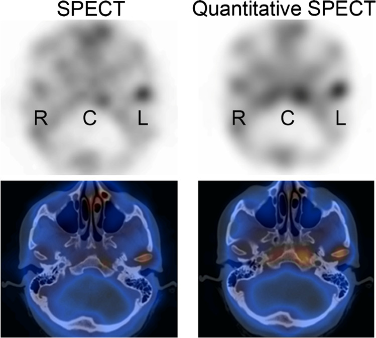
Purpose: Quantitative SPECT-CT is a modality of growing importance with initial developments in post radionuclide therapy dosimetry, and more recent expansion into bone, cardiac and brain imaging together with the concept of theranostics more generally. The aim of this document is to provide guidelines for nuclear medicine departments setting up and developing their quantitative SPECT-CT service with guidance on protocols, harmonisation and clinical use cases.

Methods: These practice guidelines were written by members of the European Association of Nuclear Medicine Physics, Dosimetry, Oncology and Bone committees representing the current major stakeholders in Quantitative SPECT-CT. The guidelines have also been reviewed and approved by all EANM committees and have been endorsed by the European Association of Nuclear Medicine.

Conclusion: The present practice guidelines will help practitioners, scientists and researchers perform high-quality quantitative SPECT-CT and will provide a framework for the continuing development of quantitative SPECT-CT as an established modality.

Keywords: Bone; Cardiology; Dosimetry; Neurology; Quantification; SPECT-CT.


Conflict of interest statement

AKK is a full-time employee at Scanomed Nuclear Medicine Center Debrecen, Hungary, a subsidiary company of Mediso Medical Imaging Systems, Budapest, Hungary. KH reports personal fees from Bayer, personal fees and other from Sofie Biosciences, personal fees from SIRTEX, non-financial support from ABX, personal fees from Adacap, personal fees from Curium, personal fees from Endocyte, grants and personal fees from BTG, personal fees from IPSEN, personal fees from Siemens Healthineers, personal fees from GE Healthcare, personal fees from Amgen, personal fees from Novartis, personal fees from ymabs, all outside the submitted work. JCD, ISA, PMG, AMD-B, JG, TvdW and L-FdG-O all declare that they do not have any conflicts of interest.

Figures

Fig. 1
Fig. 1
Example of an activity concentration recovery (ACR) curve obtained from the six hot spheres of a NEMA IEC image quality phantom filled with 10:1 contrast to background. In this example, activity concentration was measured by the maximum voxel value, which is akin to SUVmax
Fig. 2
Fig. 2
NEMA IEC image quality phantom filled with 99mTc at 10:1 contrast in three different sphere configurations reconstructed with 3 iterations and 6 subsets (upper set of images) and 20 iterations and 6 subsets (lower set of images). The corresponding recovery curves are given below for maximum voxel ACR. Note that all phantom images shown have corrections for attenuation, scatter and collimator response included and a 10 mm Gaussian filter applied
Fig. 3
Fig. 3
NEMA IEC image quality phantom filled with 99mTc at 10:1 contrast in three different sphere configurations reconstructed with 120 updates with no post-filter (upper set of images) and a 10 mm Gaussian post-filter (lower set of images). The corresponding recovery curves are given below for maximum voxel ACR
Fig. 4
Fig. 4
Bull’s eye plots of two patients in kBq/mL. The rest and stress plots are depicted pre- (left columns) and post-treatment (right columns). Moreover, the Bull’s eye plot on the right displays the difference in uptake from the subtracted (stress-rest) scans post minus pre-treatment. The patient in the top row was considered clinically deteriorated and the patient in the bottom row was reported as improved. Since the distribution of perfusion abnormalities of the patient in the top row in particular does not vary a lot, visual comparison is difficult without quantification
Fig. 5
Fig. 5
Bone SPECT-CT in a patient with mandibular growth asymmetry to the right side, showing unilateral increased uptake in the left mandibular condyle (L). The right (R) condyle and clivus (C) are shown as reference regions. In the left image SPECT-only reconstruction of counts without correction (L/Total: 60.0%; L/Clivus: 55.4%), while on the right quantitative SPECT-CT images (kBq/mL) with attenuation, scatter and resolution modelling (L/Total: 52.6%; L/Clivus: 45.5%). No partial volume correction was used. Using the traditional threshold of > 55% between the affected side and the total activity in the condyles or clivus would yield different interpretations in this example (most likely by the correction of activity in the clivus due to attenuation correction). This illustrates the need of redefining diagnostic cut-offs with novel reconstruction methods, ideally moving towards age-standardised absolute thresholds of normal condylar activity

References

    1. Bailey DL, Willowson KP. An evidence-based review of quantitative SPECT imaging and potential clinical applications. J Nucl Med. 2013;54(1):83–89. doi: 10.2967/jnumed.112.111476. - DOI - PubMed
    1. Capoccetti F, Biggi E, Rossi G, Manni C, Brianzoni E. Differentiated thyroid carcinoma: diagnosis and dosimetry using 124I PET/CT. Clin Transl Imaging. 2013;1(3):185–193. doi: 10.1007/s40336-013-0021-3. - DOI
    1. Liu T, Karlsen M, Karlberg AM, Redalen KR. Hypoxia imaging and theranostic potential of [64Cu][Cu(ATSM)] and ionic Cu(II) salts: a review of current evidence and discussion of the retention mechanisms. EJNMMI Res. 2020;10(1):33. doi: 10.1186/s13550-020-00621-5. - DOI - PMC - PubMed
    1. Blaire T, Bailliez A, Ben Bouallegue F, Bellevre D, Agostini D, Manrique A. First assessment of simultaneous dual isotope (123I/99mTc) cardiac SPECT on two different CZT cameras: a phantom study. J Nucl Cardiol. 2018;25(5):1692–1704. doi: 10.1007/s12350-017-0841-z. - DOI - PubMed
    1. Dewaraja YK, et al. MIRD pamphlet No. 24: Guidelines for quantitative 131I SPECT in dosimetry applications. J Nucl Med. 2013;54(12):2182–2188. doi: 10.2967/jnumed.113.122390. - DOI - PMC - PubMed

LinkOut - more resources