. 2023 Mar;17(1):137-150.
doi: 10.1007/s12079-022-00713-y. Epub 2022 Dec 5.

CCN2/CTGF promotes liver fibrosis through crosstalk with the Slit2/Robo signaling

Affiliations

CCN2/CTGF promotes liver fibrosis through crosstalk with the Slit2/Robo signaling

Liya Pi et al. J Cell Commun Signal. 2023 Mar.

Abstract

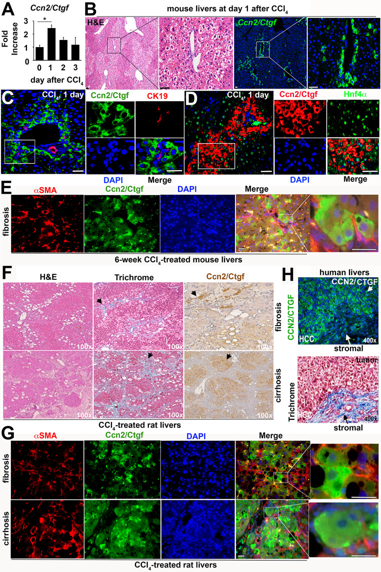
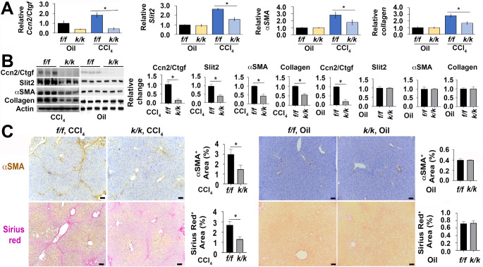
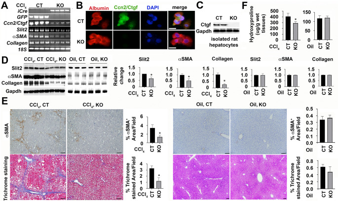
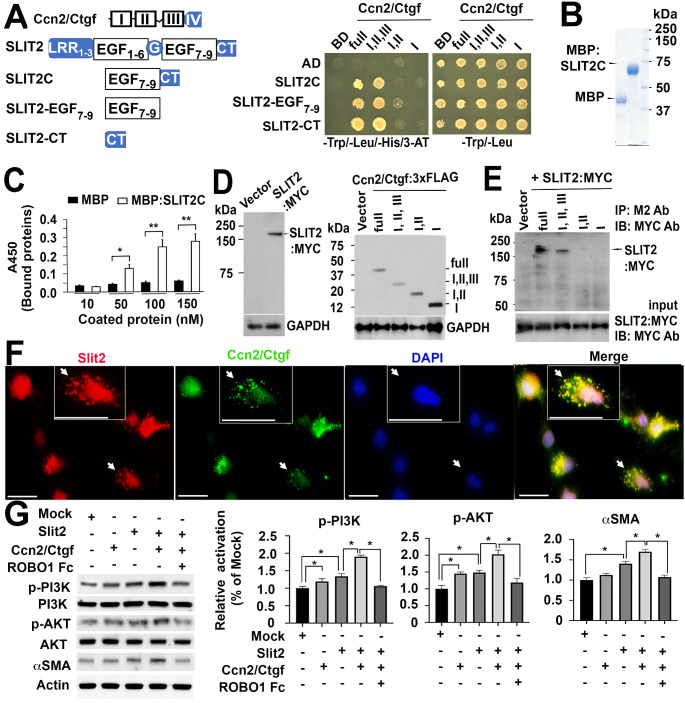
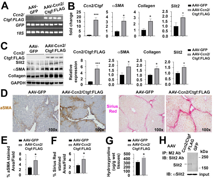
Liver fibrosis is the common outcome of many chronic liver diseases, resulting from altered cell-cell and cell-matrix interactions that promote hepatic stellate cell (HSC) activation and excessive matrix production. This study aimed to investigate functions of cellular communication network factor 2 (CCN2)/Connective tissue growth factor (CTGF), an extracellular signaling modulator of the CYR61/CTGF/Nov (CCN) family, in liver fibrosis. Tamoxifen-inducible conditional knockouts in mice and hepatocyte-specific deletion of this gene in rats were generated using the Cre-lox system. These animals were subjected to peri-central hepatocyte damage caused by carbon tetrachloride. Potential crosstalk of this molecule with a new profibrotic pathway mediated by the Slit2 ligand and Roundabout (Robo) receptors was also examined. We found that Ccn2/Ctgf was highly upregulated in periportal hepatocytes during carbon tetrachloride-induced hepatocyte damage, liver fibrosis and cirrhosis in mice and rats. Overexpression of this molecule was observed in human hepatocellular carcinoma (HCC) that were surrounded with fibrotic cords. Deletion of the Ccn2/Ctgf gene significantly reduced expression of fibrosis-related genes including Slit2, a smooth muscle actin (SMA) and Collagen type I during carbon tetrachloride-induced liver fibrosis in mice and rats. In addition, Ccn2/Ctgf and its truncated mutant carrying the first three domains were able to interact with the 7th -9th epidermal growth factor (EGF) repeats and the C-terminal cysteine knot (CT) motif of Slit2 protein in cultured HSC and fibrotic murine livers. Ectopic expression of Ccn2/Ctgf protein upregulated Slit2, promoted HSC activation, and potentiated fibrotic responses following chronic intoxication by carbon tetrachloride. Moreover, Ccn2/Ctgf and Slit2 synergistically enhanced activation of phosphatidylinositol 3-kinase (PI3K) and AKT in primary HSC, whereas soluble Robo1-Fc chimera protein could inhibit these activities. These observations demonstrate conserved cross-species functions of Ccn2/Ctgf protein in rodent livers. This protein can be induced in hepatocytes and contribute to liver fibrosis. Its novel connection with the Slit2/Robo signaling may have therapeutic implications against fibrosis in chronic liver disease.

Keywords: Carbon tetrachloride; Cellular communication network factor 2 (CCN2)/Connective tissue growth factor (CTGF); Hepatic stellate cells; Hepatocellular carcinoma; Liver fibrosis; Robo1; Slit2.


Figures

Fig. 1
Fig. 1
Ccn2/Ctgf is induced during hepatocyte damage, liver fibrosis and cirrhosis of rodent models as well as in human HCC. C57BL6 mice (n = 5) in (A-D) were subjected to IP injection of one dose of CCl4 (1ml/g body weight). (A) qRT-PCR analysis detected upregulation of Ccn2/Ctgf transcript at 24 h post CCl4 intoxication from 5 mice. Values are means ± SD from triplicate studies. *P < 0.05. (B) H&E and immunofluorescent staining detected Ccn2/Ctgf localization in periportal zones (PT) using consecutive slides. (C and D) The immunofluorescent staining labeled Ccn2/Ctgf expression in Hnf4a+ hepatocytes but not CK19+ cholangiocytes. (E) Dual staining for Ccn2/Ctgf and aSMA in fibrotic mouse livers (n = 3) that received 6-week CCl4 intoxication (0.5 ml/g body weight, twice/week). (F) Trichrome and Ccn2/Ctgf staining were performed in liver fibrosis or cirrhosis sections from Sprague Dawley (SD) rats (n = 3 per group) that were exposed to chronic CCl4 (twice a week, 0.5ml/g body weight) for 6 weeks or 3 months respectively. (G) Dual staining for Ccn2/Ctgf and aSMA was carried out on SD rats (n = 3 per group) that received chronic CCl4 for 6 weeks (upper panel) or 90 days (lower panel) respectively. (H) CCN2/CTGF and Trichrome staining were performed on consecutive sections of human HCC section (n = 2). Scale bar: 100 mm
Fig. 2
Fig. 2
Ccn2/Ctgf conditional knockouts exhibited reduced myofibroblast activation and liver fibrosis as evidenced by low production of Slit2, aSMA and Collagen type I at mRNA and protein levels in adult mice after chronic CCl4 intoxication. The conditional knockout mice (Ccn2/Ctgfk/k, n = 3) and floxed littermates (Ccn2/Ctgff/f, n = 3) were subjected to chronic intoxication by IP injection of CCl4 (0.5ul/ug body weight, twice a week) for 6 weeks. Olive oil was injected in additional experiments (n = 3 per group). (A) Downregulated transcripts of Slit2, aSMA and Collagen type I genes were found in the CCl4-treated Ccn2/Ctgfk/k livers by qRT-PCR analysis. In contrast, no difference was found compared to additional oil-treated groups. Values represent means ± SD in relation to oil treated Ccn2/Ctgff/f controls from three independent experiments. *P < 0.05. (B) Lower levels of these gene products were also detected compared to controls in Western analysis. Graphs are from densitometric analyses based on band intensity. Data are expressed as means ± SD in relation to Ccn2/Ctgff/f controls in corresponding CCl4 and oil treated Ccn2/Ctgfk/k groups from three different studies. *P < 0.05. (C) Reduced levels of aSMA and Collagen were found in IHC and Sirius Red staining of the CCl4-treated Ccn2/Ctgfk/k livers in comparison to the CCl4-treated Ccn2/Ctgff/f controls. Values were means ± SEM based on quantification of images from more than 10 fields per mouse (n = 3 mice per group). *P < 0.05. Scale bar: 500 mm in (C)
Fig. 3
Fig. 3
The hepatocyte-specific deletion of Ccn2/Ctgf gene is associated with low production of Slit2, aSMA, and Collagen type I genes during CCl4-induced liver fibrosis in rats. The hepatocyte-specific rat knockouts (KO, n = 4) were generated after tail vein injection of AAV8-iCre or AAV8-GFP as control (CT). These AAV8 infected livers received CCl4 intoxication for six weeks. Oil was also injected in parallel experiments. (A)The presence of iCre, decrease of the full-length Ccn2/Ctgf, and downregulation of Slit2, aSMA and Collagen type I genes were found in the KO livers by semi qRT-PCR analyses after chronic CCl4 intoxication. Graphs show relative changes that were expressed as means ± SD in relation to CT groups in corresponding CCl4 and oil-treated conditions (n = 3–4 per group). *P < 0.05. (B and C) Loss of Ccn2/Ctgf protein in albumin+ primary KO hepatocytes were confirmed by double staining (B) and Western blotting from total protein extracts of two different wells of the cultured primary KO hepatocytes (C). (D) Lower levels of aSMA and Collagen type I proteins were found in the KO rat livers than CT controls in Western blotting. Graphs indicate relative changes that were quantified in densiometric analyses. Values represent means ± SD in relation to CT groups in corresponding CCl4 and oil-treated conditions (n = 3–4 per group). *P < 0.05. (E) IHC and Sirius red staining detected lower levels of aSMA and Collagen in KO compared to CT group (n = 4 per group) that received chronic CCl4. Values were means ± SEM based on quantification of images from more than 10 fields per animal. *P < 0.05. (F) Decreased Collagen deposition was observed in the rat KO (n = 4) after measurement of hepatic hydroxyproline contents. Values were calculated as mg/g wet tested livers and represented means ± SD (n = 3–4 per group). *P < 0.05. Scale bar: 30 mm in (B) and 500 mm in (E)
Fig. 4
Fig. 4
Ccn2/Ctgf binds to Slit2 and potentiates HSC activation in vitro. (A) Ccn2/Ctgf interacted with SLIT2-EGF7 − 9, SLIT2-CT and SLIT2C in yeast two-hybrid analyses. LRR: leucine rich repeat domain; G: laminin G-like module; CT: C-terminal cystein knot; AD: activation domain; BD: binding domain; Trp: tryptophan; Leu: leucine; His: histidine. (B) Purified MBP and MBP fused SLIT2C were resolved using SDS-PAGE gel electrophoresis and stained with Coomassie Brilliant Blue. (C) Dose-dependent bindings of Ccn2/Ctgf to MBP:SLIT2C in solid-phase assays. Data are presented as means ± SD in triplicate wells. *P < 0.05; **P < 0.01. (D) SLIT2:MYC, Ccn2/Ctgf, and truncated mutants were expressed in CHO cells. (E) SLIT2:MYC protein was specifically detected in complexes that were immunoprecipitated with M2 antibody (Ab) and 3xFLAG tagged Ccn2/Ctgf and Ccn2/CtgfI,II,III. MYC Ab was used in immunoblotting. Equal input of SLIT2:MYC protein was added in the immunoprecipitation assays. (F) Co-localization of Slit2 and Ccn2/Ctgf proteins in primary mouse HSCs. Arrows in images and inserts indicate the same locations. Scale bar: 20 mm. (G) Western blot analyses showed that recombinant Ccn2/Ctgf protein potentiated Slit2-stimulated phosphorylation of PI3K and AKT as well as aSMA upregulation in cultured mouse HSCs, whereas presence of ROBO1-Fc chimera protein inhibited these actions mediated by Ccn2/Ctgf and Slit2. Values were means ± SD based on densitometric quantification of band intensities related to Mock controls in three independent experiments. *P < 0.05
Fig. 5
Fig. 5
Ccn2/Ctgf binds to Slit2 and potentiates HSC activation and liver fibrosis in vivo. AAV8-Ccn2/Ctgf:FLAG or GFP control was delivered into mice (n = 4 per group). Three weeks later, the mice received 6-week intoxication with CCl4. (A) Semi qRT-PCR analysis detected Ccn2/Ctgf:FLAG and co-expressed GFP in infected livers. (B and C) Ectopic expression of Ccn2/Ctgf caused upregulation of Slit2, aSMA, and Collagen type I genes as detected by qRT-PCR (B) and Western analysis (C). The band intensity in (C) was quantified by densitometry in graphs. Data in (B and C) represent means ± SD in relation to AAV8-GFP infected controls (n = 4). *P < 0.05; ***P < 0.001. (D) IHC for aSMA and Sirius red staining. (E and F) Quantification of aSMA+ areas and Sirius red stained areas was performed. Values were means ± SEM based on quantification of images from more than 10 fields per mouse (n = 4 mice per group). *P < 0.05. (G) Measurement of hepatic hydroxyproline showed increased Collagen deposition in fibrotic livers that ectopically expressed Ccn2/Ctgf:FLAG. Data are means ± SD (n = 4 mice per group). *P < 0.05. (H) Slit2 and Ccn2/Ctgf:FLAG were pulled down together in total protein lysates isolated from the AAV8-Ccn2/Ctgf:FLAG- or AAV8-GFP-infected livers (upper panel). The M2 antibody conjugated agarose was used for immunoprecipitation and a rabbit Slit2 antibody for immunoblotting. Equal input for cell lysates containing Slit2 protein in the assays were used (lower panel)

Similar articles

Cited by

References

    1. Abreu JG, Ketpura NI, Reversade B, De Robertis EM. ’connective-tissue growth factor (CTGF) modulates cell signalling by BMP and TGF-beta’. Nat Cell Biol. 2002;4:599–604. doi: 10.1038/ncb826. - DOI - PMC - PubMed
    1. Avci ME, Konu O, Yagci T. ’quantification of SLIT-ROBO transcripts in hepatocellular carcinoma reveals two groups of genes with coordinate expression’. BMC Cancer. 2008;8:392. doi: 10.1186/1471-2407-8-392. - DOI - PMC - PubMed
    1. Brigstock DR. ’strategies for blocking the fibrogenic actions of connective tissue growth factor (CCN2): from pharmacological inhibition in vitro to targeted siRNA therapy in vivo’. J Cell Commun Signal. 2009;3:5–18. doi: 10.1007/s12079-009-0043-9. - DOI - PMC - PubMed
    1. Chang J, Lan T, Li C, Ji X, Zheng L, Gou H, Ou Y, Wu T, Qi C, Zhang Q, Li J, Gu Q, Wen D, Cao L, Qiao L, Ding Y, Wang L. ’activation of Slit2-Robo1 signaling promotes liver fibrosis’. J Hepatol. 2015;63:1413–1420. doi: 10.1016/j.jhep.2015.07.033. - DOI - PubMed
    1. Coll M, Arino S, Martinez-Sanchez C, Garcia-Pras E, Gallego J, Moles A, Aguilar-Bravo B, Blaya D, Vallverdu J, Rubio-Tomas T, Lozano JJ, Pose E, Graupera I, Fernandez-Vidal A, Pol A, Bataller R, Geng JG, Gines P, Fernandez M, Sancho-Bru P (2022) Ductular reaction promotes intrahepatic angiogenesis through Slit2-Roundabout 1 signaling. Hepatology 75:353 – 68 - PMC - PubMed