. 2023 May 21;44(20):1818-1833.
doi: 10.1093/eurheartj/ehac641.

JCAD promotes arterial thrombosis through PI3K/Akt modulation: a translational study

Affiliations

JCAD promotes arterial thrombosis through PI3K/Akt modulation: a translational study

Luca Liberale et al. Eur Heart J. .

Erratum in

Abstract

Aims: Variants of the junctional cadherin 5 associated (JCAD) locus associate with acute coronary syndromes. JCAD promotes experimental atherosclerosis through the large tumor suppressor kinase 2 (LATS2)/Hippo pathway. This study investigates the role of JCAD in arterial thrombosis.

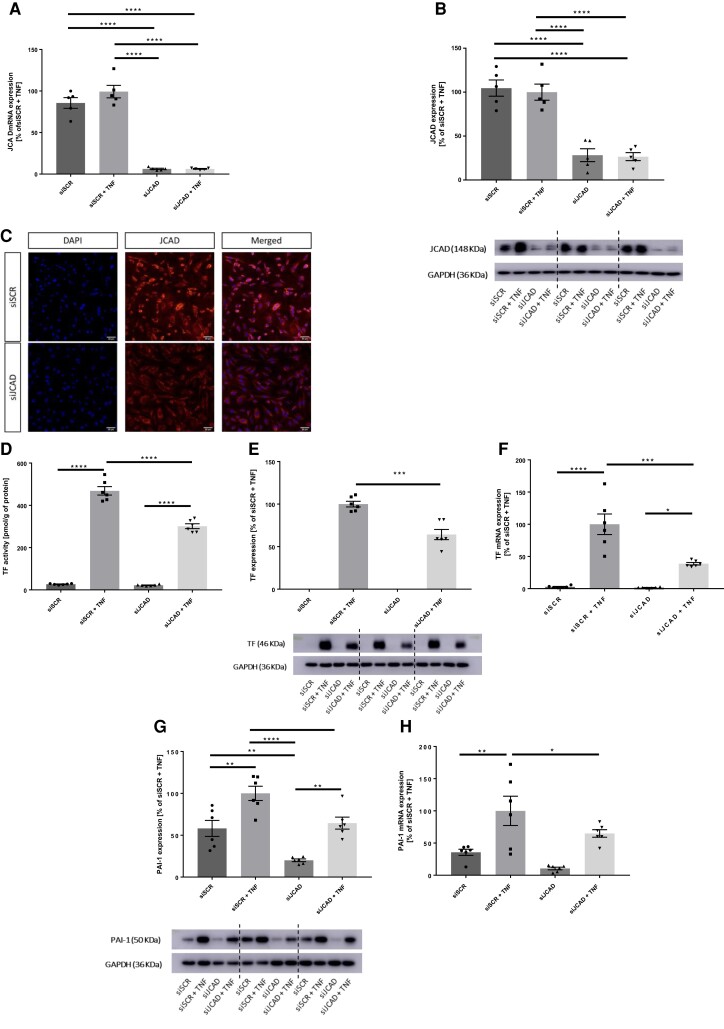
Methods and results: JCAD knockout (Jcad-/-) mice underwent photochemically induced endothelial injury to trigger arterial thrombosis. Primary human aortic endothelial cells (HAECs) treated with JCAD small interfering RNA (siJCAD), LATS2 small interfering RNA (siLATS2) or control siRNA (siSCR) were employed for in vitro assays. Plasma JCAD was measured in patients with chronic coronary syndrome or ST-elevation myocardial infarction (STEMI). Jcad-/- mice displayed reduced thrombogenicity as reflected by delayed time to carotid occlusion. Mechanisms include reduced activation of the coagulation cascade [reduced tissue factor (TF) expression and activity] and increased fibrinolysis [higher thrombus embolization episodes and D-dimer levels, reduced vascular plasminogen activator inhibitor (PAI)-1 expression]. In vitro, JCAD silencing inhibited TF and PAI-1 expression in HAECs. JCAD-silenced HAECs (siJCAD) displayed increased levels of LATS2 kinase. Yet, double JCAD and LATS2 silencing did not restore the control phenotype. si-JCAD HAECs showed increased levels of phosphoinositide 3-kinases (PI3K)/ proteinkinase B (Akt) activation, known to downregulate procoagulant expression. The PI3K/Akt pathway inhibitor-wortmannin-prevented the effect of JCAD silencing on TF and PAI-1, indicating a causative role. Also, co-immunoprecipitation unveiled a direct interaction between JCAD and Akt. Confirming in vitro findings, PI3K/Akt and P-yes-associated protein levels were higher in Jcad-/- animals. Lastly, as compared with chronic coronary syndrome, STEMI patients showed higher plasma JCAD, which notably correlated positively with both TF and PAI-1 levels.

Conclusions: JCAD promotes arterial thrombosis by modulating coagulation and fibrinolysis. Herein, reported translational data suggest JCAD as a potential therapeutic target for atherothrombosis.

Keywords: Arterial thrombosis; Cardiovascular disease; JCAD; KIAA1462; PAI-1; Tissue factor.


Conflict of interest statement

Conflict of interest: L.L. and G.G.C. are coinventors on the International Patent WO/2020/226993 filed in April 2020. The patent relates to the use of antibodies which specifically bind IL-1α to reduce various sequelae of ischemia-reperfusion injury to the central nervous system. G.G.C. is a consultant to Sovida solutions limited. T.F.L. has no conflicts related to this manuscript. Outside the work he received unrestricted research and education grants from Abbott, Amgen, Boehringer Ingelheim, Daichi-Sankyo, Novartis, Roche Diagnostics, Sanofi, Servier and Vifor. L.L. reports speaker fees outside of this work from Daichi-Sankyo. G.L. reports Consulting fees and Payment or honoraria for lectures: Novo Nordisk, Novartis, Sanofi, AstraZeneca, Boehringer, Bayer. F.C. reports speaker fees from Amgen, Astra Zeneca, Servier, BMS, other from GlyCardial Diagnostics, outside the submitted work. The other authors report no conflict of interest.

Figures

Structured Graphical Abstract
JCAD promotes arterial thrombosis through Akt modulation. PAI-1, plasminogen activator inhibitor-1; TF, tissue factor; YAP, yes-associated protein 1. Created with BioRender.com.
Figure 1
Figure 1
Effects of JCAD deletion on carotid arterial thrombosis in vivo in male mice. (A) Jcad−/− mice showed delayed time to formation of an occlusive thrombus in their carotid arteries after endothelial-specific damage as compared with Jcad+/+ littermates. (B) Representative trace of mean blood flow until occlusion (mean flow ≤ 0.1 mL for 1 min) in the two study groups. (C) JCAD deletion increased the number of episodes of thrombus embolization. (D–F) No difference in initial heart rate, blood flow and weight was reported among Jcad−/− and wild-type littermates. n = 7 different mice per group. A, CF: unpaired two-tailed Student’s t-test. *P < 0.05.
Figure 2
Figure 2
Impact of JCAD deletion on platelet count, volume and ex vivo activation and aggregation. (A and B) Jcad−/− animals did not show any difference in terms of platelet count or mean platelet volume as compared with Jcad+/+ animals. (C–E) Ex vivo collagen-induced platelet aggregation as assessed by light transmission aggregometry did not show any difference in terms of maximal aggregation, rate (slope) of aggregation and lag phase in Jcad−/− animals and WT littermates. (G–J) Ex vivo thrombin-induced platelet aggregation as assessed by light transmission aggregometry did not show any difference in terms of maximal aggregation, rate (slope) of aggregation and lag phase in Jcad−/− animals and WT littermates. (KN) FACS analysis of platelet reactivity markers after ex vivo ADP (10 µM, 20 µM) and collagen (20 µg/mL) activation in Jcad+/+ and Jcad−/− mice. Mean fluorescence intensity of CD62P was similar between groups at baseline (K and L), after ADP (K) and collagen (L) stimulation. Mean fluorescence intensity of JON/A was similar between groups at baseline (M and N), after ADP (M) and collagen (N) stimulation. n = 5 different mice per group. A-B, D–F, H–J: unpaired two-tailed Student’s t-test. K–N: paired two-tailed Student’s t-test. *P < 0.05, **P < 0.01.
Figure 3
Figure 3
Effects of JCAD deletion on extrinsic coagulation pathway and fibrinolytic system. (A) Schematic of extrinsic coagulation pathway activation (B) JCAD deletion reduced carotid artery TF activity. (C) Accordingly, arterial levels of TF were lower in Jcad−/− mice as compared with control littermates. (D) Schematic of fibrinolytic cascade (E) Plasma D-dimer concentration was significantly increased in Jcad−/− animals as compared with controls (F) Jcad−/− animals exhibited reduced arterial levels of PAI-1. (G) Phosphorylation levels of YAP were higher in Jcad−/− animals than in Jcad+/+ controls. n = 7 different mice per group. B-C, E-G: unpaired two-tailed Student’s t-test. *P < 0.05 **P < 0.01. GAPDH = glyceraldehyde 3-phosphate dehydrogenase, PAI-1 = plasminogen activator inhibitor-1; TF = tissue factor; YAP = yes-associated protein 1.
Figure 4
Figure 4
Effects of JCAD silencing in primary HACEs. (A) In HAECs, JCAD mRNA levels were significantly reduced after transfection with JCAD small interfering RNA (siJCAD) as compared with control siRNA (siSCR). (B-C) Western blotting and Immunostaining of HAECs confirmed JCAD silencing at the protein level. (D) TF activity is induced in siSCR HAECs by treatment with 10 ng/mL TNF-α, this effect is reduced upon JCAD silencing. (E) Accordingly, TF protein levels are increased in stimulated siSCR-treated cells with higher protein levels than stimulated JCAD-silenced ones. (F) TF mRNA levels follow a similar trend (G) Similarly, also TNF-α-related induction of PAI-1 is reduced in siJCAD-treated cells irrespective of TNF-α stimulation (H) PAI-1 mRNA are higher in stimulated siSCR-treated cells as compared with stimulated siJCAD ones. n = 5–6 independent experiments. A, D-H: one-way analysis of variance (ANOVA) with Tukey post hoc test (number of comparisons = 6). *P < 0.05, **P < 0.01, ***P < 0.001, ****P < 0.0001. GAPDH = glyceraldehyde 3-phosphate dehydrogenase; HAEC = human aortic endothelial cell; PAI-1 = plasminogen activator inhibitor-1; TF = tissue factor; TNF-α = tumour necrosis factor-α.
Figure 5
Figure 5
LATS2 as a mediator of JCAD in HACEs. (A) Schematic of JCAD regulation of the Hippo pathway (B) Efficacy of double JCAD and LATS2 silencing by JCAD and LATS2 small interfering RNA (siJCAD and siLATS2) as compared with control siRNA (siSCR). (C-E) Double JCAD and LATS2 silencing did not retrieve TF activity, TF expression and PAI-1 levels of siJCAD cells to those observed in siSCR-treated ones. n = 6 independent experiments. BE: one-way analysis of variance (ANOVA) with Tukey post hoc test (number of comparisons = 15 for all, exception made for D where the number of comparisons = 3). *P < 0.05, **P < 0.01, ***P < 0.001, ****P < 0.0001. GAPDH = glyceraldehyde 3-phosphate dehydrogenase; HAEC = human aortic endothelial cell; LATS-2 = large tumour suppressor kinase 2; PAI-1 = plasminogen activator inhibitor-1; TF = tissue factor; TNF-α = tumour necrosis factor-α.
Figure 6
Figure 6
PI3K/Akt pathway as a mediator of JCAD effects on HACEs. (A) JCAD silencing by mean of small interfering RNA (siJCAD) increased the levels of PI3K in HAECs stimulated with TNF-α (10 ng/mL). (B) siJCAD HAECs showed increased phosphorylation of Akt both when unstimulated or stimulated (TNF-α, 10 ng/mL) (C-E) Treatment with the PI3K/Akt inhibitor Wortmannin rescued the siSCR phenotype in siJCAD-treated HAECs in terms of TF activity, TF expression and PAI-1 levels. (F) Representative immunoblot of co-immunoprecipitation of Akt and JCAD from whole-cell lysates of stimulated and unstimulated siSCR-treated cells showing direct protein-to-protein interaction. n = 6 independent experiments. A-F: one-way analysis of variance (ANOVA) with Tukey post hoc test (number of comparisons = 6 for A-B, = 15 for C and E, = 3 for D). *P < 0.05, **P < 0.01, ***P < 0.001, ****P < 0.0001. GAPDH = glyceraldehyde 3-phosphate dehydrogenase; HAEC = human aortic endothelial cell; PAI-1 = plasminogen activator inhibitor-1; PI3K = phosphatidylinositol 3-kinase; TF = tissue factor; TNF-α = tumour necrosis factor-α.
Figure 7
Figure 7
Effect of PI3K inhibition on carotid arterial thrombosis in vivo in Jcad-/− mice. (A) JCAD deletion increases arterial levels of PI3K in mice. (B) JCAD deletion increases arterial levels of phosphorylated Akt in mice. (C) Jcad−/− mice showed delayed time to formation of an occlusive thrombus in their carotid arteries after endothelial-specific damage as compared with Jcad+/+ littermates. Such an effect was eliminated by pre-treatment with the Akt inhibitor wortmannin. (D) Pre-treatment with wortmannin associated with a trend toward a reduced number of thrombus embolization episodes in Jcad−/− mice. (E-F) No difference in terms of initial heart rate and blood flow was recorded among the three study groups. n = 8 different mice per group. A-B: unpaired two-tailed Student’s t-test. C-F: one-way analysis of variance (ANOVA) with Tukey post hoc test (number of comparisons = 3). **P < 0.01. PI3K = phosphatidylinositol 3-kinase.
Figure 8
Figure 8
JCAD, TF and PAI-1 expression in patients with STEMI. (A) Circulating JCAD levels are increased in STEMI patients as compared with CCS controls. (B-C) TF and PAI-1 protein levels are increased across JCAD tertiles. n = 22 patients for CCS and n = 39 patients for STEMI. A-C: Analysis of covariance (ANCOVA). CCS = chronic coronary syndrome; PAI-1 = plasminogen activator inhibitor-1; STEMI = ST-elevation myocardial infarction; TF = tissue factor.

Comment in

References

    1. Wang M, Hao H, Leeper NJ, Zhu L, Early Career C. Thrombotic regulation from the endothelial cell perspectives. Arterioscler Thromb Vasc Biol 2018;38:e90–e95. 10.1161/ATVBAHA.118.310367 - DOI - PMC - PubMed
    1. Breitenstein A, Camici GG, Tanner FC. Tissue factor: beyond coagulation in the cardiovascular system. Clin Sci (Lond) 2009;118:159–172. 10.1042/CS20080622 - DOI - PubMed
    1. Timmis A, Townsend N, Gale C, Grobbee R, Maniadakis N, Flather M, et al. European Society of cardiology: cardiovascular disease statistics 2017. Eur Heart J 2018;39:508–579. 10.1093/eurheartj/ehx628 - DOI - PubMed
    1. Erdmann J, Willenborg C, Nahrstaedt J, Preuss M, Konig IR, Baumert J, et al. Genome-wide association study identifies a new locus for coronary artery disease on chromosome 10p11.23. Eur Heart J 2011;32:158–168. 10.1093/eurheartj/ehq405 - DOI - PubMed
    1. Nelson CP, Goel A, Butterworth AS, Kanoni S, Webb TR, Marouli E, et al. Association analyses based on false discovery rate implicate new loci for coronary artery disease. Nat Genet 2017;49:1385–1391. 10.1038/ng.3913 - DOI - PubMed

Publication types

MeSH terms