. 2022 Dec 13;119(50):e2211018119.
doi: 10.1073/pnas.2211018119. Epub 2022 Dec 5.

A bacterium from a mountain lake harvests light using both proton-pumping xanthorhodopsins and bacteriochlorophyll-based photosystems

Affiliations

A bacterium from a mountain lake harvests light using both proton-pumping xanthorhodopsins and bacteriochlorophyll-based photosystems

Karel Kopejtka et al. Proc Natl Acad Sci U S A. .

Abstract

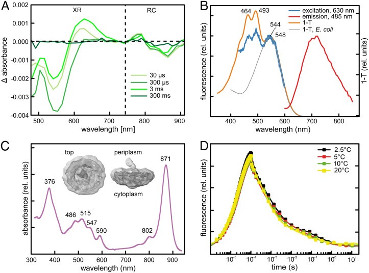
Photoheterotrophic bacteria harvest light energy using either proton-pumping rhodopsins or bacteriochlorophyll (BChl)-based photosystems. The bacterium Sphingomonas glacialis AAP5 isolated from the alpine lake Gossenköllesee contains genes for both systems. Here, we show that BChl is expressed between 4°C and 22°C in the dark, whereas xanthorhodopsin is expressed only at temperatures below 16°C and in the presence of light. Thus, cells grown at low temperatures under a natural light-dark cycle contain both BChl-based photosystems and xanthorhodopsins with a nostoxanthin antenna. Flash photolysis measurements proved that both systems are photochemically active. The captured light energy is used for ATP synthesis and stimulates growth. Thus, S. glacialis AAP5 represents a chlorophototrophic and a retinalophototrophic organism. Our analyses suggest that simple xanthorhodopsin may be preferred by the cells under higher light and low temperatures, whereas larger BChl-based photosystems may perform better at lower light intensities. This indicates that the use of two systems for light harvesting may represent an evolutionary adaptation to the specific environmental conditions found in alpine lakes and other analogous ecosystems, allowing bacteria to alternate their light-harvesting machinery in response to large seasonal changes of irradiance and temperature.

Keywords: anoxygenic photosynthesis; bacteriochlorophyll a; dual phototrophy; light energy; xanthorhodopsin.


Conflict of interest statement

The authors declare no competing interest.

Figures

Fig. 1.
Fig. 1.
MAG of a dual phototrophic Sphingomonas recovered from GKS metagenomes. (A) GKS (Left). Temperature profiles of the GKS between July 2017 and July 2018 (Right). (B) Phylogenetic position of XR and PufM from one MAG in relation to the S. glacialis AAP5 proteins. For full trees, see Dataset S1. (C) Mapping of metagenome reads to the chromosome sequence of S. glacialis AAP5. MAG, metagenome-assembled genome; CPM, counts per million reads. (D) Relative abundance of Sphingomonas XR and pufM genes in three GKS metagenomes from autumn 2018 to spring 2019.
Fig. 2.
Fig. 2.
Induction of XR- and BChl a–based phototrophy. (A) Reversed-phase chromatography of pigments from cells grown under different conditions. Cells were grown at low (7°C) or high (22°C) temperature either under continuous illumination or under 12-h dark–12-h light regime. Traces are shifted vertically. Numbers above peaks indicate: 1, retinal; 2, nostoxanthin; 3, unknown carotenoid(s); 4–6, BChl a (isomers); and 7, neurosporene. (B) Expression of the XR and pufM genes in cells grown under different temperatures. Upper panel: values for XR gene, Lower panel: values for pufM gene. The ΔCt mean values and standard deviations calculated from three parallel biological replicates for different growth phases (defined in the graphical legend) are shown. Cells were grown under 12-h dark–12-h light regime and harvested 4 h after changing the light regime to light (for XR) or to dark (for pufM).
Fig. 3.
Fig. 3.
Transcriptome dynamics at low temperature under changing light regimes. (A) Growth at 7°C under 12-h light–12-h dark regime (gray bar: light and black bar: dark). The time points for sampling are indicated by black arrows. The mean values and standard deviations from three biological replicates are shown. (B) Transcriptome dynamics of chromosomal genes during different growth phases when 6 h of light is compared with 6 h of dark. Significantly differentially expressed genes are shown as blue dots. Genes of PGC and the XR locus are highlighted in pink and orange, respectively. (C) Expression of XR locus and PGC from samples taken after 1 h and 6 h in the light compared with the previous dark period. Individual and groups of genes are defined in the graphical legend. (D) Normalized RNA-seq read mapping to the XR locus with single-nucleotide resolution. (E) Leader (rv1) and read-through (rv2) expression profile from RNA-seq (Upper panel) compared with RT-qPCR data (Lower panel). Day 4: exponential, day 5: early stationary, and day 6: late stationary phase.
Fig. 4.
Fig. 4.
PS apparatus. (A) Light-induced absorption changes elicited by a 2-ms broadband pulse in suspension of membranes covering the range where the activity of both XR and the bacterial RC can be observed. Delays are given in the legend. (B) Fluorescence excitation (blue line) and emission (red line) spectra of XR from AAP5. Orange line represents 1–T absorption spectrum. Gray line represents 1–T absorption spectrum of the heterologously expressed XR gene. Due to the presence of tetrapyrrols in the sample, the excitation spectrum was measured at 630 nm. The emission spectrum was excited by 485 nm. (C) Absorption spectrum of BChl-based RC. (Inset) Contour density of the PS complex determined by the cryo-EM. (D) BChl a fluorescence induction–relaxation kinetics recorded at various temperatures.
Fig. 5.
Fig. 5.
Impact of XR-based phototrophy. (A) Respiration activity of S. glacialis AAP5 exposed to increasing light intensity at 7°C. Orange symbols represent the activities of XR-containing cells; black symbols represent heterotrophic cells (mean values of four biological replicates). (B) ATP content in the XR-containing cells kept in the dark or exposed to white light of 500 μmol photons m−2 s−1. Three technical replicates were carried out. (C) Cultivation of S. glacialis AAP5 at 7°C under continuous light and dark. Light intensity was 500 μmol photons m−2 s−1. The mean values and standard deviations from three biological replicates are shown.
Fig. 6.
Fig. 6.
Expression regulation of dual phototrophy in S. glacialis AAP5. (A) Proposed scheme of anoxygenic photosynthesis (AP) and XR regulation by nutrients, light, and temperature. (B) Hypothetical model of an alpine lake with the main environmental factors (temperature, irradiance, and snow cover) affecting the expression of dual phototrophy during the seasons. The thin black lines indicate ice cover. Cyt, cytochrome; QH2, ubiquinol.

Comment in

References

    1. Gardiner A. T., Nguyen-Phan T. C., Cogdell R. J., A comparative look at structural variation among RC–LH1 ‘Core’ complexes present in anoxygenic phototrophic bacteria. Photosynth. Res. 145, 83–96 (2020). - PMC - PubMed
    1. Blankenship R. E., Early evolution of photosynthesis. Plant Physiol. 154, 434–438 (2010). - PMC - PubMed
    1. Yurkov V. V., Csotonyi J. T., “New light on aerobic anoxygenic phototrophs” in The Purple Phototrophic Bacteria, Hunter C. N., Daldal F., , M. C. Thurnauer, Beatty J. T., Eds. (Advances in Photosynthesis and Respiration, Springer, 2009), vol. 28, pp. 31–55.
    1. Koblížek M., Ecology of aerobic anoxygenic phototrophs in aquatic environments. FEMS Microbiol. Rev. 39, 854–870 (2015). - PubMed
    1. Kolber Z. S., et al. , Contribution of aerobic photoheterotrophic bacteria to the carbon cycle in the ocean. Science 292, 2492–2495 (2001). - PubMed

Publication types

LinkOut - more resources