. 2023 Mar;615(7950):134-142.
doi: 10.1038/s41586-022-05594-0. Epub 2022 Dec 5.

FXR inhibition may protect from SARS-CoV-2 infection by reducing ACE2

Teresa Brevini  1 Mailis Maes  2 Gwilym J Webb  3 Binu V John  4 Claudia D Fuchs  5 Gustav Buescher  6 Lu Wang  7 Chelsea Griffiths  7 Marnie L Brown  7 William E Scott 3rd  7 Pehuén Pereyra-Gerber  2 William T H Gelson  3   8 Stephanie Brown  9 Scott Dillon  9 Daniele Muraro  10 Jo Sharp  11 Megan Neary  11 Helen Box  11 Lee Tatham  11 James Stewart  12 Paul Curley  11 Henry Pertinez  11 Sally Forrest  2 Petra Mlcochova  2   4 Sagar S Varankar  9 Mahnaz Darvish-Damavandi  9   13 Victoria L Mulcahy  14 Rhoda E Kuc  15 Thomas L Williams  15 James A Heslop  9 Davide Rossetti  9 Olivia C Tysoe  9   16 Vasileios Galanakis  9 Marta Vila-Gonzalez  9 Thomas W M Crozier  2 Johannes Bargehr  9   8   17 Sanjay Sinha  9   17 Sara S Upponi  18 Corrina Fear  16 Lisa Swift  16 Kourosh Saeb-Parsy  16   19 Susan E Davies  20 Axel Wester  21 Hannes Hagström  21 Espen Melum  22   23   24   25   26 Darran Clements  9 Peter Humphreys  9 Jo Herriott  11 Edyta Kijak  11 Helen Cox  11 Chloe Bramwell  11 Anthony Valentijn  11 Christopher J R Illingworth  27   28 UK-PBC ConsortiumBassam Dahman  29 Dustin R Bastaich  29 Raphaella D Ferreira  4 Thomas Marjot  30 Eleanor Barnes  30 Andrew M Moon  31 Alfred S Barritt 4th  31 Ravindra K Gupta  2   8 Stephen Baker  2 Anthony P Davenport  15 Gareth Corbett  32 Vassilis G Gorgoulis  33   34   35 Simon J A Buczacki  9   13 Joo-Hyeon Lee  9   36 Nicholas J Matheson  2   8   31   37 Michael Trauner  5 Andrew J Fisher  7 Paul Gibbs  16   19 Andrew J Butler  16   19 Christopher J E Watson  16   19   38 George F Mells  3   14 Gordon Dougan  2 Andrew Owen  11 Ansgar W Lohse  6 Ludovic Vallier  39   40   41   42 Fotios Sampaziotis  43   44   45
Affiliations

FXR inhibition may protect from SARS-CoV-2 infection by reducing ACE2

Teresa Brevini et al. Nature. 2023 Mar.

Abstract

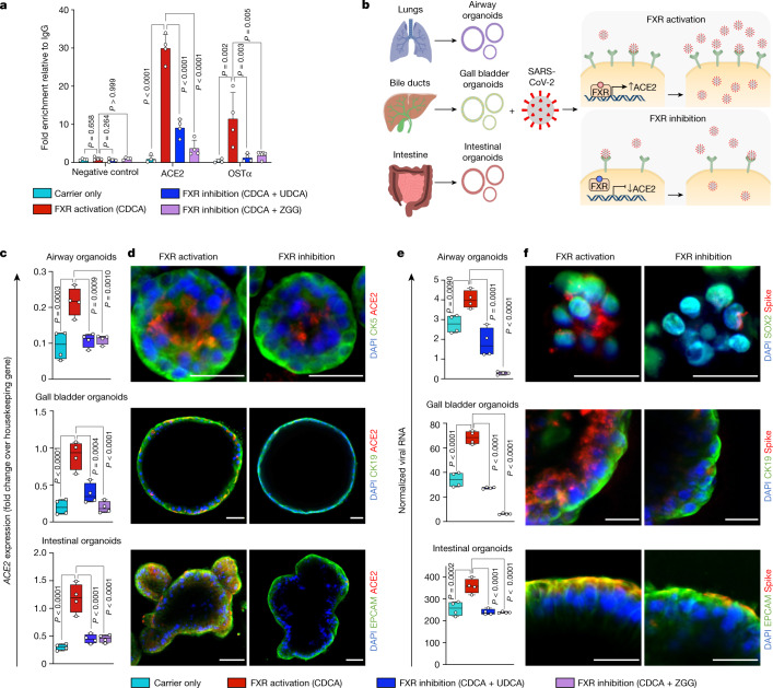
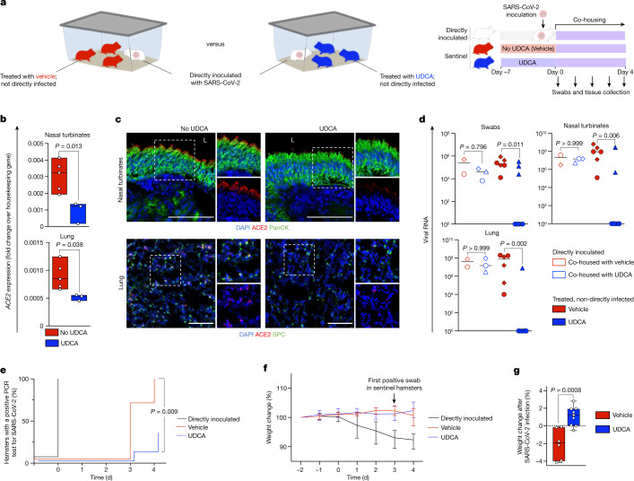
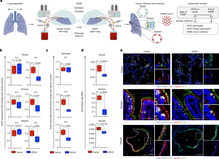
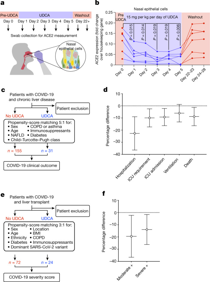
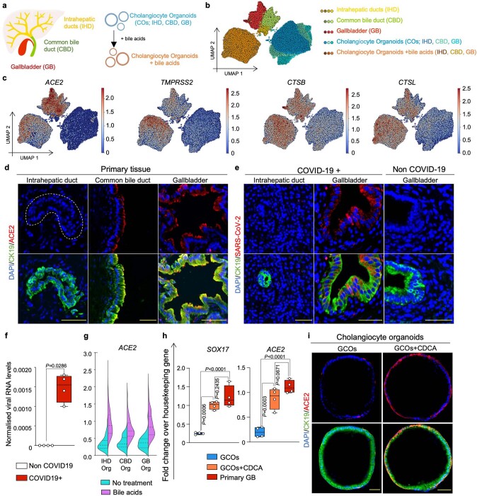
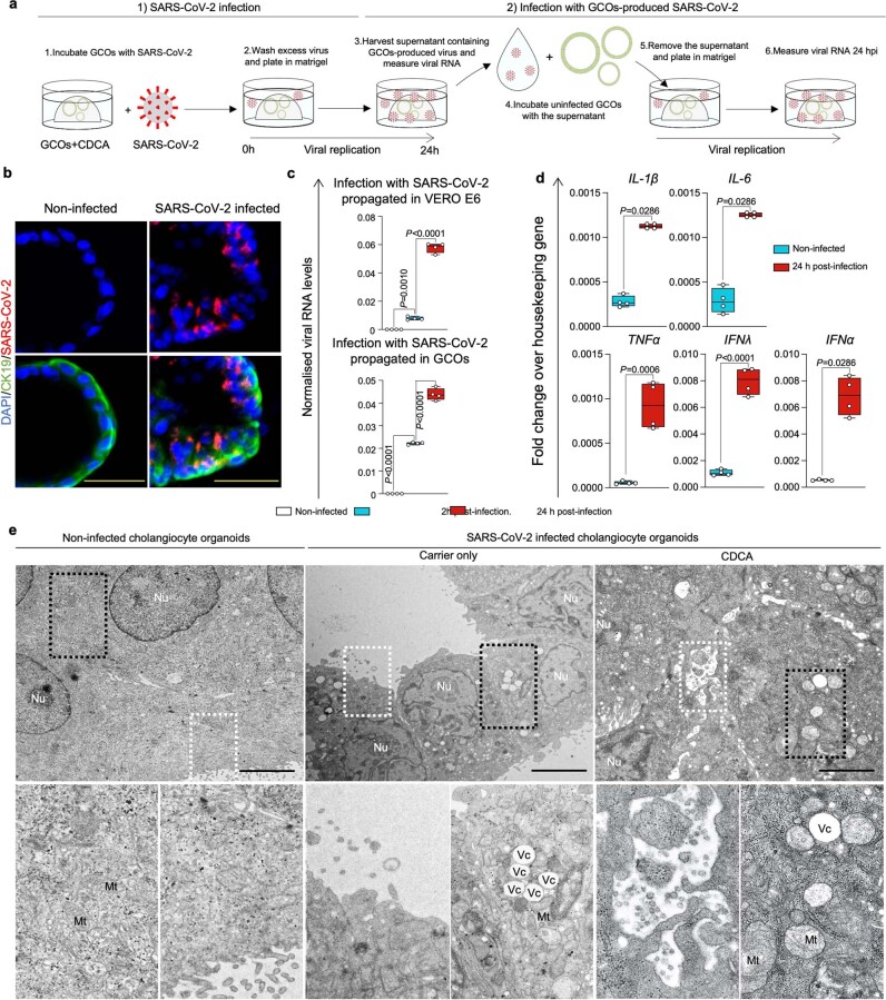
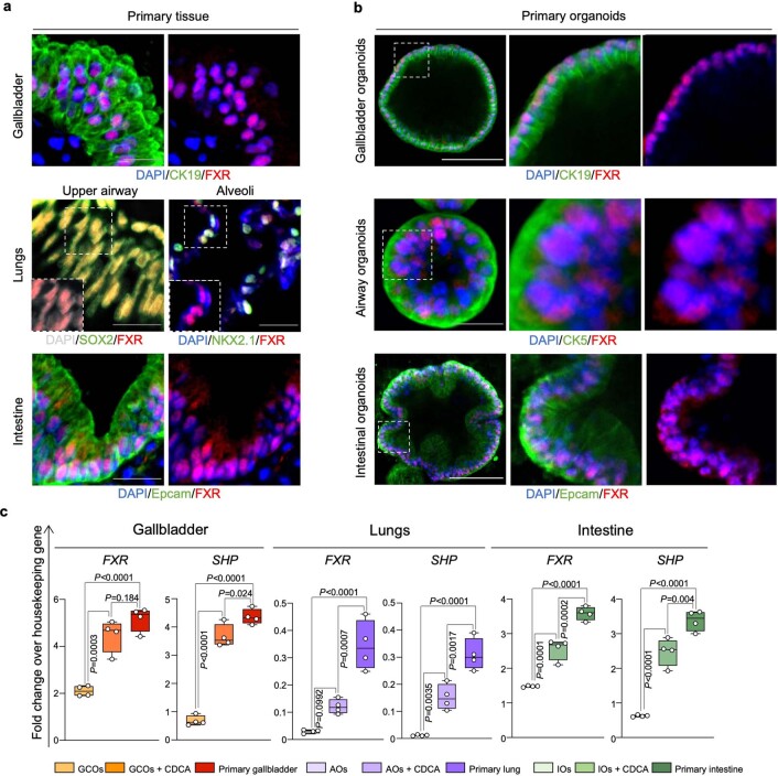
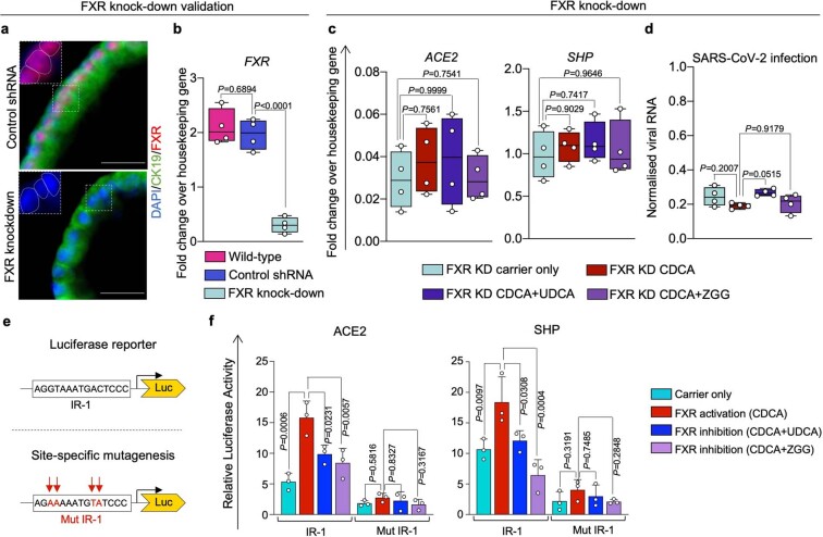
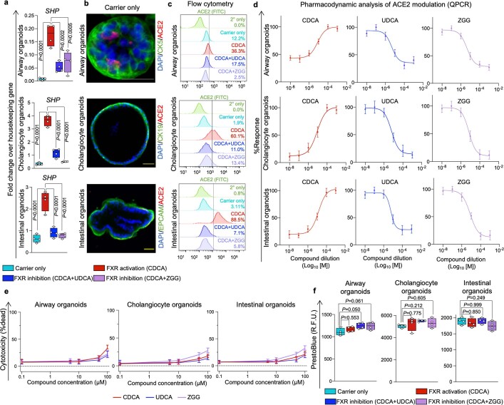
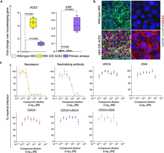
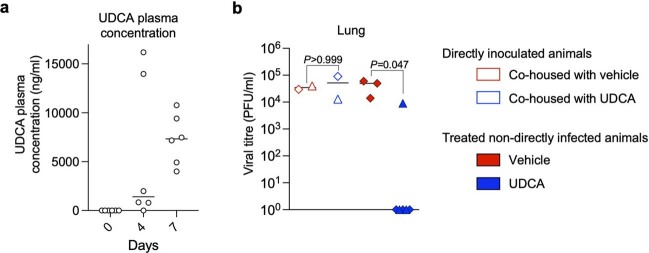
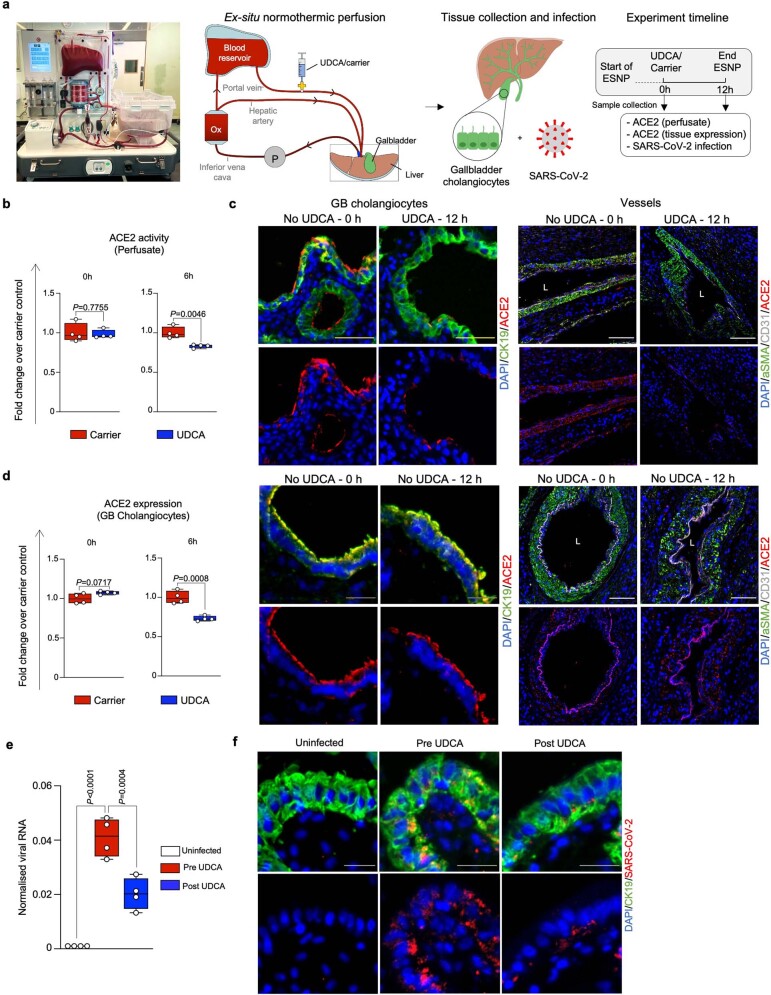
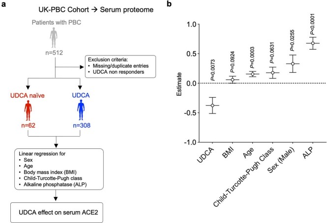
Preventing SARS-CoV-2 infection by modulating viral host receptors, such as angiotensin-converting enzyme 2 (ACE2)1, could represent a new chemoprophylactic approach for COVID-19 that complements vaccination2,3. However, the mechanisms that control the expression of ACE2 remain unclear. Here we show that the farnesoid X receptor (FXR) is a direct regulator of ACE2 transcription in several tissues affected by COVID-19, including the gastrointestinal and respiratory systems. We then use the over-the-counter compound z-guggulsterone and the off-patent drug ursodeoxycholic acid (UDCA) to reduce FXR signalling and downregulate ACE2 in human lung, cholangiocyte and intestinal organoids and in the corresponding tissues in mice and hamsters. We show that the UDCA-mediated downregulation of ACE2 reduces susceptibility to SARS-CoV-2 infection in vitro, in vivo and in human lungs and livers perfused ex situ. Furthermore, we reveal that UDCA reduces the expression of ACE2 in the nasal epithelium in humans. Finally, we identify a correlation between UDCA treatment and positive clinical outcomes after SARS-CoV-2 infection using retrospective registry data, and confirm these findings in an independent validation cohort of recipients of liver transplants. In conclusion, we show that FXR has a role in controlling ACE2 expression and provide evidence that modulation of this pathway could be beneficial for reducing SARS-CoV-2 infection, paving the way for future clinical trials.


Conflict of interest statement

F.S., L.V. and K.S.-P. are founders and shareholders of Bilitech. L.V. is a founder and shareholder of DEFINIGEN. The remaining authors declare no competing interests.

Figures

Fig. 1
Fig. 1. FXR modulates ACE2 expression and SARS-CoV-2 infection.
a, ChIP–qPCR on cholangiocyte organoids, showing that the FXR agonist CDCA promotes the binding of FXR on the ACE2 promoter, and that this is reduced by FXR inhibitors (UDCA and ZGG). OSTα as positive control; ACE2 promoter adjoining region as negative control; n = 4 independent experiments; one-way ANOVA adjusted for multiple comparisons; bars,s.d. b, Schematic representation of the suggested mechanism for FXR-mediated control of ACE2 expression and SARS-CoV-2 infection relative to e,f. c,d, qPCR (c) and immunofluorescence (d) showing the levels of ACE2 after modulation of FXR activity in primary airway, biliary and intestinal organoids. Housekeeping gene, HMBS (also known as PBGD); n = 4 independent experiments; one-way ANOVA; centre line, median; box, interquartile range (IQR); whiskers, range; bars, s.d. Yellow scale bars, 50 μm; grey scale bars, 25 μm. e, qPCR quantifying SARS-CoV-2 viral RNA 24 h after infection in primary organoids treated with physiological levels of bile acids (CDCA) in the presence or absence of FXR inhibitors (UDCA and ZGG). Housekeeping gene, GAPDH; n = 4 independent experiments; one-way ANOVA adjusted for multiple comparisons; centre line, median; box, interquartile range (IQR); whiskers, range; bars, s.d. f, Immunofluorescence images showing the presence of SARS-CoV-2 spike protein 24 hours after infection in organoids corresponding to e. Scale bars, 25 μm. CDCA, UDCA and ZGG concentration, 10 μM.
Fig. 2
Fig. 2. Inhibition of FXR reduces ACE2 expression and SARS-CoV-2 infection in vivo.
a, Schematic of the experiment performed in Syrian golden hamsters. Sentinel hamsters were not directly inoculated with virus. SARS-CoV-2 infection in sentinel hamsters was achieved through transmission from directly inoculated hamsters after co-housing. b, qPCR showing that treatment with UDCA reduces the levels of ACE2 in hamster nasal turbinates and lungs. Housekeeping gene, Gapdh; n = 5 vehicle (no UDCA) group versus n = 3 UDCA group; unpaired two-tailed t-test; centre line, median; box, interquartile range; whiskers, range; bars, s.d. c, Immunofluorescence images showing the levels of ACE2 in nasal and respiratory epithelium of hamsters receiving UDCA versus vehicle. n = 3 hamsters per group. Scale bars, 100 μm. d, qPCR showing the levels of SARS-CoV-2 RNA in swabs, nasal turbinates and lungs of directly inoculated hamsters and sentinel hamsters treated with UDCA or vehicle and co-housed with infected hamsters. Samples were collected after four days of co-housing. SARS-CoV-2 nucleocapsid RNA quantification relative to 18s rRNA. n = 3 hamsters per group; n = 9 UDCA, n = 6 vehicle hamsters; hamsters from each experiment are represented with different symbols; Kruskal–Wallis test adjusted for multiple comparisons. e, Kaplan–Meier curve showing the percentage of hamsters with a PCR-positive swab for SARS-CoV-2 over the course of the experiment outlined in a. n = 9 UDCA, n = 6 vehicle, n = 5 directly inoculated hamsters; log-rank Mantel–Cox test comparing UDCA versus vehicle. f, Percentage weight change from the start of the experiment outlined in a. Bars, range. Day 0 corresponds to the start of co-housing. g, Percentage weight change after SARS-CoV-2 infection in sentinel hamsters. The time of infection was defined as the earliest day on which a sentinel hamster had a positive swab (day 3 for both UDCA and vehicle groups). n = 3 independent experiments; n = 9 UDCA, n = 6 vehicle, n = 5 directly inoculated hamsters; unpaired two-tailed t-test; centre line, median; box, interquartile range; whiskers, range; bars, s.d. Source data
Fig. 3
Fig. 3. FXR inhibition reduces ACE2 levels and SARS-CoV-2 infection in a human lung ex vivo.
a, Schematic representation of the lung ESNP experiment, including type of samples and timeline. 0 h: baseline sample collection and UDCA or carrier administration. The 0-h samples were collected before administration of UDCA. 6 h: 6 h after UDCA or carrier administration. For each time point, four independent tissue samples were obtained from the lung parenchyma (alveoli), the airways and the vessels for each lung and used for ACE2 measurement and viral infection (n = 4 lung parenchyma, n = 4 airway and n = 4 pulmonary vessel samples per lung per time point). b, qPCR showing that treatment with UDCA reduces the levels of ACE2 in human alveoli, airway and pulmonary vessels perfused ex situ. Housekeeping gene, GAPDH; n = 4 independent samples; unpaired two-tailed t-test; centre line, median; box, interquartile range (IQR); whiskers, range; error bars, s.d. c, ACE2 enzymatic activity in the perfusate, showing that UDCA reduces ACE2. n = 4 independent samples; unpaired two-tailed t-test; centre line, median; box, interquartile range (IQR); whiskers, range; bars, s.d. d, qPCR showing that 6 h of ESNP with UDCA reduces SARS-CoV-2 infection in human alveoli, airway and pulmonary vessels ex vivo. Housekeeping gene, GAPDH. n = 4 independent samples; unpaired two-tailed t-test. e, Immunofluorescence staining for ACE2 and SARS-CoV-2 in human alveoli, airway and pulmonary vessels after ESNP (6 h) with UDCA or carrier. n = 4 independent samples. White scale bars, 100 μm, yellow scale bars, 50 μm. UDCA concentration, 2,000 ng ml−1. AcTub, acetylated α-tubulin; L, lumen.
Fig. 4
Fig. 4. UDCA is associated with lower levels of ACE2 and a better clinical outcome in patients with COVID-19.
a,b, Schematic representation of the study design. Six healthy individuals received 15 mg per kg per day of UDCA for 5 days. ACE2 levels were measured by qPCR in nasal epithelial cells collected with nasopharyngeal swabs. Day 0 corresponds to samples collected immediately before starting UDCA treatment. Samples were collected daily during drug administration and again at day 22–23 and 24–28 to assess the washout of UDCA. b, qPCR measurement of the levels of ACE2 in nasal epithelial cells collected with nasopharyngeal swabs. Each dot represents one individual measurement; lines connect dots from the same individual (n = 6). Housekeeping gene, GAPDH; n = 6 individuals; one-way ANOVA with Geisser–Greenhouse correction. See Supplementary Table 5 for participant characteristics. c, Schematic overview of the analysis performed in the exploratory cohort corresponding to d (see Methods). d, Propensity-score-matched analyses showing major outcomes after SARS-CoV-2 infection in patients taking UDCA compared to control individuals not taking UDCA. n = 155 patients not on UDCA; n = 31 patients on UDCA. See Supplementary Tables 6 and 7 for patient characteristics. Bars, 95% CI. ICU, intensive care unit. e, Schematic overview of the analysis performed in the validation cohort corresponding to f (see Methods). f, Propensity-score-matched analyses showing disease severity after SARS-CoV-2 infection in patients taking UDCA compared to control individuals not taking UDCA, using the NIH COVID-19 severity score. Moderate +, moderate, severe or critical disease; severe +, severe or critical disease. n = 72 patients not on UDCA; n = 24 patients on UDCA. See Supplementary Table 8 for patient characteristics. Bars, 95% CI.
Extended Data Fig. 1
Extended Data Fig. 1. Expression of SARS-CoV-2 entry genes in cholangiocytes.
(a) Schematic illustration of different primary human cholangiocyte populations corresponding to different areas of the biliary tree and COs derived from different areas of the biliary tree grown in absence or presence of the bile acids. (b) UMAP plot illustrating different cholangiocyte populations from (a) analysed by scRNA-seq. (c) UMAP plots showing that viral entry related genes are predominantly expressed in extrahepatic cholangiocytes and COs treated with bile acids. (d–e) Immunofluorescence illustrating that ACE2 is expressed in extrahepatic cholangiocytes (d) and that SARS-CoV-2 infects gall bladder cholangiocytes of patients with COVID-19 but not intrahepatic cholangiocytes (e). N = 4 independent samples. Scale bars 50 μm. (f) QPCR confirming detection of SARS-CoV-2 RNA in bile of patients with COVID-19. Housekeeping gene, HMBS; n = 4; two-tailed Mann–Whitney test; centre line, median; box, interquartile range (IQR); whiskers, range; bars, standard deviation. (g) Violin plot of scRNA-seq data from (b) showing that COs upregulate ACE2 when treated with bile acids regardless of their region of origin. (h) QPCR validating that upon treatment with the bile acid CDCA COs assume a gall bladder identity expressing the gall bladder marker SOX17 and upregulating ACE2 at levels comparable to primary gall bladder. Housekeeping gene, HMBS; n = 4 independent experiments; one-way ANOVA adjusted for multiple comparisons; ns, non-significant; centre line, median; box, interquartile range (IQR); whiskers, range; bars, standard deviation. (i) Immunofluorescence showing that CDCA induces ACE2 expression in GCOs. N = 4 independent experiments. Scale bars 50 μm.
Extended Data Fig. 2
Extended Data Fig. 2. CDCA-treated GCOs can be infected with SARS-CoV-2.
(a) Schematic representation of the methodology used to infect GCOs with SARS-CoV-2 and test the capacity of SARS-CoV-2 virions produced in GCOs to infect new (uninfected) cells. (b) Immunofluorescence validating SARS-CoV-2 infection in GCOs. N = 4 independent experiments. Scale bars 50 μm. (c) QPCR confirming infection of CDCA-treated GCOs with SARS-CoV-2 propagated in VERO E6 cells (top panel) and with SARS-CoV-2 propagated in GCOs treated with CDCA (bottom panel), illustrating that SARS-CoV-2 produced in GCOs+CDCA retains its infectious capacity. Scale bars 50 μm; housekeeping gene, GAPDH; n = 4 independent experiments; Kruskal–Wallis test adjusted for multiple comparisons; centre line, median; box, interquartile range (IQR); whiskers, range; bars, standard deviation. (d) QPCR showing upregulation of innate immune and antiviral response genes in GCOs+CDCA following SARS-CoV-2 infection. Housekeeping gene, GAPDH; n = 4, 2 biological and 2 technical replicates; two-tailed Mann–Whitney test (IL-1β, IL-6, IFNα) and two-tailed unpaired t-test (TNF, IFNλ). (CDCA concentration, 10 μM); centre line, median; box, interquartile range (IQR); whiskers, range; bars, standard deviation. (e) Transmission electron micrograph of uninfected COs (left panel) and COs infected with SARS-CoV-2 in the absence (central panel) or presence of CDCA (right panel) showing key morphological features of viral infection and cell death, such as production of viral particles, formation of pathologic vacuoles (Vc) and swollen mitochondria (Mt). N = 3 independent experiments. Scale bars 5 μm.
Extended Data Fig. 3
Extended Data Fig. 3. FXR is present and active in tissues affected by COVID-19 and their corresponding CDCA-treated organoids.
(a–b) Immunofluorescence images confirming expression of FXR in primary human gall bladder, lungs and intestinal tissue (a) and in corresponding primary organoids treated with physiological levels of bile acids (CDCA, 10 μM) (b). N = 4 independent experiments. White scale bars 100 μm; grey scale bar 25 μm. (c) QPCR analysis validating expression of FXR and its downstream effector SHP in primary tissue and corresponding organoids in presence or absence of physiological levels of bile acids (CDCA). CDCA treatment increases FXR and SHP expression in organoids to levels that are closer to primary tissue. Housekeeping gene, HMBS; n = 4 independent experiments; one-way ANOVA adjusted for multiple comparisons; centre line, median; box, interquartile range (IQR); whiskers, range; bars, standard deviation. AOs, airway organoids; IOs, intestinal organoids.
Extended Data Fig. 4
Extended Data Fig. 4. UDCA and ZGG require FXR to reduce ACE2 and SARS-CoV-2 infection.
(a–b) Immunofluorescence (a) and QPCR (b) showing downregulation of FXR expression following FXR knockdown (KD) in COs. Scale bars 25 μm. Housekeeping gene HMBS. One-way ANOVA adjusted for multiple comparisons; n = 4 independent experiments; centre line, median; box, interquartile range; whiskers, range; bars, standard deviation. (c) QPCR on FXR KD COs showing no change in the expression of ACE2 and the FXR downstream effector SHP following treatment with CDCA, UDCA or ZGG, demonstrating that FXR is indispensable for regulating ACE2 and SHP through these compounds. ACE2 and SHP expression in wild-type organoids shown in Fig. 1c and Extended Data Fig. 5a. Housekeeping gene, HMBS; n = 4; one-way ANOVA adjusted for multiple comparisons; ns, non-significant; centre line, median; box, interquartile range; whiskers, range; bars, standard deviation. (d) QPCR quantifying SARS-CoV-2 RNA 24 h post infection in FXR KD COs treated with CDCA, UDCA or ZGG. SARS-CoV-2 infection in the presence of FXR is shown in Fig. 1e. Housekeeping gene, GAPDH; n = 4 independent experiments; one-way ANOVA adjusted for multiple comparisons; centre line, median; box, interquartile range; whiskers, range; bars, standard deviation. (CDCA, UDCA and ZGG concentration, 10 μM). (e) Schematic illustrating the luciferase reporter construct containing the FXRE IR-1 identified in Fig. 1a and the mutagenesis strategy used in panel (f). (f) Luciferase reporter assay in HEK293 cells showing the transcriptional activity associated with the IR-1 located in the ACE2 promoter upon treatment with CDCA, UDCA or ZGG. Site-directed mutagenesis on IR-1 abolishes FXR binding/transactivation confirming the specificity of FXR binding on the ACE2 IR-1. IR-1 located in the SHP promoter used as positive control. N = 3 independent experiments; one-way ANOVA adjusted for multiple comparisons; bars, standard deviations. (CDCA, UDCA and ZGG concentration, 50 μM).
Extended Data Fig. 5
Extended Data Fig. 5. FXR modulation in biliary, airway and intestinal organoids.
(a) QPCR analysis in primary airway, biliary and intestinal organoids demonstrating that CDCA activates FXR, whereas UDCA and ZGG inhibit it, as evidenced by corresponding changes in the expression of the FXR downstream target SHP. Housekeeping gene, HMBS; n = 4 independent experiments; one-way ANOVA adjusted for multiple comparisons; centre line, median; box, interquartile range (IQR); whiskers, range; bars, standard deviation. (b) Immunofluorescence showing ACE2 expression levels in primary organoids in absence of bile acids. The panel is complementary to Fig. 1d showing the modulation of ACE2 following FXR activation (CDCA) and inhibition (UDCA/ZGG). N = 4 independent experiments. Yellow scale bars 50 μm; grey scale bar 25 μm. (c) Flow cytometry histograms showing changes in ACE2 levels upon modulation of FXR activity in primary airway, biliary and intestinal organoids. n = 3 independent experiments. (CDCA, UDCA and ZGG concentration, 10 μM). (d) Dose–response curves showing the effect of 0.01 μM – 1 mM of CDCA, UDCA and ZGG on the expression of ACE2 in primary airway, biliary and intestinal organoids (n = 3 independent experiments). Response defined as percentage of the maximal ACE2 expression level for each condition via QPCR. Bars, SEM. (e) Percentage of non-viable cells following treatment of airway, biliary and intestinal organoids with CDCA, UDCA and ZGG at a range of 0.1 μM – 100 μM showing that these compounds cause minimal cell death within the tested range. N = 3 independent experiments. Bars, range. (f) Resazurin assay (PrestoBlue) showing that treatment with 10 μM of CDCA, UDCA or ZGG does not affect cellular viability. N = 4 independent experiments; one-way ANOVA adjusted for multiple comparisons; ns, non-significant; centre line, median; box, interquartile range (IQR); whiskers, range; bars, standard deviation.
Extended Data Fig. 6
Extended Data Fig. 6. ACE2 downregulation is required for the UDCA- or ZGG-mediated reduction in SARS-CoV-2 infection.
(a-b) QPCR analysis (a) and immunofluorescence (b) illustrating ACE2 and FXR expression in wild-type HEK293 cells and HEK293T cells stably expressing ACE2. Primary human airway tissue used as positive control. Housekeeping gene, HMBS; n = 4; one-way ANOVA adjusted for multiple comparisons; centre line, median; box, interquartile range (IQR); whiskers, range; bars, standard deviation. Scale bars 100 μm. (c) SARS-CoV-2 infection of HEK293T cells genetically engineered to stably express ACE2. Cells were treated with the indicated doses of CDCA, UDCA or ZGG, infected with SARS-CoV-2 at an MOI of 0.01, and analysed after 24 h. The SARS-CoV-2 RdRp inhibitor remdesivir and a neutralizing antibody cocktail blocking the interaction between SARS-CoV-2 spike and ACE2 (REGN-COV2) were included as positive controls. N = 3; one-way ANOVA adjusted for multiple comparisons; mean values ± SEM.
Extended Data Fig. 7
Extended Data Fig. 7. FXR inhibition reduces ACE2 levels in vivo.
(a) Schematic representation of the experiment performed. (b) QPCR showing that treatment with UDCA in FVB/N mice reduces ACE2 levels in lung, gall bladder and intestinal tissue. Housekeeping gene, GAPDH; n = 4 animals per group (UDCA vs. no UDCA control group, see Methods); unpaired two-tailed t-test; centre line, median; box, interquartile range (IQR); whiskers, range; bars, standard deviation. (c) Immunofluorescence images showing ACE2 levels upon treatment with UDCA in respiratory, biliary and intestinal epithelium in FVB/N mice. N = 4 mice per group. White scale bars 100 μm; yellow scale bars 50 μm. (d) QPCR showing that treatment with UDCA in Syrian golden hamsters reduces ACE2 levels in the gall bladder and intestinal tissue. Housekeeping gene, GAPDH; n = 5 vehicle/No UDCA group vs n = 3 UDCA group; unpaired two-tailed t-test; centre line, median; box, interquartile range (IQR); whiskers, range; bars, standard deviation. (e) Immunofluorescence images showing ACE2 levels upon treatment with UDCA in biliary and intestinal epithelium in Syrian golden hamsters. N = 3 hamsters per group. White scale bars 100 μm. Source data
Extended Data Fig. 8
Extended Data Fig. 8. UDCA plasma concentration in vivo.
(a) UDCA concentration in the plasma of hamsters over 7 days of treatment with 416 mg/kg/day of UDCA. N = 6 animals/group; line, median. (b) Viral titre showing levels of infectious virus as measured by plaque assay in lungs in directly inoculated hamsters and sentinel animals treated with UDCA or vehicle and co-housed with infected animals. Samples were collected after 4 days of co-housing. N = 6 UDCA vs n = 3 vehicle animals; Kruskal–Wallis test with Dunn’s correction for multiple comparisons. Animals from each experiment are represented with different symbols; line, median. Source data
Extended Data Fig. 9
Extended Data Fig. 9. FXR inhibition reduces SARS-CoV-2 infection in human organs ex vivo.
(a) Photograph and schematic of the liver ex situ normothermic perfusion (ESNP) experiment performed; including type of samples and timeline. 0 h: 0 h baseline sample collection and UDCA/carrier administration. The 0 h samples were collected immediately prior to UDCA administration. 12 h: 12 h after UDCA/carrier administration. For each time point, 4 independent tissue samples were obtained from the grafts’ gall bladder and used for ACE2 measurement and viral infection (n = 4 gall bladder tissue samples per time point). (b) ACE2 enzymatic activity measurement showing that ESNP with UDCA reduces ACE2 activity in the circulating perfusate, compared to carrier only control. N = 4 independent samples; unpaired two-tailed t-test; centre line, median; box, interquartile range (IQR); whiskers, range; bars, standard deviation. (c) Immunofluorescence images showing ACE2 expression in gall bladder cholangiocytes and cells of the vasculature (smooth muscle and endothelial cells) in human livers before and after ESNP with UDCA/carrier. N = 4 independent samples. White scale bars 100 μm; yellow scale bars 50 μm, grey scale bars 25 μm. (d) QPCR demonstrating that ESNP with UDCA reduces ACE2 levels in gall bladder cholangiocytes, compared to carrier only control. Housekeeping gene, HMBS; n = 4; unpaired two-tailed t-test; centre line, median; box, interquartile range (IQR); whiskers, range; bars, standard deviation. (e-f) QPCR (e) and immunofluorescence (f) showing that UDCA reduces SARS-CoV-2 infection in human gall bladder ex vivo. Housekeeping gene, GAPDH; n = 4; one-way ANOVA adjusted for multiple comparisons; centre line, median; box, interquartile range (IQR); whiskers, range; bars, standard deviation. Scale bars 25 μm. (UDCA, concentration, 2,000 ng/ml).
Extended Data Fig. 10
Extended Data Fig. 10. UDCA is associated with lower levels of ACE2 in patients with PBC.
(a) Schematic illustrating the patient cohorts compared in (b). (b) Multiple linear regression analysis of serum ACE2 demonstrates that UDCA correlates with lower ACE2 levels in patients with primary biliary cholangitis (PBC) receiving UDCA (n = 308) vs. patients with PBC who were naïve to treatment (n = 62). Values plotted are β coefficients. (BMI, body mass index; ALP, alkaline phosphatase). Bars, 95% CI.

Comment in

References

    1. Gaziano L, et al. Actionable druggable genome-wide Mendelian randomization identifies repurposing opportunities for COVID-19. Nat. Med. 2021;27:668–676. doi: 10.1038/s41591-021-01310-z. - DOI - PMC - PubMed
    1. World Health Organization. WHO Guidelines: Drugs to Prevent COVID-19https://www.who.int/publications/i/item/WHO-2019-nCoV-prophylaxes-2021-1 (WHO, 2021). - PubMed
    1. Bartoszko JJ, et al. Prophylaxis against covid-19: living systematic review and network meta-analysis. Br. Med. J. 2021;373:n949. doi: 10.1136/bmj.n949. - DOI - PMC - PubMed
    1. World Health Organization. Therapeutics and COVID-19: living guideline (version 9.3, 3 March 2022) (WHO, 2022). - PubMed
    1. The RECOVERY Collaborative Group. Dexamethasone in hospitalized patients with Covid-19. N. Engl. J. Med. 2021;384:693–704. doi: 10.1056/NEJMoa2021436. - DOI - PMC - PubMed

Publication types

MeSH terms