. 2023 Jan:226:109338.
doi: 10.1016/j.exer.2022.109338. Epub 2022 Dec 5.

The TGM2 inhibitor cysteamine hydrochloride does not impact corneal epithelial and stromal wound healing in vitro and in vivo

Affiliations

The TGM2 inhibitor cysteamine hydrochloride does not impact corneal epithelial and stromal wound healing in vitro and in vivo

A L Minella et al. Exp Eye Res. 2023 Jan.

Abstract

Corneal wound healing is integral for resolution of corneal disease or for post-operative healing. However, corneal scarring that may occur secondary to this process can significantly impair vision. Tissue transglutaminase 2 (TGM2) inhibition has shown promising antifibrotic effects and thus holds promise to prevent or treat corneal scarring. The commercially available ocular solution for treatment of ocular manifestations of Cystinosis, Cystaran®, contains the TGM2 inhibitor cysteamine hydrochloride (CH). The purpose of this study is to assess the safety of CH on corneal epithelial and stromal wounds, its effects on corneal wound healing, and its efficacy against corneal scarring following wounding. Quantitative polymerase chain reaction (qPCR) and immunohistochemistry (IHC) were first used to quantify and localize TGM2 expression in the cornea. Subsequently, (i) the in vitro effects of CH at 0.163, 1.63, and 16.3 mM on corneal epithelial cell migration was assessed with an epithelial cell migration assay, and (ii) the in vivo effects of application of 1.63 mM CH on epithelial and stromal wounds was assessed in a rabbit model with ophthalmic examinations, inflammation scoring, color and fluorescein imaging, optical coherence tomography (OCT), and confocal biomicroscopy. Post-mortem assessment of corneal tissue post-stromal wounding included biomechanical characterization (atomic force microscopy (AFM)), histology (H&E staining), and determining incidence of myofibroblasts (immunostaining against α-SMA) in wounded corneal tissue. TGM2 expression was highest in corneal epithelial cells. Application of the TGM2 inhibitor CH did not affect in vitro epithelial cell migration at the two lower concentrations tested. At 16.3 mM, decreased cell migration was observed. In vivo application of CH at 57 mM was well tolerated and did not adversely affect wound healing. No difference in corneal scarring was found between CH treated and vehicle control eyes. This study shows that the TGM2 inhibitor CH, at the FDA-approved dose, is well tolerated in a rabbit model of corneal wound healing and does not adversely affect epithelial or stromal wound healing. This supports the safe use of this medication in Cystinosis patients with open corneal wounds. CH did not have an effect on corneal scarring in this study, suggesting that Cystaran® administration to patients with corneal wounds is unlikely to decrease corneal fibrosis.

Keywords: Corneal wound healing; Cystaran; Cysteamine hydrochloride; Fibrosis; Phototherapeutic keratectomy; Scarring; Tissue transglutaminase 2.


Conflict of interest statement

Declaration of competing interest The authors have no conflicts to disclose.

Figures
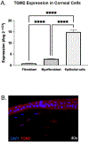
Fig. 1.
Fig. 1.
TGM2 is strongly expressed in the cornea during health and healing. A. TGM2 expression by TaqMan qPCR in healthy human corneal cells shows expression in fibroblasts and myofibroblasts, and strong expression in epithelial cells; n=3 per cell type. Samples normalized to GAPDH (**** = P < 0.0001). B. Immunohistochemistry shows strong TGM2 expression in the cytoplasm of epithelial cells and lesser expression in stroma in rabbit cornea 7 days post-wounding (n = 3).
Fig. 2.
Fig. 2.
Cysteamine hydrochloride does not adversely affect epithelial cell migration or epithelial wound healing at therapeutic doses. A. In vitro assessment of epithelial cell migration shows cell death at the highest concentration (16.3 mM) but no significant affect on viability or migration at the lowest dose (0.163 mM) or the in vitro dose corresponding to the Cystaran therapeutic dose (1.63 mM, P > 0.05; Bars = means, error bars = standard deviation). B. Representative images of epithelial cell migration assay showing significantly impaired migration at the highest concentration of CH tested, with no significant difference in migration between low and moderate concentrations and media negative control (n= 3 eyes per group, p > 0.05 for all timepoints). C. In vivo epithelial wound healing in a rabbit model was comparable between control and CH treated eyes. D. Representative fluoresceine images of control versus CH treated rabbit eyes illustrating the comparable epithelial healing times. All ulcers healed within 2 days (n = 3 eyes per group).
Fig. 3.
Fig. 3.
In vivo application of cysteamine hydrochloride at 6.5 mg/mL is safe in a rabbit model of corneal stromal wound healing. A. Application in a rabbit corneal stromal wound model showed no significant effect of CH application on in vivo stromal wound healing (P > 0.05 for all timepoints). B. In vivo confocal microscopy showed no toxic effect to corneal endothelial cells with no significant effect on cell density or cellular morphology between treated and control eyes. (P = 0.9972 Day 28, P = >0.9999 Day 42). C. Assessment of inflammation by scoring of H&E sections as well as by clinical SPOTS scoring, showed no significant difference between CH treated and control eyes. SPOTS score, when divided by extraocular inflammation (conjunctivitis score) and intraocular inflammation (uveitis score), remained insignificant (P > 0.05 for all timepoints and scores, n= 8 eyes per group to day 28, n -= 4 eyes per group to day 42).
Fig. 4.
Fig. 4.
In vivo application of cysteamine hydrochloride at 6.5 mg/mL does not affect corneal scarring. A. Representative clinical, slit beam, and OCT images illustrating the area of corneal haze at final day 42 and graphic representation of clinical haze scores. Corneal haze was present but not significantly different between control and CH treated eyes (n= 8 eyes per group to day 28, n = 4 eyes per group to day 42, P > 0.05 for all timepoints). B. Corneal fibrosis, as scored based on H&E histology sections, was not significantly different between treated and control eyes (n = 7 per group, P = 0.3473). C. IHC for α-SMA expression showed no difference in expression between treated and control eyes, suggesting no appreciable change in KFM transformation (n = 7 per group, P = 0.2821). D. Corneal thickness as measured by OCT, was not different between control and treated eyes suggesting no difference in amount of scarring (n= 8 eyes per group to day 28, n = 4 eyes per group to day 42, p > 0.05 for all timepoints). E. Atomic force microscopy of sections from the area of wounded cornea showed no difference in stiffness between control and treated eyes (n= 4 eyes per time point, P = 0.99 at Day 28, P = 0.96 at Day 42).

References

    1. Aragona P, Aguennouz M, Rania L, Postorino E, Sommario MS, Roszkowska AM, et al., 2015. Matrix metalloproteinase 9 and transglutaminase 2 expression at the ocular surface in patients with different forms of dry eye disease. Ophthalmology 122 (1), 62–71. - PubMed
    1. Barathi VA, Weon SR, Tan QS, Lin KJ, Tong L, Beuerman RW, 2011. Transglutaminases (TGs) in ocular and periocular tissues: effect of muscarinic agents on TGs in scleral fibroblasts. PLoS One 6 (4), e18326. - PMC - PubMed
    1. Boneham GC, Collin HB, 1995. Steroid inhibition of limbal blood and lymphatic vascular cell growth. Curr. Eye Res 14 (1), 1–10. - PubMed
    1. Buss DG, Sharma A, Giuliano EA, Mohan RR, 2010. Efficacy and safety of mitomycin C as an agent to treat corneal scarring in horses using an in vitro model. Vet. Ophthalmol 13 (4), 211–218. - PMC - PubMed
    1. Dreier B, Thomasy SM, Mendonsa R, Raghunathan VK, Russell P, Murphy CJ, 2013. Substratum compliance modulates corneal fibroblast to myofibroblast transformation. Invest. Ophthalmol. Vis. Sci 54 (8), 5901–5907. - PMC - PubMed

Publication types

MeSH terms

Substances