Review
. 2023 Jan;46(1):75-90.
doi: 10.1016/j.tins.2022.11.003. Epub 2022 Nov 16.

Olfactory dysfunction in COVID-19: new insights into the underlying mechanisms

Affiliations
Review

Olfactory dysfunction in COVID-19: new insights into the underlying mechanisms

Rafal Butowt et al. Trends Neurosci. 2023 Jan.

Abstract

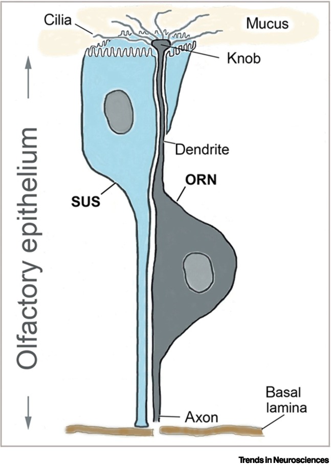
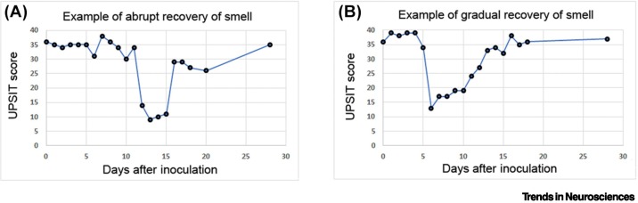
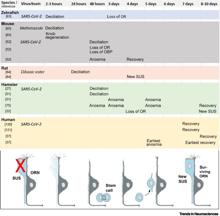
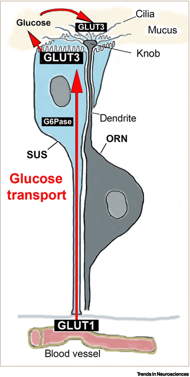
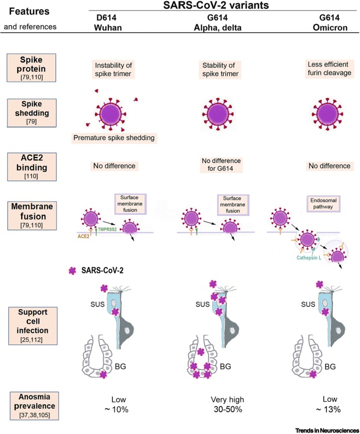
The mechanisms of olfactory dysfunction in COVID-19 are still unclear. In this review, we examine potential mechanisms that may explain why the sense of smell is lost or altered. Among the current hypotheses, the most plausible is that death of infected support cells in the olfactory epithelium causes, besides altered composition of the mucus, retraction of the cilia on olfactory receptor neurons, possibly because of the lack of support cell-derived glucose in the mucus, which powers olfactory signal transduction within the cilia. This mechanism is consistent with the rapid loss of smell with COVID-19, and its rapid recovery after the regeneration of support cells. Host immune responses that cause downregulation of genes involved in olfactory signal transduction occur too late to trigger anosmia, but may contribute to the duration of the olfactory dysfunction.

Keywords: SARS-CoV-2; anosmia; olfactory epithelium; parosmia; smell loss; sustentacular cell.


Conflict of interest statement

Declaration of interests The authors declare no competing interests.

Figures

Figure 1
Figure 1
Intimate relationship between the olfactory receptor neuron (ORN) and its support cell, the sustentacular cell (SUS), in the olfactory epithelium. The sense of smell in humans depends on 10 million ORNs and a similar number of SUS. The ORN has an apical dendritic process that projects a thickening (knob) into the lumen above the olfactory epithelium (nasal cavity) from which 10–15 cilia extend that bear the odorant receptors. The olfactory epithelium is covered by mucus to protect the dendritic processes. Odorant molecules inhaled in the nasal cavity dissolve in the mucus to bind to odorant receptors on the olfactory cilia. The basal process of the bipolar neuron is an axon that penetrates the basal lamina and the cribriform plate and forms synaptic contacts in a specific glomerulus in the olfactory bulb of the brain. Each mature ORN is tightly wrapped by its SUS [44], as shown. The SUS extends throughout the olfactory epithelium, with its basal process reaching the basal lamina, while the apical surface of the SUS is covered with microvilli, which intermingle with the cilia of the ORNs. The SUS provides structural support to the epithelium, and is thought to have multiple important functions. Notably, the SUS, as well as cells in the Bowman glands (not shown) secrete the mucus, and likely provide energy (glucose) to the cilia [57., 58., 59.] (for details, see Figure 4), so that the cilia can conduct the energy-consuming olfactory signal transduction. Additional important functions include regulation of the ionic composition of the mucus [117], detoxification and odorant clearance [118], and expression of odorant-binding proteins [119].
Figure 2
Figure 2
Key features of anosmia in coronavirus disease 2019 (COVID-19): rapid onset, relatively short duration, and usually rapid recovery. Examples of two typical cases of anosmia, according to scores obtained with the University of Pennsylvania Smell Identification Test (UPSIT) on volunteers inoculated with the G614 variant of severe acute respiratory syndrome coronavirus 2 (SARS-CoV-2) on day 0 [36]. (A) Example of a case with abrupt onset and abrupt recovery of smell. (B) Example of a case with abrupt onset and gradual recovery of the sense of smell. Note the relatively short duration of anosmia (mean of 7.8 days; range of 4–21 days for transient anosmia [36]), which is consistent with a similar time-course reported in meta-analyses that examined epidemiological studies of the same variant and populations in similar geographic regions [37,38]. Modified from [36].
Figure 3
Figure 3
Timetable of the events that ensue when the sustentacular support cell (SUS) is damaged or eliminated. This figure summarizes the events when the SUS is damaged by either a toxin or due to virus infection. Notably, damage of the SUS causes within 2–3 h a physical separation of the SUS from the olfactory receptor neuron (ORN), swelling of the knob and its degeneration, and retraction of the cilia [27,51,63., 64., 65.]. Deciliation continues from 2 to 48 h. The ORN resumes an immature stage of its dendritic extension, with focus on growth of its processes rather than on neurotransmission and sensory transduction. Gene expression of odorant receptors (ORs) is downregulated at 2 days (mouse [52]), 3 days (zebrafish [63]), and 4 days (hamster [22]) after inoculation. Odorant-binding proteins (OBPs) and receptor transporting protein 1 (RTP1) are also reduced [52]. In most animal models, loss of smell is evident as early as 2 days after lesion of SUS (mouse) and, depending on the animal model, anosmia lasts from 2 to 8 days. In humans, it lasts from 7 to 10 days (mean values) after infection with severe acute respiratory syndrome coronavirus 2 (SARS-CoV-2) [37]. The stem cells in the basal layers begin to divide at 3 days after SUS damage [66], and the first newly regenerated SUS appear at 4–8 days [22,64]. Recovery of smell begins at 4 days after SUS lesion in the mouse, at 8 days in the hamster [75], and the olfactory epithelium appears fully intact in the hamster at 7 days [74] or 14 days [51]. Human data on recovery of smell are according to pertinent studies [36,37,120,121].
Figure 4
Figure 4
Illustration of how a sustentacular support cell (SUS) provides the cilia of its olfactory receptor neuron (ORN) with glucose. The sustentacular cell (SUS) takes up glucose from its basal process, close to the blood vessels, via the glucose transporter 1 (GLUT1). Glucose or glucose-6-phosphate (G6P) diffuses along its gradient from the basal to the apical end, glucose-6 phosphatase (G6Pase) converts the G6P back to glucose, and secretes it through the glucose transporter 3 (GLUT3) into the mucus. Within the mucus, olfactory cilia, which lack mitochondria, uptake glucose via their GLUT3 and generate ATP by glycolysis. Likewise, the SUS-related cells in the Bowman glands also traffic glucose similarly from the blood supply to the mucus (not shown). The glucose-trafficking mechanisms are summarized as previously reported [57., 58., 59.]. The glucose support function may be the most important and acute way by which SUS and Bowman gland cells maintain ORN function.
Figure 5
Figure 5
Illustration of the molecular mechanisms that can explain why severe acute respiratory syndrome coronavirus 2 (SARS-CoV-2) variants cause different amounts of olfactory dysfunction. This figure summarizes how the properties of three SARS-CoV-2 virus variants (D614, G614, and omicron) differ in ways that likely determine to what extent sustentacular support cells (SUS) in the olfactory epithelium become infected and whether their loss will lead to anosmia. The original D614 (Wuhan) virus results in premature spike shedding, lower spike density, and, therefore, less effective virus entry [79]. This may cause less infection of SUS and, therefore, results in a low prevalence of anosmia (~10%) [37]. The G614 variant has the D614G mutation, which stabilizes the spike trimer and prevents premature spike shedding; the higher spike density allows the G614 variant to infect SUS cells effectively [79], resulting in a high (30–50%) anosmia prevalence [38]. All three variants bind to the virus entry protein angiotensin-converting enzyme 2 (ACE2), expressed by SUS, and with no significant differences in binding affinity to ACE2 [21,79,80]; thus, this cannot explain differences in anosmia. The first two variants, D614 and G614, both enter host cells by using surface membrane fusion mediated by the protease TMPRSS2 [79,110]. The new mutations in the omicron variant cause a less efficient furin cleavage, resulting in reduced surface membrane fusion mediated by TMPRSS2 [79,80,111]. Therefore, omicron prefers an endosomal route that is less efficient for SUS infection, possibly because many host cells have developed defenses for the endosomal entry [78,79,111]. As a result, the omicron variant, despite retaining the D614G mutation, is associated with a lower anosmia prevalence of ~13% [105].

References

    1. Dhurvey V., et al. Two worst pandemics- Spanish Flu and COVID-19: a review. Magna Sci. Adv. Biol. Pharm. 2021;4:1–12.
    1. Haehner A., et al. SARS-CoV-2 leads to significantly more severe olfactory loss than other seasonal cold viruses. Life (Basel) 2022;12:461. - PMC - PubMed
    1. Edwards S.N. Understanding the present through the past: a comparison of Spanish news coverage of the 1918 flu and COVID-19 pandemics. J. Mass. Commun. Q. 2022;99:12–43.
    1. Leyden E., Guttmann S., editors. Auftrage des Vereins für Innere Medicin in Berlin. Verlag J.F. Bergmann; 1892.
    1. Hwang C.S. Olfactory neuropathy in severe acute respiratory syndrome: report of a case. Acta Neurol. Taiwanica. 2006;15:26–28. - PubMed

Publication types