

Cortical and subcortical morphological alterations in motor subtypes of Parkinson's disease
- PMID: 36470900
- PMCID: PMC9723125
- DOI: 10.1038/s41531-022-00435-3
Cortical and subcortical morphological alterations in motor subtypes of Parkinson's disease
Abstract
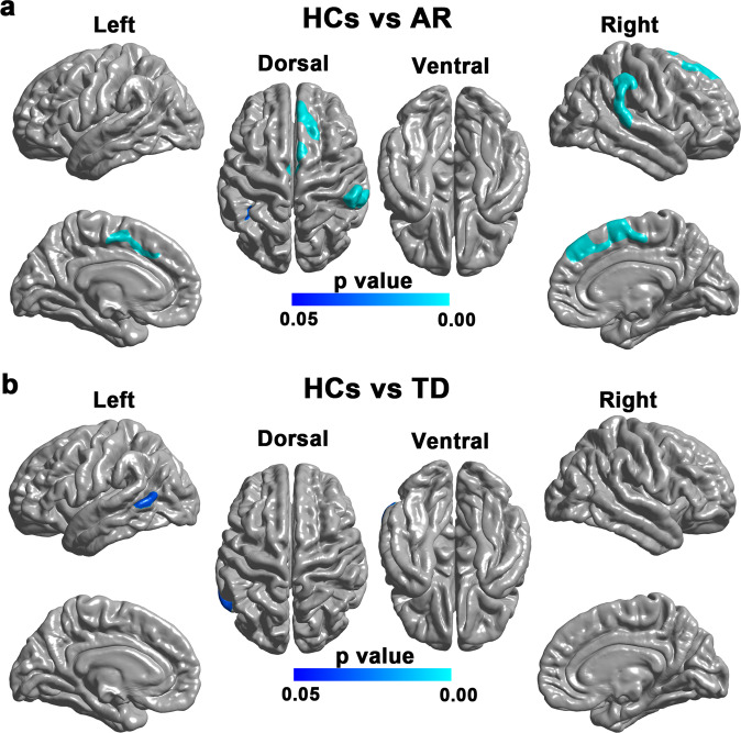
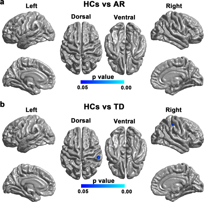
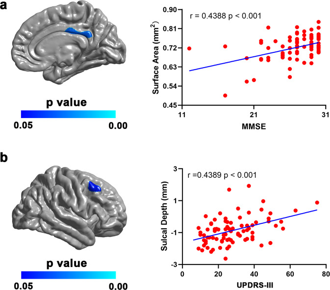
Parkinson's disease (PD) can be classified into an akinetic-rigid (AR) and a tremor-dominant (TD) subtype based on predominant motor symptoms. Patients with different motor subtypes often show divergent clinical manifestations; however, the underlying neural mechanisms remain unclear. This study aimed to characterize the cortical and subcortical morphological alterations in motor subtypes of PD. T1-weighted MRI images were obtained for 90 patients with PD (64 with the AR subtype and 26 with the TD subtype) and 56 healthy controls (HCs). Cortical surface area, sulcal depth (measured by Freesurfer's Sulc index), and subcortical volume were computed to identify the cortical and subcortical morphological alterations in the two motor subtypes. Compared with HCs, we found widespread surface area reductions in the AR subtype yet sparse surface area reductions in the TD subtype. We found no significant Sulc change in the AR subtype yet increased Sulc in the right supramarginal gyrus in the TD subtype. The hippocampal volumes in both subtypes were lower than those of HCs. In PD patients, the surface area of left posterior cingulate cortex was positively correlated with Mini-Mental State Examination (MMSE) score, while the Sulc value of right middle frontal gyrus was positively correlated with severity of motor impairments. Additionally, the hippocampal volumes were positively correlated with MMSE and Montreal Cognitive Assessment scores and negatively correlated with severity of motor impairments and Hoehn & Yahr scores. Taken together, these findings may contribute to a better understanding of the neural substrates underlying the distinct symptom profiles in the two PD subtypes.
© 2022. The Author(s).
Conflict of interest statement
The authors declare no competing interests.
Figures
Similar articles
-
Different functional connectivity modes of the right fronto-insular cortex in akinetic-rigid and tremor-dominant Parkinson's disease.Neurol Sci. 2021 Jul;42(7):2937-2946. doi: 10.1007/s10072-020-04917-1. Epub 2020 Nov 24. Neurol Sci. 2021. PMID: 33236247
-
Association of Gyrification Pattern, White Matter Changes, and Phenotypic Profile in Patients With Parkinson Disease.Neurology. 2021 May 11;96(19):e2387-e2394. doi: 10.1212/WNL.0000000000011894. Epub 2021 Mar 25. Neurology. 2021. PMID: 33766988
-
Comparison of two motor subtype classifications in de novo Parkinson's disease.Parkinsonism Relat Disord. 2018 Sep;54:74-78. doi: 10.1016/j.parkreldis.2018.04.021. Epub 2018 Apr 18. Parkinsonism Relat Disord. 2018. PMID: 29703644
-
Neuroimaging Detectable Differences between Parkinson's Disease Motor Subtypes: A Systematic Review.Mov Disord Clin Pract. 2020 Nov 6;8(2):175-192. doi: 10.1002/mdc3.13107. eCollection 2021 Feb. Mov Disord Clin Pract. 2020. PMID: 33553487 Free PMC article. Review.
-
Post mortem studies in Parkinson's disease--is it possible to detect brain areas for specific symptoms?J Neural Transm Suppl. 1999;56:1-29. doi: 10.1007/978-3-7091-6360-3_1. J Neural Transm Suppl. 1999. PMID: 10370901 Review.
Cited by
-
Auto-Classification of Parkinson's Disease with Different Motor Subtypes Using Arterial Spin Labelling MRI Based on Machine Learning.Brain Sci. 2023 Oct 29;13(11):1524. doi: 10.3390/brainsci13111524. Brain Sci. 2023. PMID: 38002484 Free PMC article.
-
Association between neutrophil-to-lymphocyte ratio and motor subtypes in idiopathic Parkinson's disease: a prospective observational study.BMC Neurol. 2024 Oct 8;24(1):379. doi: 10.1186/s12883-024-03887-7. BMC Neurol. 2024. PMID: 39379829 Free PMC article.
-
Surface-Based Morphometry Analysis of the Cerebral Cortex in Patients With Probable Idiopathic Rapid Eye Movement Sleep Behavior Disorder.Brain Behav. 2024 Oct;14(10):e70057. doi: 10.1002/brb3.70057. Brain Behav. 2024. PMID: 39344375 Free PMC article.
-
Grey matter volume differences across Parkinson's disease motor subtypes in the supplementary motor cortex.Neuroimage Clin. 2025;45:103724. doi: 10.1016/j.nicl.2024.103724. Epub 2024 Dec 10. Neuroimage Clin. 2025. PMID: 39673940 Free PMC article.
-
Cognitive Impairment-Associated Risk Factors of Parkinson's Disease: A Hospital-Based Study in a Cohort of Upper Egypt Parkinson's Patients.Brain Sci. 2025 Apr 27;15(5):459. doi: 10.3390/brainsci15050459. Brain Sci. 2025. PMID: 40426630 Free PMC article.
References
Grants and funding
- 82071883/National Natural Science Foundation of China (National Science Foundation of China)
- 61971017/National Natural Science Foundation of China (National Science Foundation of China)
- 62171106/National Natural Science Foundation of China (National Science Foundation of China)
- 82071883/National Natural Science Foundation of China (National Science Foundation of China)
- 82071883/National Natural Science Foundation of China (National Science Foundation of China)
- 82071883/National Natural Science Foundation of China (National Science Foundation of China)
- 82071883/National Natural Science Foundation of China (National Science Foundation of China)
- 82071883/National Natural Science Foundation of China (National Science Foundation of China)
- 82071883/National Natural Science Foundation of China (National Science Foundation of China)
- 82071883/National Natural Science Foundation of China (National Science Foundation of China)
- 82071883/National Natural Science Foundation of China (National Science Foundation of China)
LinkOut - more resources
Full Text Sources
Research Materials
