. 2022 Dec 5;12(1):20987.
doi: 10.1038/s41598-022-24772-8.

Helical stability of the GnTV transmembrane domain impacts on SPPL3 dependent cleavage

Affiliations

Helical stability of the GnTV transmembrane domain impacts on SPPL3 dependent cleavage

Alkmini A Papadopoulou et al. Sci Rep. .

Abstract

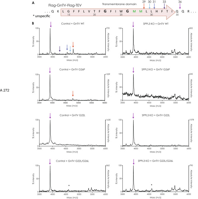
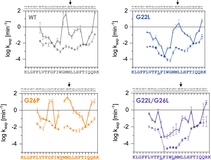
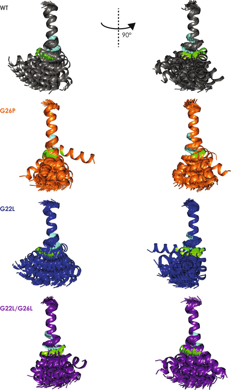
Signal-Peptide Peptidase Like-3 (SPPL3) is an intramembrane cleaving aspartyl protease that causes secretion of extracellular domains from type-II transmembrane proteins. Numerous Golgi-localized glycosidases and glucosyltransferases have been identified as physiological SPPL3 substrates. By SPPL3 dependent processing, glycan-transferring enzymes are deactivated inside the cell, as their active site-containing domain is cleaved and secreted. Thus, SPPL3 impacts on glycan patterns of many cellular and secreted proteins and can regulate protein glycosylation. However, the characteristics that make a substrate a favourable candidate for SPPL3-dependent cleavage remain unknown. To gain insights into substrate requirements, we investigated the function of a GxxxG motif located in the transmembrane domain of N-acetylglucosaminyltransferase V (GnTV), a well-known SPPL3 substrate. SPPL3-dependent secretion of the substrate's ectodomain was affected by mutations disrupting the GxxxG motif. Using deuterium/hydrogen exchange and NMR spectroscopy, we studied the effect of these mutations on the helix flexibility of the GnTV transmembrane domain and observed that increased flexibility facilitates SPPL3-dependent shedding and vice versa. This study provides first insights into the characteristics of SPPL3 substrates, combining molecular biology, biochemistry, and biophysical techniques and its results will provide the basis for better understanding the characteristics of SPPL3 substrates with implications for the substrates of other intramembrane proteases.


Conflict of interest statement

The authors declare no competing interests.

Figures

Figure 1
Figure 1
(A) Amino acids of the GnTV TM domain and its flanking regions are depicted using the single letter code together with the respective nucleotide triplets of the DNA coding region. The predicted TMD is annotated within the red arrow, which also depicts the N- to C-terminal orientation of the protein. The potentially α-helix destabilizing glycines are marked in bold. Below the sequence of GnTV the respective amino acid sequences of the three sets of mutations are shown. The mutated amino acids are marked in red. (B) Intracellular and secreted GnTV levels of the same sample were analysed. As lysates were analysed on different Western Blots than the corresponding supernatants, GnTV WT was used as a control and levels of sGnTV were always normalised to GnTV WT. To compare the turnover of all GnTV mutants, the log2 of soluble GnTV (sGnTV) divided by the full length GnTV (FL GnTV) is depicted in a linear scale. GnTV FL comprises the sum of GnTVim and GnTVmat. To confirm the reproducibility of the experiments, three (n = 3) biological replicates were seeded and analysed on different weeks for each of the mutants, each biological replicate comprised three technical replicates. The values are quantified from Western blot analysis (Supplementary Fig. 1) and are normalized to the secretion of GnTV WT (0). Mean + SEM, multiple unpaired, two-tailed t tests with Holm-Sidak multiple comparisons correction. (C) For each mutant secretion of sGnTV by both SPPL3 and unknown protease(s) in HEK293 cells (Control) was set to 100% and the secretion of GnTV of the same mutant by only the unknown protease(s) in SPPL3 KO cells is depicted. n = 3 biological replicates with 3 technical replicates each, mean + SEM, two-tailed t test for each pair separately (D) Using the results of 1C, SPPL3 dependent secretion was calculated by subtracting percentage of secretion by the unknown protease from the secretion by both proteases. (E) On the x axis, the total secretion of each mutant compared to the WT is plotted. The results from Fig. 1B are being used, but instead of the log2 results the linear numbers with secretion of GnTV WT normalised to 1 were used. The y axis depicts the SPPL3 dependent secretion in percentage. Results from Fig. 1D where used. The values of GnTV WT are marked with the dotted lines. n.s. = non significant, *p < 0.05, **p < 0.01.
Figure 2
Figure 2
(A) HEK293 cells were stably transfected with N-terminally Flag and C-terminally V5-tagged GnTV WT, GnTV G26P, GnTV G22L or GnTV G22L/G26L and were co-stained with V5 to visualize GnTV and a trans-Golgi marker (TGN46). DAPI was used to stain the nuclei. White bar = 10 μm. (B) SPPL3 KO cells were stably transfected with either SPPL3 WT or catalytically inactive SPPL3 D272A (D/A). Expression levels are shown in Western Blot on the right (uncropped blots in supplements). Note that expression of the catalytically inactive variant is stronger than that of SPPL3 WT. The indicated GnTV variants were transiently expressed and secretion of GnTV (sGnTV) was quantified relative to intracellular GnTV (FL GnTV). Secretion was normalized to the secretion of the respective GnTV mutants in cells with endogenous expression of SPPL3 (Control). n = 3 biological replicates; mean + SEM, two-tailed t test for each pair separately. n.s. = non significant, *p < 0.05, **p < 0.01, ***p < 0.001.
Figure 3
Figure 3
(A) Amino acid sequences of the TM domain of the GnTV peptide used in the mass spectrometric analysis. Small arrows indicate the peptides detected. (B) C-terminal cleavage sites of GnTV. Mass spectrometric analysis of fragments generated in presence (Control) or absence (SPPL3 KO) of SPPL3, respectively. Numbers indicate the position of the most C-terminal amino acid of the respective cleavage product. *marks unspecific peak. The intensities were normalized with the highest peak set at 100%. On the right side of each graph, the absolute intensity of the highest peak is indicated.
Figure 4
Figure 4
Conformational flexibility of GnTV TM helices probed by DHX. DHX rate constants of individual amides. Within regions of biphasic DHX, more positive values denote kexp,A and the sizes of the data points approximate the deuteron populations A and B. Values characterizing single H-bond openings (kexp, kexp,A) are connected by thick lines. Error bars correspond to the errors of fit in kexp determination from n = 3 independent DHX reactions. The main cleavage site is indicated by an arrow.
Figure 5
Figure 5
Structural bundles aligned on residues G15–G/L22. The 50 lowest energy structures out of 200 are shown. Green: M28, indicating the major SPPL3 cleavage site; light blue G/L22 and G/L/P26.
Figure 6
Figure 6
Spread of the structural bundles superimposed on L14-F21. Shown is the position of the C-terminal helix end (at residue 35) when the N-terminal is aligned along the z-axis thus pointing out of the plane of the figure. The position of residues L14–W25 are depicted by the central helical wheel.

References

    1. Steiner H, Fluhrer R, Haass C. Intramembrane proteolysis by gamma-secretase. J. Biol. Chem. 2008;283:29627–29631. doi: 10.1074/jbc.R800010200. - DOI - PMC - PubMed
    1. Rawson RB, et al. Complementation cloning of S2P, a gene encoding a putative metalloprotease required for intramembrane cleavage of SREBPs. Mol. Cell. 1997;1:47–57. doi: 10.1016/S1097-2765(00)80006-4. - DOI - PubMed
    1. Langosch D, Steiner H. Substrate processing in intramembrane proteolysis by gamma-secretase - the role of protein dynamics. Biol. Chem. 2017;398:441–453. doi: 10.1515/hsz-2016-0269. - DOI - PubMed
    1. Grigorenko AP, Moliaka YK, Korovaitseva GI, Rogaev EI. Novel class of polytopic proteins with domains associated with putative protease activity. Biochem. Biokhimiia. 2002;67:826–835. doi: 10.1023/A:1016365227942. - DOI - PubMed
    1. Ponting CP, et al. Identification of a novel family of presenilin homologues. Hum. Mol. Genet. 2002;11:1037–1044. doi: 10.1093/hmg/11.9.1037. - DOI - PubMed

Publication types

MeSH terms