. 2023 Mar;19(3):334-345.
doi: 10.1038/s41589-022-01198-x. Epub 2022 Dec 5.

Structure of SpoT reveals evolutionary tuning of catalysis via conformational constraint

Affiliations

Structure of SpoT reveals evolutionary tuning of catalysis via conformational constraint

Hedvig Tamman et al. Nat Chem Biol. 2023 Mar.

Abstract

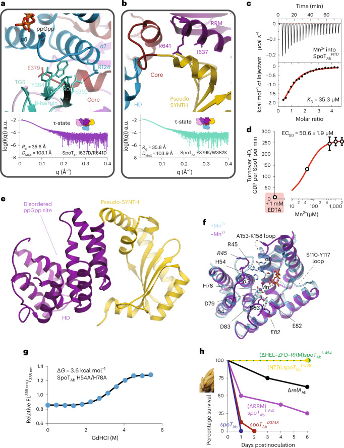
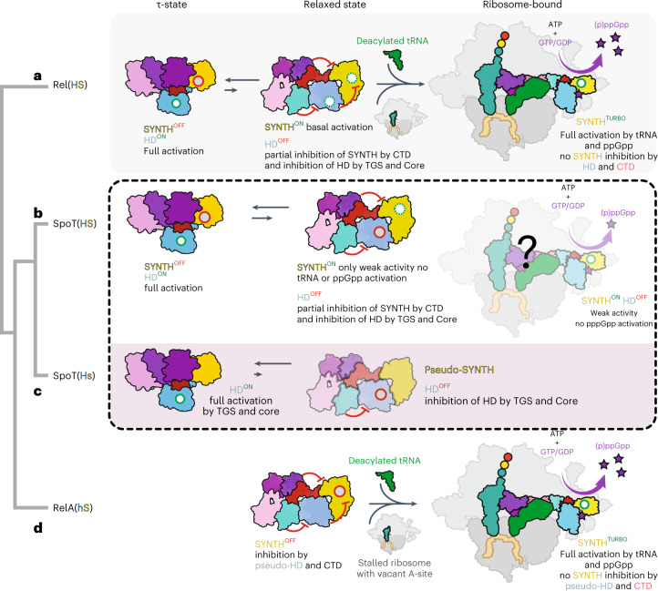
Stringent factors orchestrate bacterial cell reprogramming through increasing the level of the alarmones (p)ppGpp. In Beta- and Gammaproteobacteria, SpoT hydrolyzes (p)ppGpp to counteract the synthetase activity of RelA. However, structural information about how SpoT controls the levels of (p)ppGpp is missing. Here we present the crystal structure of the hydrolase-only SpoT from Acinetobacter baumannii and uncover the mechanism of intramolecular regulation of 'long'-stringent factors. In contrast to ribosome-associated Rel/RelA that adopt an elongated structure, SpoT assumes a compact τ-shaped structure in which the regulatory domains wrap around a Core subdomain that controls the conformational state of the enzyme. The Core is key to the specialization of long RelA-SpoT homologs toward either synthesis or hydrolysis: the short and structured Core of SpoT stabilizes the τ-state priming the hydrolase domain for (p)ppGpp hydrolysis, whereas the longer, more dynamic Core domain of RelA destabilizes the τ-state priming the monofunctional RelA for efficient (p)ppGpp synthesis.


Conflict of interest statement

A.G.-P. is cofounder and stockholder of Santero Therapeutics, a company that aims to develop inventions described in this paper. The other authors declare no competing interests.

Figures

Fig. 1
Fig. 1. A. baumannii SpoT is a monofunctional alarmone HD.
a, Evolution of long RSHs in Proteobacteria. Duplication of the ancestral bifunctional RSH Rel in Beta- and Gammaproteobacterial lineages gave rise to RelA and SpoT, leading to subfunctionalization of RelA as monofunctional SYNTH-only alarmone synthetase and SpoT as a predominantly HD RSH. In the Moraxellaceae family of Gammaproteobacteria, SpoT has undergone further subfunctionalization, evolving into a monofunctional HD-only alarmone HD. b, Alignment of SYNTH-critical regions in long RSHs highlights the sequence divergence in Moraxellaceae SpoTs. c, Coexpression of SpoTAb counteracts the growth defect in ppGpp0relA ΔspoT) E. coli caused by RelA expression. This demonstrates that SpoTAb is HD active in the E. coli host. d, While the SYNTH activity of ectopically expressed SpoTEc is essential and sufficient for promoting the growth of ppGpp0 E. coli on M9 minimal medium, SpoTAb fails to promote the growth of ΔrelA ΔspoT E. coli on M9. This demonstrates that, unlike SpoTEc, which is SYNTH-active, SpoTAb is SYNTH-inactive.
Fig. 2
Fig. 2. Full-length monomeric A. baumannii SpoT adopts a compact ‘mushroom’-shaped HD-active τ-state.
a, Structure of ‘mushroom’-shaped SpoTAb–ppGpp complex in the τ-state. The domain organization, from N to C terminus: NTDs HD, pseudo-synthetase (pseudo-SYNTH) and Core domains, and CTDs, TGS, HEL, ZFD and RRM. The ppGpp alarmone is in red. b, Cartoon representation the SpoTAb. The ‘stem’ of the mushroom is formed by the enzymatic HD domain and the ‘cap’ by the regulatory domains: NTD pseudo-SYNTH domain and the CTD. c, Ribbon representation of the SpoTAb–ppGpp complex. The α6/α7 motif is held in the hydrolysis-compatible position by the folded Core domain and the TGS β-hairpin, with the Core domain communicating allosteric signals to HD from the regulatory domains. d, The HD activity of SpoTAb is insensitive to the addition of E. coli 70S ribosomes, and nonspecifically weakly inhibited by both aminoacylated and deacyated E. coli tRNAVal. e, Analytical SEC of SpoTAb supports its monomeric nature in solution. a.u., arbitrary units. f, Experimental SAXS analysis of SpoTAb at 8 mg ml−1 further confirms the monomeric nature of SpoTAb. The analysis of the normalized Kratky plot (insert) of the SAXS curve reveals folded globular shape of SpoTAb. g, Ab initio envelope of SpoTAb reconstructed from the experimental SAXS data superimposed on the crystal structure. Comparison of both models shows that in solution the enzyme adopts the same conformation as observed in the crystal. Error bars represent a s.d. of three or more independent samples examined over three independent experiments.
Fig. 3
Fig. 3. Defining features of A. baumannii SpoT catalysis.
a, Surface representation of SpoTAb in the τ-state. The active site cavity in the HD domain is boxed in dashed lines. b, Zoom into the HD-active site of the SpoTAb–ppGpp complex. The acidic half of the interface (residues R45, Y51, E82, D83 and K140) and the Mn2+ ion activate the water molecule for nucleophilic attack of the pyrophosphate bond pf ppGpp, while the basic half of the interface (K46, K158 and R161) stabilizes the 3′ and 5′ phosphates of the alarmone substrate. c, Ribbon representation of the active site of SpoTAb revealing the residues involved in coordination of ppGpp. The unbiased mFo-DFc electron density map corresponding to these residues is shown in dark blue. d, Effects of Ala substitutions in the ppGpp binding site on the HD activity of SpoTAb. The residues for substitution were selected as per c. e, The HD functionality test of truncated versions of SpoTAb. SpoTAb variants (each labeled on the figure) were coexpressed expressed with RelAAb in ∆relAspoT Ptac::relA A. baumannii (AB5075). The ability of SpoTAb to promote the growth is reflective of its HD competence. f, HD activity of SpoTAb and the C-terminally truncated SpoTAb variants. Turnovers corresponding to each protein variant are colored as per the domain color code in e. Error bars represent s.d. of three or more independent samples examined over three independent experiments.
Fig. 4
Fig. 4. The CTD controls the hydrolysis activity of SpoT by controlling the equilibrium between HD-active τ-state and HD-inactive relaxed conformations.
a, Circos plot of long-distance interactions (ten residues or more) within SpoTAb. Each domain is defined and colored as in Fig. 2a. The thickness of connecting lines represent the number of contacts between two domains. b, Cartoon representation of the allosteric network defined by the Core domain connecting the domains of the enzyme in the τ-state. The key interface residues are shown as sticks and labeled. c, HD activity of crucial Core residues involved in interactions with other domains of SpoTAb (A384R contacting SYNTH, A351K contacting RRM, L356D contacting HD, L373G/D374G contacting ZFD, Y375G contacting the TGS). The TGS:HD interface is also probed with the E379K/W382K point mutant and ΔTGS–HEL versions. The τ-state stabilizing substitutions D374R and I637D/R641D increase the HD activity. d,e, SAXS curves of L356D in the τ-state (d) or relaxed state (e). f, Pseudo-atomic model of the relaxed state of SpoTAb calculated with Dadimodo using the experimental SAXS data from e. g, Cartoon representation of experimentally observed conformational states as well as particle dimensions of long-RSH enzymes. Error bars represent s.d. of three or more independent samples examined over three independent experiments.
Fig. 5
Fig. 5. The Core domain of SpoT transduces the allosteric signal from the regulatory CTD and pseudo-SNTH to the enzymatic HD domain.
a, Effects of substitutions at the α6–α7:Core–TGS interface. Interactions stabilizing the α6–α7 motif of the HD-active site with the Core wrapping around α7 and the TGS β-hairpin stabilizing α6. Experimental SAXS curve of SpoTAbE379K/W382K shows it remains in the τ-state. b, Effects of substitutions at the HD–Core–RRM interface with RRM locked in place via the Core and supporting interaction provided by pseudo-SYNTH, indeed the SAXS curve of SpoTAbI637D/R641D is consistent with the dimensions of the τ-state. c, ITC titration of Mn2+ into apo-SpoTAb. d, HD activity of apo-SpoTAb as a function of increasing concentrations of Mn2+. e, Structure of the Mn2+-free SpoTAbNTD. The HD domain is in purple, and the pseudo-SYNTH is in yellow. The disordered active site is labeled. f, Superposition of the HD domain of SpoTAb complexed with ppGpp (in light blue) onto Mn2+-free SpoTAb (in purple). The key differences in conformation of catalytically crucial active site residues and the structural elements α3, α4 and α8 are highlighted with dashed arrows and shown in bold, respectively. g, Thermal denaturation profile monitored by far UV circular dichroism (CD) spectrum at 222 nm of SpoTAbH54A/H78A, which cannot bind Mn2+ in the HD domain. h, Virulence assays in the G. mellonella infection model demonstrate the essentiality of intact allosteric regulation of SpoTAb for virulence. G. mellonella larvae were injected with roughly 2 × 10 CFU of A. baumannii (AB5075) strains (10 µl at roughly 2 × 107 CFU per ml), eight larvae were inoculated per strain and incubated at 37 °C in the dark. The viability of the larvae was scored every 24 h. Error bars represent s.d. of three or more independent samples examined over three independent experiments.
Fig. 6
Fig. 6. The enzymatic output of subfunctionalized RelA and SpoT RSH enzymes is evolutionarily tuned through constrains of the conformational landscape.
a, Off ribosomes the ancestral bifunctional Rel[HS] assumes a τ-state with the CTD organizing the HD-active site and promoting the HD activity. This, in turn, strongly inhibits SYNTH activity via intra-NTD regulation. On amino acid starvation, Rel is recruited to starved ribosomal complexes. The ribosome-bound Rel assumes an extended conformation in which the auto-inhibitory effect of the CTD region on the SYNTH activity is released. The full activation of SYNTH is achieved on binding (p)ppGpp to an allosteric site within the NTD releasing the SYNTH inhibition by the HD domain. Thus, the full activation of either SYNTH or HD requires allosteric signaling from CTD to NTD enzymatic domains. b, Evolution of SpoT as a predominantly HD involved the loss of the allosteric control of the NTD by (p)ppGpp. In the bifunctional SpoT[HS] present in most Gamma- and Betaproteobacteria, the enzyme is capable of inefficient (p)ppGpp synthesis in the relaxed state despite the equilibrium strongly favoring the HD-active τ-state; it remains to be determined whether or not synthesis occurs on the ribosome. c, Subfunctionalization of SpoT in Moraxellaceae resulted in the monofunctional HD SpoT[Hs], which naturally populates only the compact τ-state, it is not control by pppGpp or starved ribosomes and is SYNTH-inactive. d, Subfunctionalization of Beta- and Gammaproteobacterial RelA[hS] constitutes the other extreme case of evolutionary restriction of the conformational dynamics of the ancestral Rel[HS]. While losing its HD activity, RelA retains all the allosteric elements of Rel involved in the regulation of (p)ppGpp synthesis and off the ribosome it does not assume the τ-state. Instead, it predominantly populates the functionally frustrated relaxed conformation, primed to switch to the elongated ribosome-associated state triggered by the 70S ribosome, uncharged tRNA and alarmones during stringency. Red circles represent inhibited catalytic centers, green circles represent fully activated catalytic centers and dashed green circles represent idling catalytic centers.

References

    1. Atkinson GC, Tenson T, Hauryliuk V. The RelA/SpoT homolog (RSH) superfamily: distribution and functional evolution of ppGpp synthetases and hydrolases across the tree of life. PLoS ONE. 2011;6:e23479. doi: 10.1371/journal.pone.0023479. - DOI - PMC - PubMed
    1. Hauryliuk V, Atkinson GC, Murakami KS, Tenson T, Gerdes K. Recent functional insights into the role of (p)ppGpp in bacterial physiology. Nat. Rev. Microbiol. 2015;13:298–309. doi: 10.1038/nrmicro3448. - DOI - PMC - PubMed
    1. Irving SE, Corrigan RM. Triggering the stringent response: signals responsible for activating (p)ppGpp synthesis in bacteria. Microbiology. 2018;164:268–276. doi: 10.1099/mic.0.000621. - DOI - PubMed
    1. Ronneau, S. & Hallez, R. Make and break the alarmone: regulation of (p)ppGpp synthetase/hydrolase enzymes in bacteria. FEMS Microbiol. Rev. 43, 389–400 (2019). - PMC - PubMed
    1. Anderson, B. W., Fung, D. K. & Wang, J. D. Regulatory themes and variations by the stress-signaling nucleotide alarmones (p)ppGpp in bacteria. Annu. Rev. Genet.23, 115–133 (2021). - PubMed

Publication types