. 2022 Dec 5;14(1):168.
doi: 10.1186/s13148-022-01379-4.

Quantification of DNA methylation for carcinogenic risk estimation in patients with non-alcoholic steatohepatitis

Affiliations

Quantification of DNA methylation for carcinogenic risk estimation in patients with non-alcoholic steatohepatitis

Junko Kuramoto et al. Clin Epigenetics. .

Abstract

Background: In recent years, non-alcoholic steatohepatitis (NASH) has become the main cause of hepatocellular carcinoma (HCC). As a means of improving the treatment of NASH-related HCCs based on early detection, this study investigated the feasibility of carcinogenic risk estimation in patients with NASH.

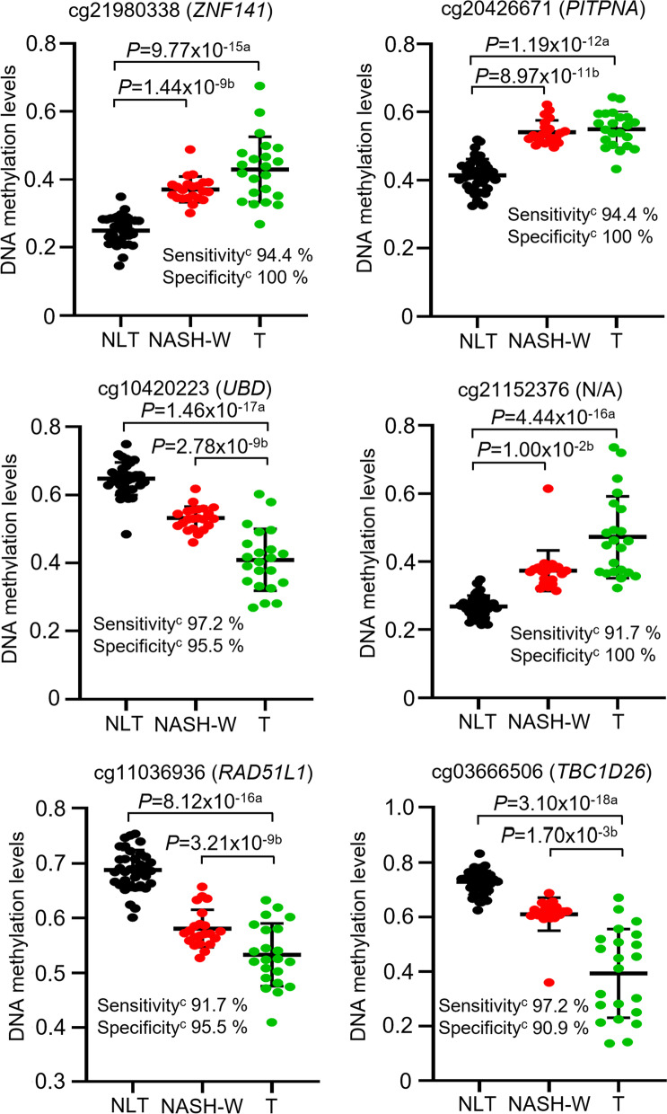
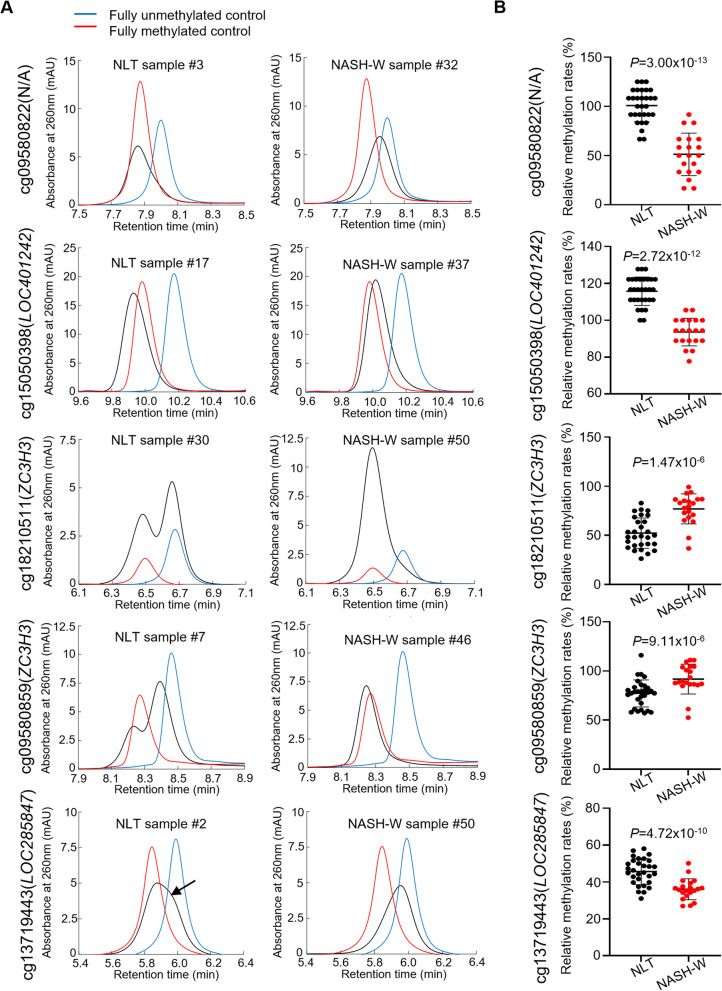
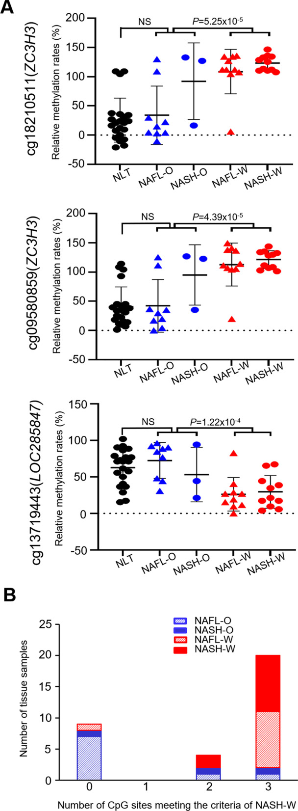
Results: Normal liver tissue (NLT), non-cancerous liver tissue showing histological findings compatible with non-alcoholic fatty liver from patients without HCC (NAFL-O), non-cancerous liver tissue showing NASH from patients without HCC (NASH-O), non-cancerous liver tissue showing non-alcoholic fatty liver from patients with HCC (NAFL-W), non-cancerous liver tissue showing NASH from patients with HCC (NASH-W) and NASH-related HCC were analyzed. An initial cohort of 171 tissue samples and a validation cohort of 55 tissue samples were used. Genome-wide DNA methylation screening using the Infinium HumanMethylation450 BeadChip and DNA methylation quantification using high-performance liquid chromatography (HPLC) with a newly developed anion-exchange column were performed. Based on the Infinium assay, 4050 CpG sites showed alterations of DNA methylation in NASH-W samples relative to NLT samples. Such alterations at the precancerous NASH stage were inherited by or strengthened in HCC samples. Receiver operating characteristic curve analysis identified 415 CpG sites discriminating NASH-W from NLT samples with area under the curve values of more than 0.95. Among them, we focused on 21 CpG sites showing more than 85% specificity, even for discrimination of NASH-W from NASH-O samples. The DNA methylation status of these 21 CpG sites was able to predict the coincidence of HCC independently from histopathological findings such as ballooning and fibrosis stage. The methylation status of 5 candidate marker CpG sites was assessed using a HPLC-based system, and for 3 of them sufficient sensitivity and specificity were successfully validated in the validation cohort. By combining these 3 CpG sites including the ZC3H3 gene, NAFL-W and NASH-W samples from which HCCs had already arisen were confirmed to show carcinogenic risk with 95% sensitivity in the validation cohort.

Conclusions: After a further prospective validation study using a larger cohort, carcinogenic risk estimation in liver biopsy specimens of patients with NASH may become clinically applicable using this HPLC-based system for quantification of DNA methylation.

Keywords: Carcinogenic risk estimation; DNA methylation; Hepatocellular carcinoma; High-performance liquid chromatography; Infinium assay; Non-alcoholic steatohepatitis.


Conflict of interest statement

None of the authors have any competing interests to disclose.

Figures

Fig. 1
Fig. 1
Scattergrams of DNA methylation levels obtained by the Infinium assay for representative CpG sites showing area under the curve values of more than 0.95 in receiver operating characteristic curve analysis for discrimination between non-cancerous liver tissue showing non-alcoholic steatohepatitis (NASH) derived from partial hepatectomy specimens from patients with hepatocellular carcinoma (HCC) (NASH-W) (n = 22, red) and normal liver tissue (NLT) samples (n = 36, black). The Infinium probe ID and gene symbol are shown at the top of each panel. N/A, not annotated (designed in the intergenic regions). DNA methylation alteration at the NASH stage compared to NLT was inherited by or strengthened in NASH-related HCC (T) samples (n = 22, green). Using each CpG site and its cutoff value described in Additional file 1: Table S3, NASH-W samples were discriminated from NLT samples with sufficient sensitivity and specificity (90.9–100%). aP values by Welch’s t test. bP values by Jonckheere–Terpstra trend test. cSensitivity was defined as the ratio of the number of tissue samples diagnosed as NASH-W based on the criteria described in Additional file 1: Table S3 relative to the exact number of NASH-W samples, and specificity was defined as the ratio of the number of tissue samples not diagnosed as NASH-W based on the criteria relative to the exact number of NLT samples
Fig. 2
Fig. 2
High-performance liquid chromatography (HPLC) analysis using the newly developed anion-exchange column for specimens of normal liver tissue (NLT) (n = 30) and non-cancerous liver tissue showing non-alcoholic steatohepatitis (NASH) derived from partial hepatectomy specimens from patients with hepatocellular carcinoma (HCC) (NASH-W) (n = 20) in the initial cohort. A Chromatograms of PCR products encompassing the marker CpG sites (cg09580822, g15050398, cg18210511, cg09580859 and cg13719443) for representative tissue samples (black). Gene names are shown on the left side. N/A: not annotated (the probe was located within the intergenic region). Chromatograms of fully unmethylated and fully methylated control DNAs are shown in blue and red, respectively. For cg09580822 and cg15050398, chromatograms with a “single peak” were obtained in both NLT and NASH-W. For cg18210511 and cg09580859, NLT showed a “bimodal peak” pattern, whereas a “single peak” pattern was observed in NASH-W. For cg15050398, NLT showed a “single peak with a shoulder” (indicated by an arrow) pattern. B Scattergrams of the relative DNA methylation rates obtained by HPLC analysis for cg09580822, g15050398, cg18210511, cg09580859 and cg13719443. Such rates are described in “Results” section and differed significantly between NLT (black) and NASH-W (red) samples. P values were obtained by Welch’s t test
Fig. 3
Fig. 3
High-performance liquid chromatography (HPLC) analysis using the newly developed anion-exchange column for specimens of normal liver tissue (NLT) (n = 22, black), non-cancerous liver tissue (N) showing non-alcoholic fatty liver (NAFL) derived from partial hepatectomy specimens from patients without hepatocellular carcinoma (HCC) (NAFL-O) (n = 9, blue), N samples showing non-alcoholic steatohepatitis (NASH) from patients without HCC (NASH-O) (n = 3, blue), N samples showing NAFL from patients with HCC (NAFL-W) (n = 10, red), and N samples showing NASH from patients with HCC (NASH-W) (n = 11, red) in the validation cohort. A Scattergrams of the relative DNA methylation rates obtained by HPLC analysis for cg18210511, cg09580859 and cg13719443 whose estimation ability was validated in the validation cohort. Such rates were obtained as described in “Results” section. Such rates for both NAFL-W and NASH-W samples differed significantly from both NAFL-O and NASH-O samples regardless of histological features (i.e., regardless of NAFL or NASH), whereas the rates for both NAFL-O and NASH-O samples did not differ from those of NLT samples. P values were obtained by Welch’s t test. B Histograms showing the number of CpG sites satisfying the criteria for cg18210511, cg09580859 and cg13719443 based on the cutoff values shown in Table 4. If the tissue sample was estimated to have carcinogenic risk when two marker CpG sites or more satisfied the criteria for cg18210511, cg09580859 and cg13719443, regardless of histological features (i.e., regardless of NAFL or NASH), the liver tissue specimens from which HCCs had originated (9 NAFL-W samples and all 11 NASH-W samples) were considered to possess carcinogenic risk with 95% sensitivity. The possibility that patients from whom 2 NAFL-O and 2 NASH-O samples were obtained, which satisfied the criteria for two of more markers, would develop HCCs in the future could not be ruled out

Similar articles

Cited by

References

    1. Younossi ZM, Golabi P, de Avila L, Paik JM, Srishord M, Fukui N, et al. The global epidemiology of NAFLD and NASH in patients with type 2 diabetes: a systematic review and meta-analysis. J Hepatol. 2019;71(4):793–801. doi: 10.1016/j.jhep.2019.06.021. - DOI - PubMed
    1. Sheka AC, Adeyi O, Thompson J, Hameed B, Crawford PA, Ikramuddin S. Nonalcoholic steatohepatitis: a review. JAMA. 2020;323(12):1175–1183. doi: 10.1001/jama.2020.2298. - DOI - PubMed
    1. Younossi Z, Anstee QM, Marietti M, Hardy T, Henry L, Eslam M, et al. Global burden of NAFLD and NASH: trends, predictions, risk factors and prevention. Nat Rev Gastroenterol Hepatol. 2018;15(1):11–20. doi: 10.1038/nrgastro.2017.109. - DOI - PubMed
    1. Kim HS, El-Serag HB. The epidemiology of hepatocellular carcinoma in the USA. Curr Gastroenterol Rep. 2019;21(4):17. doi: 10.1007/s11894-019-0681-x. - DOI - PubMed
    1. Eguchi Y, Wong G, Lee IH, Akhtar O, Lopes R, Sumida Y. Hepatocellular carcinoma and other complications of non-alcoholic fatty liver disease and non-alcoholic steatohepatitis in Japan: a structured review of published works. Hepatol Res. 2021;51(1):19–30. doi: 10.1111/hepr.13583. - DOI - PubMed

Publication types