Review
. 2023;11(1):1-11.
doi: 10.1007/s40134-022-00405-w. Epub 2022 Dec 1.

Aging-Related Findings of the Respiratory System in Chest Imaging: Pearls and Pitfalls

Affiliations
Review

Aging-Related Findings of the Respiratory System in Chest Imaging: Pearls and Pitfalls

Elisa Baratella et al. Curr Radiol Rep. 2023.

Abstract

Purpose of review: The purpose of this review is to describe the main features of the aging chest, studied through different imaging modalities.

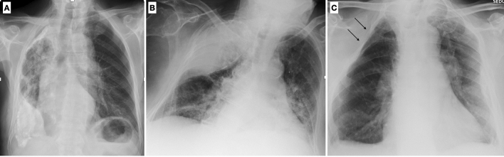
Recent findings: Aging-related changes of the respiratory system are inevitable. Therefore, it is mandatory to be familiar with the para-physiological changes that occurs, in order to avoid inappropriate interpretation of radiological findings that put patients at risk of over or undertreatment.

Summary: The role of the radiologist is fundamental in evaluating aging-related processes affecting the respiratory system and in distinguishing them from frank diseases.

Keywords: Aging chest; Artificial intelligence; Chest CT; Chest X-ray; Digital tomosynthesis.


Conflict of interest statement

Conflict of interestFor all authors none were declared.

Figures

Fig. 1
Fig. 1
Main alterations of thoracic structures in the geriatric population
Fig. 2
Fig. 2
Chest X-ray in postero-anterior (A) and lateral projection (B) shows typical chest wall morphologic changes in shape and dimension: a reduction in the lateral diameter and an increase in the sagittal diameter. On the lateral view a shortening of the thoracic spine is evident, along with the deformation of vertebral bodies and increasing of dorsal kyphosis
Fig. 3
Fig. 3
On chest X-ray of the elderly, a common finding is the elevation of the left hemidiaphragm related to cardiothoracic surgery (A); on the contrary a lowering of the left hemidiaphragm due to increasing heart volume can be frequently seen as well (B). On sagittal MPR reconstruction, typings of the diaphragm caused by chronic obstructive pulmonary disease should not be misinterpreted as pathological findings (C)
Fig. 4
Fig. 4
Three-dimensional images of thoracic wall show chondro-sternal ossification, typically located centrally in females (A) and peripherally in males (B)
Fig. 5
Fig. 5
Using commercial software, it is possible to quantify the volume and the density of paraspinal muscles, commonly atrophic in elderly patients
Fig. 6
Fig. 6
Chest X-ray of a 74-year-old female shows diffuse tracheobronchial calcifications (A), better visualized on coronal MPR reconstruction (B)
Fig. 7
Fig. 7
Axial high-resolution CT image in inspiratory phase of a 78-year-old male with a severe COPD shows a saber-sheath trachea characterized by a reduction in coronal diameter with an increase in sagittal diameter (tracheal index < 0.67). Moreover, ossification of tracheal ring can be noticed
Fig. 8
Fig. 8
Axial high-resolution CT shows bronchial wall thickening and mild dilatation in the lower lobes in an asymptomatic 75-year-old female with normal functional pulmonary tests (A). A normal bronchoarterial ratio requires the bronchus and the adjacent pulmonary artery to have the same diameter (D diameter, T thickness of the bronchial wall) (B)
Fig. 9
Fig. 9
Chest-X-ray in two projections (postero-anterior and latero-lateral) shows a typical example of lung emphysema with pulmonary hyperinflation, flattened hemidiaphragms, and increased lung (A) and retrosternal (B) radiolucency
Fig. 10
Fig. 10
Different types of emphysema: centrilobular, in which the central part of the acinus is predominantly affected (A); paraseptal, in which the distal part of the acinus is predominantly involved (B), and panlobular, in which the whole acinus is affected (C)
Fig. 11
Fig. 11
Quantitative analysis of airways and pulmonary emphysema can be done with commercial software, in order to quantify the extent of the disease and to differentiate the contribution of small airways and emphysema in COPD (Courtesy of Lucio Calandriello)
Fig. 12
Fig. 12
Axial CT scan shows a focal ground glass opacity (arrow) close to an osteophyte of the thoracic spine, representing a focal area of atelectasis
Fig. 13
Fig. 13
HRCT demonstrates subpleural basal reticulations in a never-smoker 78-year-old man (arrows) (A). Axial image in a 67-year-old man with a diagnosis of idiopathic pulmonary fibrosis shows diffuse irregular septal thickening, traction bronchiectasis, and honeycombing (B)
Fig. 14
Fig. 14
Chest X-ray shows a doubtful nodule in the right lung (arrow). DTS demonstrates that this opacity is due to a bony island of sclerosis in the costal arch (enostosis) (B). Enostosis is a typical imaging finding which can mimic a pulmonary opacity in the elderly
Fig. 15
Fig. 15
A baseline CT scan shows an incidental lung nodule in the right apex (A); 13 months later a follow-up CT scan was performed (B). By applying a CAD system, the diameter, the volume, and the doubling time can be calculated
Fig. 16
Fig. 16
Axial CT image shows bilateral pleural plaques in a 76-year-old male with previous asbestos exposure (A). Three-dimensional reconstructions show the typical distribution of the pleural plaques (B, C)
Fig. 17
Fig. 17
Chest X-ray shows diffuse calcified pleural plaques on the right hemithorax due to the presence of extensive and coarse unilateral calcified pleural thickening (fibrothorax) (A). Chest X-ray shows an extensive opacity of the right upper lung with smooth internal margins and an obtuse angle with the lateral chest wall due to the sequelae of an extra-pleural artificial pneumothorax (B). Chest X-ray of an 83-year-old female shows multiple ribs resection following surgical treatment of TB (thoracoplasty) (C)

References

    1. World Population Prospects 2022, Summary of Results. https://www.un.org/development/desa/pd/sites/www.un.org.development.desa....
    1. Singh S, Bajorek B. Defining 'elderly' in clinical practice guidelines for pharmacotherapy. Pharm Pract (Granada) 2014;12(4):489. doi: 10.4321/S1886-36552014000400007. - DOI - PMC - PubMed
    1. Copley SJ, Wells AU, Hawtin KE, Gibson DJ, Hodson JM, Jacques AE, et al. Lung morphology in the elderly: comparative CT study of subjects over 75 years old versus those under 55 years old. Radiology. 2009;251(2):566–573. doi: 10.1148/radiol.2512081242. - DOI - PubMed
    1. Hochhegger B, Meirelles GS, Irion K, Zanetti G, Garcia E, Moreira J, et al. The chest and aging: radiological findings. J Bras Pneumol. 2012;38(5):656–665. doi: 10.1590/S1806-37132012000500016. - DOI - PubMed
    1. Gossner J, Nau R. Geriatric chest imaging: when and how to image the elderly lung, age-related changes, and common pathologies. Radiol Res Pract. 2013;2013:584793. - PMC - PubMed