Case Reports
. 2022 Nov 26:2022:2091676.
doi: 10.1155/2022/2091676. eCollection 2022.

Noncontiguous Multifocal Spondylodiscitis in 3 Regions of the Spine

Affiliations
Case Reports

Noncontiguous Multifocal Spondylodiscitis in 3 Regions of the Spine

Fernando González González et al. Case Rep Orthop. .

Abstract

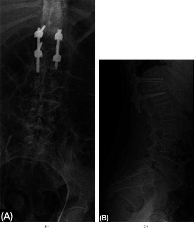
Spondylodiscitis is an uncommon infectious disease of the spine, usually presenting in 1 or 2 contiguous levels, associated with risk factors such as diabetes, intravenous drugs, corticosteroids, and invasive procedures. The most common presentation is pain with nonspecific systemic manifestations. Diagnosis relies on clinical suspicion, laboratories, and imaging studies. Urgent treatment is important due to the high morbid mortality associated with sepsis or a fulminant disease course. We report the case of a 39-year-old female diagnosed with noncontiguous multifocal spondylodiscitis, in the cervical, thoracic, and lumbar spine. The patient initially presented with back pain, inability to walk and severe neurological deficit in the upper and lower extremities, upon diagnosis broad-spectrum antibiotics were initiated. A staged surgical approach was performed in the 3 spine segments. During the 6 month follow-up, the patient presented walking with assistance, with the recovery of strength in the upper and lower extremities.


Conflict of interest statement

All authors certify that they have no affiliations with or involvement in any organization or entity with any financial interest or nonfinancial interest in the subject matter or materials discussed in this manuscript.

Figures

Figure 1
Figure 1
MRI showing the cervical, thoracic, and lumbar spine. (a, b) At the cervical spine, loss of the lordotic angle is demonstrated; destruction of the 6th cervical vertebrae is noted. In C3-C4, there is a collapse of the disc with changes in the signal intensity of the vertebral endplates, which are hyperintense in STIR T2 and isointense in T1 as well as an epidural abscess of 27 × 4.5 mm and encompasses the entire topography of C3 and C4, displacing and compressing the spinal cord. (c, d) At the level of the T12 and L1 vertebral bodies, respectively, in their lower and upper endplates, there is an erosive lesion of 17 × 18 mm involving the intervertebral disc, hyperintense on T2, and hypointense on T1, presenting wall enhancement following contrast. (e) The morphology and height of vertebral bodies T6 and T7 presented morphology alterations due to the destruction of the lower and upper vertebral endplates, respectively, associated with an expansive mass including the intervertebral disc, noted as hypointense on T1 and enhanced on T1 FatSat with gadolinium, and these changes are also observed in the epidural space, surrounding the spinal cord.
Figure 2
Figure 2
Postoperative x-ray of the thoracic spine: (a, b) postoperative changes are visualized at the level of the bony structures by left lateral instrumentation, with an intersomatic cage prosthesis at the level of T6-T7; (c) bone structures with postsurgical changes due to instrumentation in the thoracic spine, observing fixation screws and bar from the body of T4 to the body of T7.
Figure 3
Figure 3
(a) Pathology report of T6-T7 intervertebral disc shows extensive fibrosis with mixed inflammatory infiltration, bone and cartilage fragments, and reactive-looking atypia. There was no evidence of granulomas. (b) Cultures isolated Escherichia coli extended-spectrum beta-lactamases (ESBL) and Gram-positive cocci Staphylococcus aureus.
Figure 4
Figure 4
Postoperative x-ray of the cervical spine: (a) surgical material consisting of a 64 mm titanium plate and transcortical locked screws placed from C3 to C7; (b) intersomatic cages at C3-C4 and C6-C7 levels. The amplitude of the intervertebral spaces C2-C3, C4-C5, and C5-C6 is reduced.
Figure 5
Figure 5
Postoperative x-ray of the lumbar spine: (a) transpedicular screws and rod are seen between T12 and L1. The form and height of vertebral bodies are preserved; (b) appropriate amplitude of inter somatic space. A lumbar angle of 57° indicates physiological lordosis.

References

    1. Petkova A., Zhelyazkov C., Kitov B. Spontaneous spondylodiscitis-epidemiology, clinical features, diagnosis, and treatment. Folia Medica . 2017;59(3):254–260. doi: 10.1515/folmed-2017-0024. - DOI - PubMed
    1. Teo M., Trivedi R., Murphy M. Non-contiguous multifocal Staphylococcus aureus discitis involves the cervical, thoracic and lumbar spine. Acta Neurochirurgica . 2010;152(3):471–474. doi: 10.1007/s00701-009-0355-9. - DOI - PubMed
    1. Kulkarni A., Hee H. Adjacent level discitis after anterior cervical discectomy and fusion (ACDF): a case report. European Spine Journal . 2006;15(S5):559–563. doi: 10.1007/s00586-005-0003-x. - DOI - PMC - PubMed
    1. Lam K., Webb J. Discitis. Hospital Medicine . 2004;65(5):280–286. doi: 10.12968/hosp.2004.65.5.13703. - DOI - PubMed
    1. Li J., Yan D., Duan L., Zhang Z., Zhu H., Zhang Z. Percutaneous discectomy and drainage for postoperative intervertebral discitis. Archives of Orthopaedic and Trauma Surgery . 2011;131(2):173–178. doi: 10.1007/s00402-010-1115-2. - DOI - PubMed

Publication types

LinkOut - more resources