

Immunohistochemical Characterization of Gingival Fibromas
- PMID: 36472794
- PMCID: PMC10293518
- DOI: 10.1007/s12105-022-01493-y
Immunohistochemical Characterization of Gingival Fibromas
Abstract
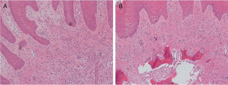
Purpose: Gingival fibromas (GFs) are fibrous lesions of the gingiva that are not well defined in the literature. They are histologically similar to peripheral ossifying fibromas (POFs), both being characterized as cellular proliferations of dense fibrous tissue, with POFs differing in that they demonstrate foci of calcification. This study aims to expand upon the immunohistochemical characterization of GFs, and to confirm their osteoblastic phenotype.
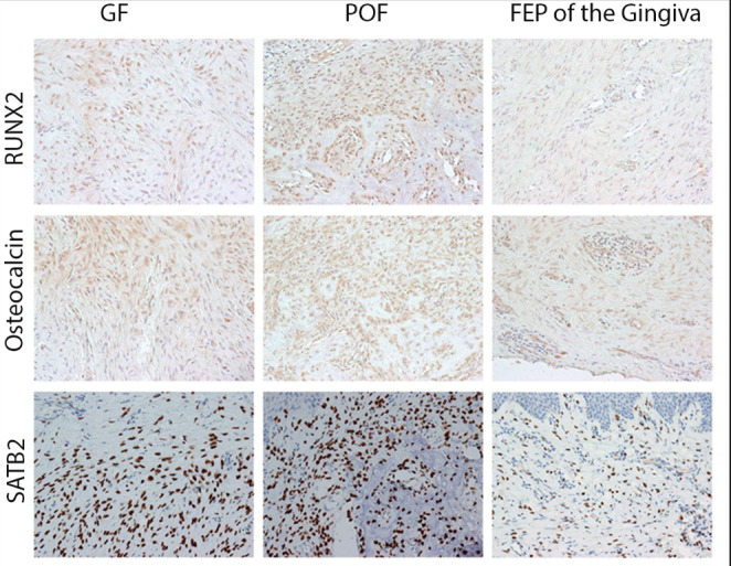
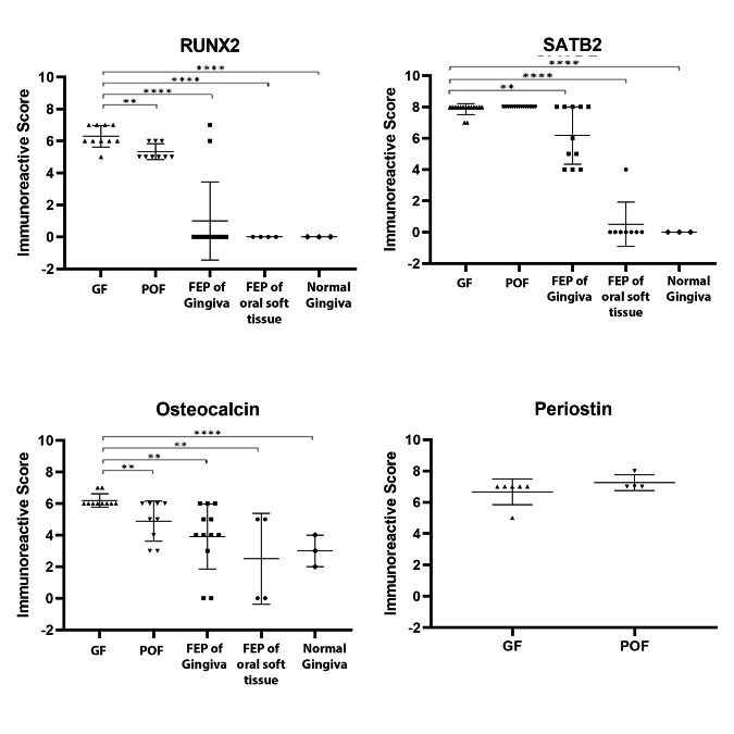
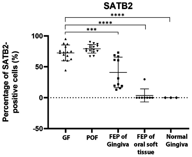
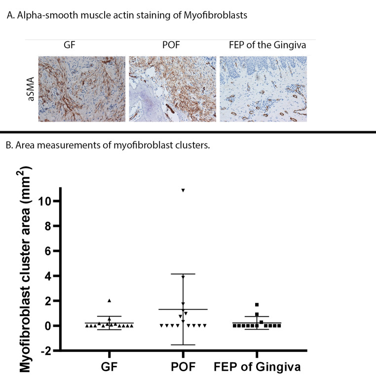
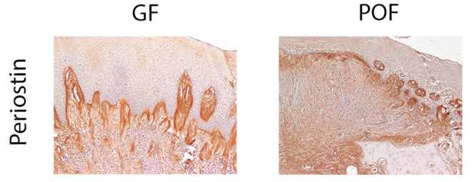
Methods: Formalin fixed, paraffin embedded GFs, POFs and fibroepithelial polyps (FEPs) of the gingiva were examined. Immunohistochemical staining was performed for special AT-rich sequence binding protein 2 (SATB2), runt-related transcription factor 2 (RUNX2), osteocalcin and alpha-smooth muscle actin (αSMA). Sections were evaluated by light microscopy and the immunohistochemical staining patterns were assigned immunoreactive scores (IRS) based on percentage of stained cells and intensity of staining.
Results: GFs, POFs, and FEPs of the gingiva expressed osteoblastic markers SATB2, RUNX2 and osteocalcin. GFs and POFs expressed αSMA while FEPs of the gingiva did not. GFs and POFs had similar staining patterns of SATB2, RUNX2 and αSMA.
Discussion: These findings demonstrate that GFs and POFs exhibit a similar immunohistochemical profile, and supports a theory that GFs are osteoblastic lesions possibly related to POFs.
Keywords: Fibroepithelial polyps of the gingiva; Gingival fibroma; Gingival lesions; Peripheral ossifying fibroma.
© 2022. The Author(s), under exclusive licence to Springer Science+Business Media, LLC, part of Springer Nature.
Conflict of interest statement
The authors have no relevant financial or non-financial interests to disclose. The authors have no competing interests to declare that are relevant to the content of this article.
Figures
References
-
- Rossmann JA. Reactive Lesions of the Gingiva: Diagnosis and Treatment Options. Open Pathol J Bentham Science Publishers Ltd. 2011;5:23–32.
-
- Buchner A, Shnaiderman-Shapiro A, Vered M. Relative frequency of localized reactive hyperplastic lesions of the gingiva: a retrospective study of 1675 cases from Israel. J Oral Pathol Med. 2010;39:631–8. Available from: https://pubmed.ncbi.nlm.nih.gov/20456619/. - PubMed
-
- Hunasgi S, Koneru A, Vanishree M, Manvikar V. Assessment of reactive gingival lesions of oral cavity: A histopathological study. J Oral Maxillofac Pathol. 2017;21:180. Available from: http://www.ncbi.nlm.nih.gov/pubmed/28479713. - PMC - PubMed
-
- Buchner A, Hansen LS. The histomorphologic spectrum of peripheral ossifying fibroma. Oral Surg Oral Med Oral Pathol. 1987;63:452–61. Available from: https://pubmed.ncbi.nlm.nih.gov/3472146/. - PubMed
-
- Ganatra S, Summerlin D-J, Zunt S, Abdelsayed R. The Gingival Fibroma: A Distinct Clinicopathologic Entity. Oral Surg Oral Med Oral Pathol. Oral Radiol. 1999. p. 202.
MeSH terms
Substances
Grants and funding
LinkOut - more resources
Full Text Sources
