. 2022 Dec 21;14(50):55320-55331.
doi: 10.1021/acsami.2c11946. Epub 2022 Dec 6.

Stimuli-Responsive Membrane Anchor Peptide Nanofoils for Tunable Membrane Association and Lipid Bilayer Fusion

Affiliations

Stimuli-Responsive Membrane Anchor Peptide Nanofoils for Tunable Membrane Association and Lipid Bilayer Fusion

Vignesh Udyavara Nagaraj et al. ACS Appl Mater Interfaces. .

Abstract

Self-assembled peptide nanostructures with stimuli-responsive features are promising as functional materials. Despite extensive research efforts, water-soluble supramolecular constructs that can interact with lipid membranes in a controllable way are still challenging to achieve. Here, we have employed a short membrane anchor protein motif (GLFD) and coupled it to a spiropyran photoswitch. Under physiological conditions, these conjugates assemble into ∼3.5 nm thick, foil-like peptide bilayer morphologies. Photoisomerization from the closed spiro (SP) form to the open merocyanine (MC) form of the photoswitch triggers rearrangements within the foils. This results in substantial changes in their membrane-binding properties, which also varies sensitively to lipid composition, ranging from reversible nanofoil reformation to stepwise membrane adsorption. The formed peptide layers in the assembly are also able to attach to various liposomes with different surface charges, enabling the fusion of their lipid bilayers. Here, SP-to-MC conversion can be used both to trigger and to modulate the liposome fusion efficiency.

Keywords: lipid bilayer fusion; liposomes; membrane activity; peptide bilayer; self-assembly; spiropyran.


Conflict of interest statement

The authors declare no competing financial interest.

Figures

Scheme 1
Scheme 1. Schematic Description of 1-GLFD
SP form (ring closed) and MC form (ring opened) of the spiropyran interconverting under UV–vis irradiation (UV λ = 365 nm).
Figure 1
Figure 1
Reversible formation of 1-GLFD assemblies monitored by the induced CD (ICD) signals. Stepwise addition of (a) NaCl and (b) GdmCl to 1-GLFD results in a gradual increase of the ICD signal at ∼380 nm. CD spectra of 1-GLFD (300 μM in PBS buffer) of (c) L and (d) S forms. The black line displays the CD spectrum of the original morphology. The blue dashed line displays the CD spectrum after irradiation with UV light at 365 nm for 5 min. The green line displays the CD spectrum after subsequent irradiation by visible light for 5 min. Insets: ICD values for several UV–vis cycles indicate reversibility of both morphologies. UV–vis irradiation was repeated three times.
Figure 2
Figure 2
Morphology of 1-GLFD nanofoils. (a) AFM color-mapped height image of a typical 1-GLFD assembly on a Si(100) wafer substrate (for details, see the Supporting Information and Figure S5). (b) TEM image of the obtained nanofoils, with normal, overlapping, and partially wrapped-up assemblies, indicating significant flexibility for these systems (for size distribution analysis, see Figure S2 and Table S1). (c) Cross-sectional height profile of the overlapping foils along the red line displayed in panel (a). The height profiles obtained provide an ∼6.2 nm average thickness per layer, which likely includes a hydration shell with sodium ions between the negatively charged foils (for further details, see the Supporting Information). (d) Schematic description of the flexible foils observed in panel (b). (e) Schematic description of the SP-to-MC conversion depicted within a small section of a nanofoil based on NMR and CD investigations.
Figure 3
Figure 3
Change of ICD peak intensities upon UV–vis irradiation cycles for (a) L and (b) S morphologies in the presence of various liposomes. ICD values for neutral PC (100%) (blue, squares) and cationic PC–DOTAP (80:20%) (green, triangles) are displayed by solid lines. Negatively charged PG (100%) (gray, hollow squares) and PC–PG (80:20%) (red, circles) liposomes are displayed by dashed lines. Each irradiation was performed for 5 min, and maximum values of the corresponding ICD peaks are displayed. UV–vis irradiation was repeated three times.
Figure 4
Figure 4
Inner structure and membrane behavior of the formed peptide nanofoils. (a) Flow-LD spectra of the studied L and S morphologies in solution and in the presence of lipid bilayers. Without liposomes, both S (solid lines) and L (dashed lines) display negative LD spectra, preserved throughout a UV–vis irradiation cycle. A significant increase in the intensity of the negative LD peaks suggests that the inner orientation of SPs within L is improved by irradiation. In the presence of liposomes, the LD peaks change the sign for both forms, indicating that the inner orientation of the systems changes significantly when bound to the lipid bilayer surface. (b) Schematic description of the obtained peptide bilayers forming the nanofoils. The direction of the electronic transition dipole moments (TDMs) corresponding to the main peaks at ∼270 and ∼360 nm is displayed as a cyan arrow. (c) LD spectra of L in the presence of PC–DOTAP liposomes during UV–vis irradiation cycles. Note that upon the second UV irradiation (UV2), the appearance of the band at 546 nm indicates the membrane-bound form of individual merocyanine moieties. (d) Schematic mechanism of the stepwise membrane adsorption of L, controlled by irradiation steps, to the membrane surface of model liposomes. The estimated relative changes between free and membrane-bound states are displayed on a small subsection of the peptide bilayer. Based on the obtained LD spectra, 1-GLFD molecules are assumed to become preferentially parallel to the membrane surface when bound to the lipid bilayer (for more details, see the Supporting Information and Figure S19). UV–vis irradiation was repeated two times.
Figure 5
Figure 5
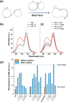
Nanofoil-assisted lipid bilayer mixing. (a) Schematic depiction of a plausible lipid membrane fusion process in the presence of nanofoils under applied shear force. Labeled lipids are highlighted by yellow and pink stars, outlining the lipid mixing observed during the employed FRET assays. (b, c) FRET efficiency of L and S nanofoils with labeled (NBD-PE/Rh-PE/PC) and nonlabeled (PC) liposomes in a 50 wt % sucrose buffer. The samples were rotated for 20 min with a shear rate of 3100 s–1. As a control, labeled liposomes were mixed with nonlabeled liposomes; L and S were added to control and shear flow was applied as above. All spectra were recorded using two accumulations. A single UV irradiation was applied prior to rotation for L and S. For UValt irradiation was applied for L and S prior to adding sucrose to the samples. (d) Bar graph representing the fluorescence intensities of controls (labeled and nonlabeled liposomes) with L and S, followed by UV and UValt irradiations (see Figures S11–S17 for before and after shearing with rotation of respective samples).

Similar articles

Cited by

References

    1. Lossada F.; Hoenders D.; Guo J.; Jiao D.; Walther A. Self-Assembled Bioinspired Nanocomposites. Acc. Chem. Res. 2020, 53, 2622–2635. 10.1021/acs.accounts.0c00448. - DOI - PubMed
    1. Tao K.; Levin A.; Adler-Abramovich L.; Gazit E. Fmoc-Modified Amino Acids and Short Peptides: Simple Bio-Inspired Building Blocks for the Fabrication of Functional Materials. Chem. Soc. Rev. 2016, 45, 3935–3953. 10.1039/c5cs00889a. - DOI - PubMed
    1. Chan K. H.; Xue B.; Robinson R. C.; Hauser C. A. E. Systematic Moiety Variations of Ultrashort Peptides Produce Profound Effects on Self-Assembly, Nanostructure Formation, Hydrogelation, and Phase Transition. Sci. Rep. 2017, 7, 1289710.1038/s41598-017-12694-9. - DOI - PMC - PubMed
    1. Loo Y.; Zhang S.; Hauser C. A. E. From Short Peptides to Nanofibers to Macromolecular Assemblies in Biomedicine. Biotechnol. Adv. 2012, 30, 593–603. 10.1016/j.biotechadv.2011.10.004. - DOI - PubMed
    1. Sun B.; Tao K.; Jia Y.; Yan X.; Zou Q.; Gazit E.; Li J. Photoactive Properties of Supramolecular Assembled Short Peptides. Chem. Soc. Rev. 2019, 48, 4387–4400. 10.1039/c9cs00085b. - DOI - PMC - PubMed