. 2023 Apr;123(4):380-392.
doi: 10.1055/a-1993-4193. Epub 2022 Dec 6.

Transglutaminase Activities of Blood Coagulant Factor XIII Are Dependent on the Activation Pathways and on the Substrates

Affiliations

Transglutaminase Activities of Blood Coagulant Factor XIII Are Dependent on the Activation Pathways and on the Substrates

Rameesa D Syed Mohammed et al. Thromb Haemost. 2023 Apr.

Abstract

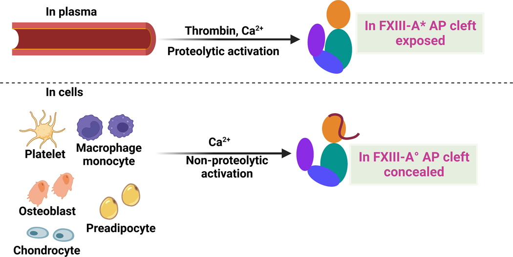
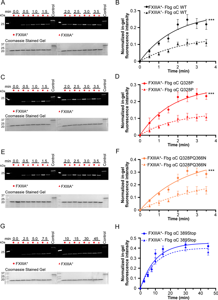
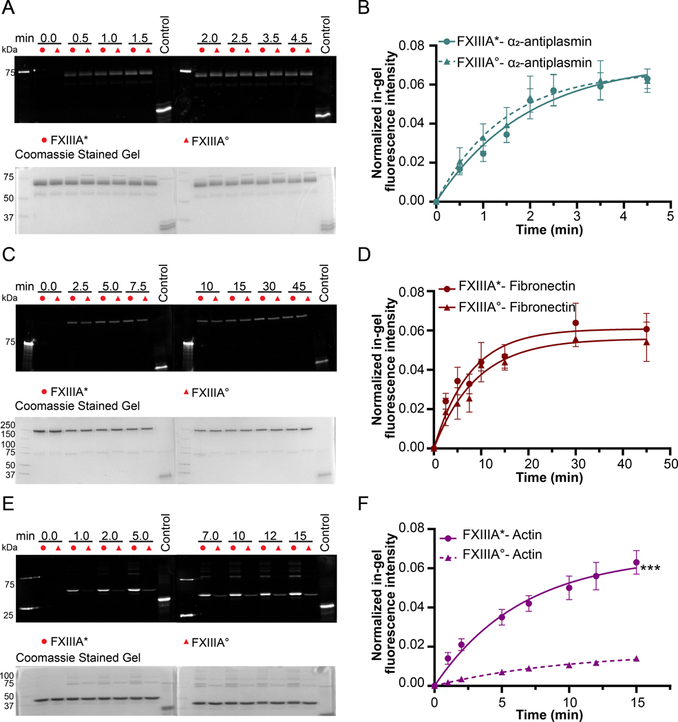
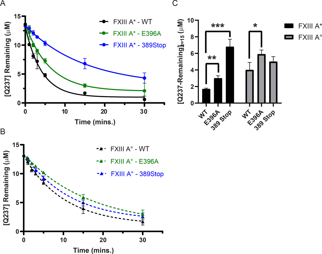
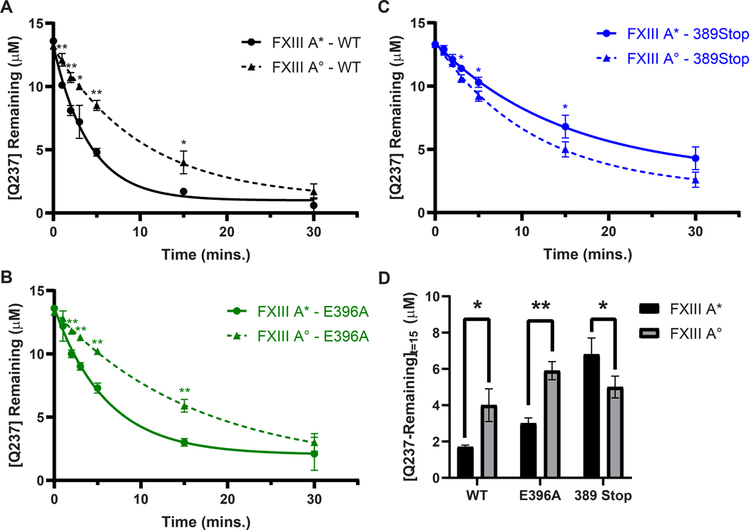
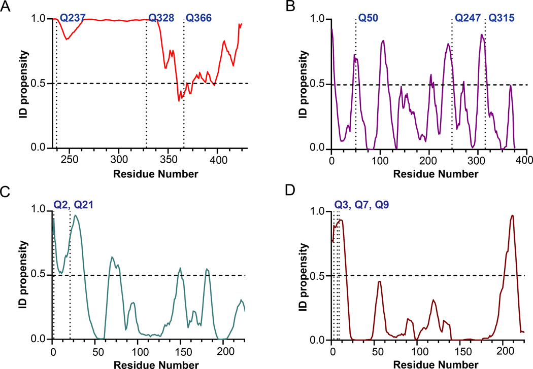
Factor XIII (FXIII) catalyzes formation of γ-glutamyl-ε-lysyl crosslinks between reactive glutamines (Q) and lysines (K). In plasma, FXIII is activated proteolytically (FXIII-A*) by the concerted action of thrombin and Ca2+. Cellular FXIII is activated nonproteolytically (FXIII-A°) by elevation of physiological Ca2+ concentrations. FXIII-A targets plasmatic and cellular substrates, but questions remain on correlating FXIII activation, resultant conformational changes, and crosslinking function to different physiological substrates. To address these issues, the characteristics of FXIII-A* versus FXIII-A° that contribute to transglutaminase activity and substrate specificities were investigated. Crosslinking of lysine mimics into a series of Q-containing substrates were measured using in-gel fluorescence, mass spectrometry, and UV-Vis spectroscopy. Covalent incorporation of fluorescent monodansylcadaverine revealed that FXIII-A* exhibits greater activity than FXIII-A° toward Q residues within Fbg αC (233-425 WT, Q328P Seoul II, and Q328PQ366N) and actin. FXIII-A* and FXIII-A° displayed similar activities toward α2-antiplasmin (α2AP), fibronectin, and Fbg αC (233-388, missing FXIII-binding site αC 389-402). Furthermore, the N-terminal α2AP peptide (1-15) exhibited similar kinetic properties for FXIII-A* and FXIII-A°. MALDI-TOF mass spectrometry assays with glycine ethyl ester and Fbg αC (233-425 WT, αC E396A, and truncated αC (233-388) further documented that FXIII-A* exerts greater benefit from the αC 389-402 binding site than FXIII-A°. Conformational properties of FXIII-A* versus A° are proposed to help promote transglutaminase function toward different substrates. A combination of protein substrate disorder and secondary FXIII-binding site exposure are utilized to control activity and specificity. From these studies, greater understandings of how FXIII-A targets different substrates are achieved.


Conflict of interest statement

None declared.

Figures

Fig. 1
Fig. 1. Cartoon models highlighting FXIII locations and activation strategies.
pFXIII undergoes proteolytic activation by thrombin to form active FXIII-A* in which AP is removed and AP cleft is exposed. cFXIII is expressed in a variety of cells including platelets, monocytes, macrophages, chondrocytes, osteoblasts and preadipocytes. cFXIII undergoes nonproteolytic activation to form FXIII-A° in which AP is still associated with A subunit. In resting platelets and monocytes, FXIII is of cytoplasmic localization, however upon activation of these cells, FXIII-A is exposed on the surface of these cells. Cartoon model is created with BioRender.com.
Fig. 2
Fig. 2. Incorporation of monodansyl cadaverine (MDC) into Fbg αC (233 – 425) WT and variants using FXIII-A* and FXIII-A°.
The crosslinking reaction between Fbg αC (233 – 425) WT and variants (Fbg αC Q328P, Fbg αC Q328PQ366N, and Fbg αC 389Stop) and MDC was initiated by adding 100 nM proteolytically or non-proteolytically activated FXIII-A (FXIII-A*/FXIII-A°). The positive control used was MDC crosslinked into N, N-dimethylated β-casein. The quantification of the gels was performed as described in the methods section. The curves represent one phase exponential association fits of the data as a function of time. Data were reported as mean ± SD (N=3). The gels for the MDC incorporation and the quantification curves include Fbg αC (233 – 425) WT (A and B), Fbg αC (233 – 425) Q328P (C and D), Fbg αC (233 – 425) Q328PQ366N (E and F), and Fbg αC (233 – 388) 389Stop (G and H). t-tests analysis showed that activity differences between FXIII-A* and FXIII-A° are significant (***P < 0.001) for all substrates except Fbg αC (233 – 388) 389Stop.
Fig. 3
Fig. 3. Incorporation of monodansyl cadaverine (MDC) into α2 -antiplasmin, fibronectin and actin using FXIII-A* and FXIII-A°.
The addition of 100 nM FXIII-A* or FXIII-A° initiated the incorporation of MDC (1 mM) into α2 -antiplasmin and actin (2 μM). For fibronectin (1 μM), 200 nM FXIII-A*/FXIII-A° were used. The positive control used was MDC crosslinked into N, N-dimethylated β-casein. The quantification of the gels was performed as described in the methods section. The curves represent one phase exponential association fits of the data as a function of time. Data were reported as mean ± SD (N=3). The gels for the MDC incorporation and the quantification curves include α2 -antiplasmin (A and B), fibronectin (C and D), and actin (E and F). t-tests analysis showed that activity differences between FXIII-A* and FXIII-A° are significant (***P < 0.001) for actin and not for (P > 0.05) α2 -antiplasmin and fibronectin.
Fig. 4
Fig. 4. Transglutaminase activity of FXIII-A* is more affected by Fbg αC binding site mutations than FXIII-A°.
FXIII-catalyzed Q237-GEE crosslinking assays between WT Fbg αC (233 – 425, black) and the FXIII binding site variants E396A (green) and 389 Stop (blue) were performed using 50 nM of either (A) FXIII-A* (circles, solid line), or (B) FXIII-A° (triangles, dashed line). Reactions were monitored by measuring the concentration of uncrosslinked Q237 remaining at each time point. Plots represent one phase exponential decay fits of uncrosslinked Q237 concentrations as a function of time. Data were reported as mean ± SD (N = 3). (C) FXIII GEE-crosslinking activity after 15 minutes of reaction time was compared between Fbg. αC WT, E396A, and 389 Stop. Shown here is the mean uncrosslinked Q237 concentration ± SD in μM after 15 minutes of reaction time (N = 3). Statistical significance was determined using the Student’s t-test (*P < 0.05, **P < 0.01, ***P < 0.001).
Fig. 5
Fig. 5. Transglutaminase activities between FXIII-A* and FXIII-A° on Fbg. αC Q237 are significantly different.
FXIII-catalyzed Q237-GEE crosslinking assays between (A) WT Fbg αC (233 – 425, black) and the FXIII binding site variants (B) E396A (green) and (C) 389 Stop (blue) were performed using 50 nM of either FXIII-A* (circles, solid lines), or FXIII-A° (triangles, dashed lines). Reactions were monitored by measuring the concentration of uncrosslinked Q237 remaining at each time point. Plots represent one phase exponential decay fits of uncrosslinked Q237 concentrations as a function of time. Data were reported as mean ± SD (N = 3). (D) FXIII GEE-crosslinking activity after 15 minutes of reaction time was compared between Fbg. αC WT, E396A, and 389 Stop. Shown here is the mean uncrosslinked Q237 concentration ± SD in μM after 15 minutes of reaction time (N = 3). Statistical significance was determined using the Student’s t-test (*P < 0.05, **P < 0.01).
Fig. 6
Fig. 6. PONDR Analysis of Fbg αC (233–425), actin, and α2-antiplasmin and fibronectin for order-disorder characteristics.
Intrinsic disorder propensity (ID propensity) of the amino acid sequences of (A) Fbg αC (233–425) (B) actin (C) α2-antiplasmin (1–225) and (D) Fibronectin (1–225) were calculated with the PONDR VLXT algorithm (www.pondr.com). The amino acid sequences of the proteins were accessed from the UniProt database (Uniprot ID for actin (P68135), α2-antiplasmin (P08697), and Fibronectin (P02751)). ID propensity value equal to or greater than 0.5 indicated disorder. The major and other potential reactive glutamines are labeled in the graph. For α2-antiplasmin and fibronectin only 225 amino acid residues from the N-terminus were used for plotting since the major reactive Qs are located in the said region.

References

    1. Richardson VR, Cordell P, Standeven KF, Carter AM. Substrates of Factor XIII-A: roles in thrombosis and wound healing. Clin Sci (Lond). 2013;124(3):123–37 - PubMed
    1. Schroeder V, Kohler HP. Factor XIII: Structure and Function. Semin Thromb Hemost. 2016;42(4):422–8 - PubMed
    1. Muszbek L, Bereczky Z, Bagoly Z, Komaromi I, Katona E. Factor XIII: a coagulation factor with multiple plasmatic and cellular functions. Physiol Rev. 2011;91(3):931–72 - PubMed
    1. Byrnes JR, Wolberg AS. Newly-Recognized Roles of Factor XIII in Thrombosis. Semin Thromb Hemost. 2016;42(4):445–54 - PMC - PubMed
    1. Mitchell JL, Mutch NJ. Let’s cross-link: diverse functions of the promiscuous cellular transglutaminase factor XIII-A. J Thromb Haemost. 2019;17(1):19–30 - PubMed