. 2022 Nov 18:2022:6256384.
doi: 10.1155/2022/6256384. eCollection 2022.

Knockdown of lncRNA XIST Ameliorates IL-1 β-Induced Apoptosis of HUVECs and Change of Tissue Factor Level via miR-103a-3p/HMGB1 Axis in Deep Venous Thrombosis by Regulating the ROS/NF- κ B Signaling Pathway

Affiliations

Knockdown of lncRNA XIST Ameliorates IL-1 β-Induced Apoptosis of HUVECs and Change of Tissue Factor Level via miR-103a-3p/HMGB1 Axis in Deep Venous Thrombosis by Regulating the ROS/NF- κ B Signaling Pathway

Guangxin Cao et al. Cardiovasc Ther. .

Abstract

Background: The effect of lncRNA X inactive-specific transcript (XIST) inducing cardiovascular diseases on deep vein thrombosis (DVT) and its mechanism has not been reported. In this study, we uncovered the mystery that lncRNA XIST causes DVT with HUVEC dysfunction.

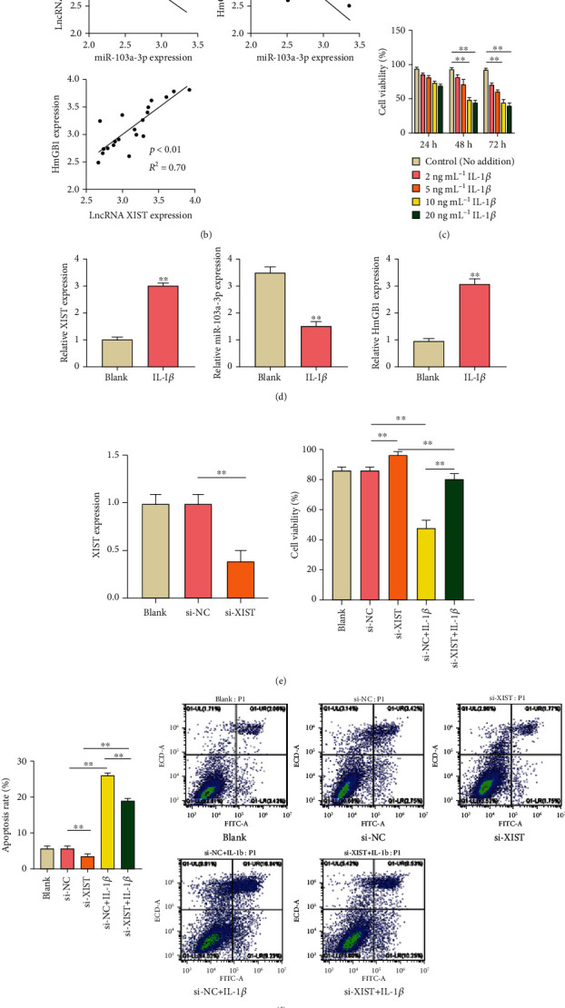
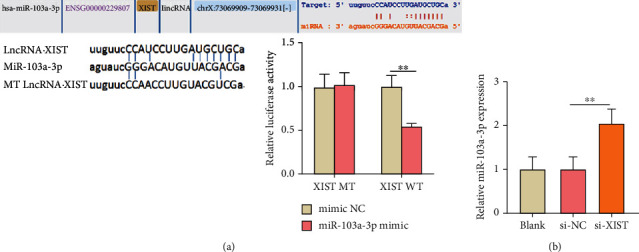
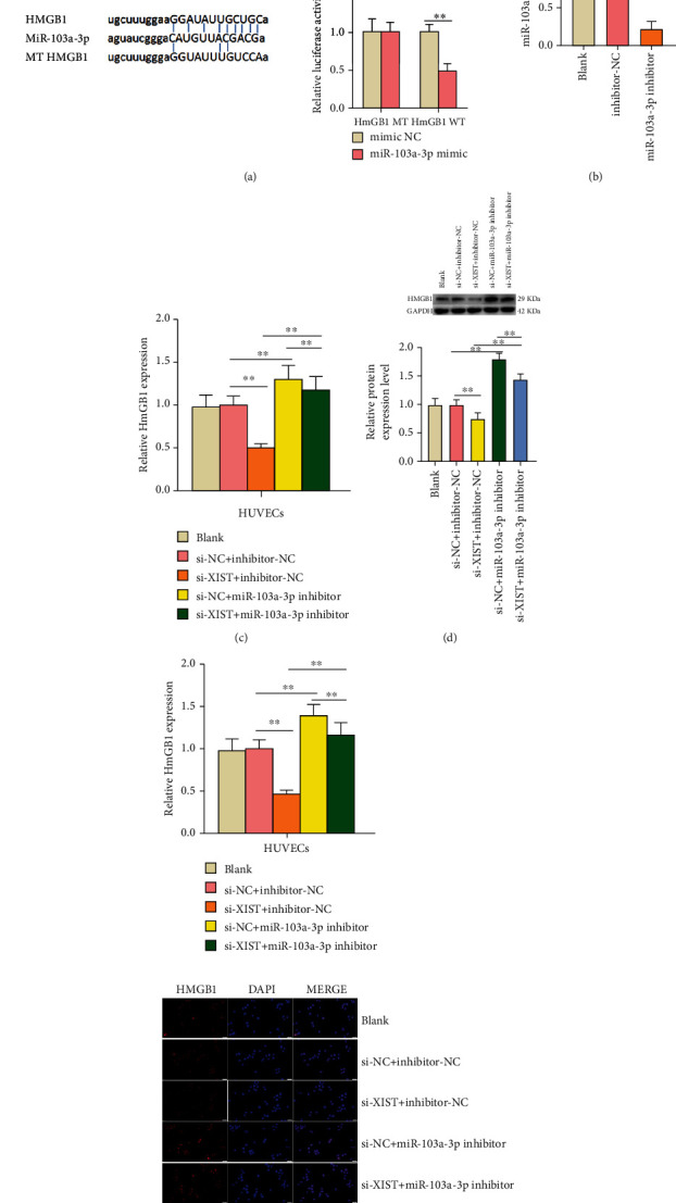
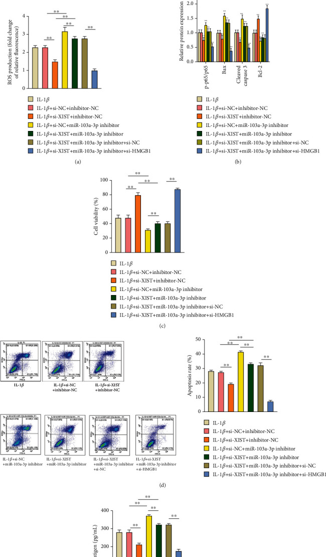
Method: The expression levels of lncRNA XIST and miR-103a-3p were detected by qRT-PCR, and HMGB1 expression was determined by qRT-PCR and western blot. The correlations among the expression levels of lncRNA XIST, miR-103a-3p, and HMGB1 were determined by Spearman's rank-order correlation test. XIST siRNA (si-XIST) was transfected into HUVECs to knock down the intrinsic expression of lncRNA XIST. The influences of si-XIST on interleukin-1 beta- (IL-1β-) treated HUVEC viability and apoptosis and the level of tissue factor (TF) were detected by MTT, flow cytometry, and ELISA kit, respectively. The relationships between lncRNA XIST, miR-103a-3p, and HMGB1 were predicted by the Encyclopedia of RNA Interactomes (ENCORI) database and verified by dual luciferase reporter assay. The effects of lncRNA XIST and miR-103a-3p on HMGB1 expression were detected by qRT-PCR, western blot, and immunofluorescence analysis. The levels of ROS/NF-κB pathway-related proteins were detected to study the regulatory mechanism of lncRNA XIST/miR-103a-3p/HMGB1 on IL-1β-treated HUVECs apoptosis and change of TF level.

Results: The upregulated expression levels of lncRNA XIST and HMGB1 and downregulated level of miR-103a-3p were found in the plasma of DVT patients and IL-1β-treated HUVECs. Si-XIST promoted cell viability and inhibited HUVEC apoptosis and ameliorated the change of TF level triggered by IL-1β. lncRNA XIST sponged miR-103a-3p and miR-103a-3p targeted HMGB1. Si-XIST inhibited the ROS/NF-κB pathway to suppress HUVEC apoptosis and ameliorate the change of TF level induced by IL-1β via the miR-103a-3p/HMGB1 axis.

Conclusion: lncRNA XIST sponged miR-103a-3p improving HMGB1 expression to exacerbate DVT by activating the ROS/NF-κB signaling pathway. Our findings indicated that lncRNA XIST can be used as a potential therapeutic target in DVT.


Conflict of interest statement

The authors declare no conflict of interest.

Figures

Figure 1
Figure 1
Knockdown of lncRNA XIST attenuated the apoptosis of HUVECs and resulted in a decrease of IL-1β-induced TF level in vivo. (a) The expression levels of lncRNA XIST and miR-103a-3p were detected by qRT-PCR, and the expression level of HMGB1 was analyzed by qRT-PCR and western blot. (b) The correlations among the expression levels of lncRNA XIST, miR-103a-3p, and HMGB1 were calculated by Spearman's rank-order correlation test. (c) The cell viability of HUVECs under IL-1β addition was determined by MTT assay. (d) The expression levels of lncRNA XIST, miR-103a-3p, and HMGB1 were measured under IL-1β treatment by qRT-PCR. (e) The expression level of lncRNA XIST in transfected HUVECs was analyzed by qRT-PCR, and the cell viability in transfected cells was detected by MTT assay. (f) The cell apoptosis was analyzed by Annexin V-FITC/PI double staining kit. (g) The expression levels of proteins related to apoptosis were detected by western blot. (h) The concentration of TF antigen was determined by the ELISA kit. The nontransfected HUVECs served as the blank group. P < 0.05 and ∗∗P < 0.01.
Figure 2
Figure 2
lncRNA XIST sponged miR-103a-3p in HUVECs. (a) Bioinformatic analysis using ENCORI database showed the interaction between lncRNA XIST with miR-103a-3p, and the binding relationship of lncRNA XIST and miR-103a-3p was verified by luciferase reporter gene assays. MT: mutant type; WT: wild type. (b) The expression of miR-103a-3p in transfected HUVECs was detected by qRT-PCR assays. P < 0.05 and ∗∗P < 0.01.
Figure 3
Figure 3
The expression of HMGB1 sponged by miR-103a-3p was improved by adding lncRNA XIST to sponge miR-103a-3p. (a) Bioinformatic analysis using ENCORI database shows the interaction of miR-103a-3p with HMGB1, and the binding relationship of miR-103a-3p and HMGB1 was proved by luciferase reporter gene assays. (b) The expression of miR-103a-3p in transfected HUVECs was detected by qRT-PCR. (c) The expression of HMGB1 in cotransfected cells was analyzed by qRT-PCR. (d) The expression level of HMGB1 protein in cotransfected cells was analyzed by western blot. (e) The expression level of HMGB1 protein in cotransfected cells was detected by immunofluorescence analysis (scale bar: 20 μm). The nontrasfected HUVECs served as the blank group. P < 0.05 and ∗∗P < 0.01.
Figure 4
Figure 4
ROS-induced apoptosis and change of TF level based on adding IL-1β were regulated via lncRNA XIST/miR-103a-3p/HMGB1. (a) The ROS production in cotransfected cells was analyzed by DCFH-DA staining. (b) The expression levels of proteins related to the NF-κB pathway and apoptosis in cotransfected cells were analyzed by western blot. (c) The viability of cotransfected cells was determined by MTT assay. (d) The apoptosis of cotransfected cells was tested by Annexin V-FITC/PI double staining kit. (e) The concentration of TF antigen in cotransfected cells was determined by the ELISA kit. P < 0.05 and ∗∗P < 0.01.

Similar articles

Cited by

References

    1. Gutmann C., Siow R., Gwozdz A. M., Saha P., Smith A. Reactive oxygen species in venous thrombosis. International Journal of Molecular Sciences . 2020;21(6, article 1918) doi: 10.3390/ijms21061918. - DOI - PMC - PubMed
    1. Chen L., Wang J., Wang B., et al. miR-126 inhibits vascular endothelial cell apoptosis through targeting PI3K/Akt signaling. Annals of Hematology . 2016;95(3):365–374. doi: 10.1007/s00277-015-2567-9. - DOI - PubMed
    1. Patterson B. O., Hinchliffe R., Loftus I. M., Thompson M. M., Holt P. J. E. Indications for catheter-directed thrombolysis in the management of acute proximal deep venous thrombosis. Arteriosclerosis, Thrombosis, and Vascular Biology . 2010;30(4):669–674. doi: 10.1161/ATVBAHA.109.200766. - DOI - PubMed
    1. Miao W., Zhao K., Deng W., Teng J. Coagulation factor hyperfunction after subarachnoid hemorrhage induces deep venous thrombosis. World Neurosurgery . 2018;110:e46–e52. doi: 10.1016/j.wneu.2017.09.200. - DOI - PubMed
    1. Quinn J. J., Chang H. Y. Unique features of long non-coding RNA biogenesis and function. Nature Reviews Genetics . 2016;17(1):47–62. doi: 10.1038/nrg.2015.10. - DOI - PubMed