

Atypical Bronchial Carcinoid Tumor Revealed by Liver Biopsy
- PMID: 36475219
- PMCID: PMC9719747
- DOI: 10.7759/cureus.31104
Atypical Bronchial Carcinoid Tumor Revealed by Liver Biopsy
Abstract
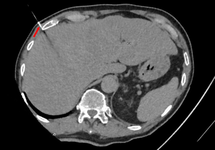
Bronchial carcinoid tumors develop from the Kulchitsky cells of the bronchial epithelium, which are stem cells with neuroendocrine properties. These tumors are divided into two types: typical forms and atypical forms, the latter being much rarer, more aggressive, and having a much higher probability of recurrence and distant metastasis. We report herein a rare case of an atypical lung carcinoid tumor metastatic to the liver. The patient is a 79-year-old woman who presented with purely digestive symptoms evolving for two years, with loss of appetite and deterioration of her general condition. The radiological assessment showed a pulmonary lesion with secondary hepatic and osseous nodules. A hepatic biopsy was performed and morphological and immunohistochemical results were compatible with an atypical bronchial carcinoid tumor, metastatic to the liver and bone.
Keywords: atypical; carcinoid tumor; case report; lung cancer; typical.
Copyright © 2022, Hayoune et al.
Conflict of interest statement
The authors have declared that no competing interests exist.
Figures


Similar articles
-
Disseminated typical bronchial carcinoid tumor.Vojnosanit Pregl. 2013 May;70(5):516-21. doi: 10.2298/vsp1305516n. Vojnosanit Pregl. 2013. PMID: 23789293
-
Atypical carcinoid localized at the bronchus accompanied by diffuse idiopathic pulmonary neuroendocrine cell hyperplasia in the distal lung: a rare case report.J Thorac Dis. 2017 Sep;9(9):E774-E778. doi: 10.21037/jtd.2017.08.75. J Thorac Dis. 2017. PMID: 29221341 Free PMC article.
-
Bronchial carcinoid tumor: A case report.Int J Surg Case Rep. 2020;77:349-352. doi: 10.1016/j.ijscr.2020.11.043. Epub 2020 Nov 11. Int J Surg Case Rep. 2020. PMID: 33212309 Free PMC article.
-
Bronchial carcinoid tumors metastatic to the sella turcica and review of the literature.Pituitary. 2012 Jun;15(2):160-5. doi: 10.1007/s11102-012-0388-6. Pituitary. 2012. PMID: 22485018 Review.
-
Atypical bronchial carcinoid with postobstructive mycobacterial infection: case report and review of literature.BMC Pulm Med. 2019 Feb 15;19(1):41. doi: 10.1186/s12890-019-0806-x. BMC Pulm Med. 2019. PMID: 30767776 Free PMC article. Review.
References
-
- Two different types of carcinoid tumors of the lung: immunohistochemical and ultrastructural investigation and their histogenetic consideration. Min KW. https://doi.org/10.3109/01913123.2012.707962. Ultrastruct Pathol. 2013;37:23–35. - PubMed
-
- Pulmonary neuroendocrine/carcinoid tumors: a review article. Bertino EM, Confer PD, Colonna JE, Ross P, Otterson GA. https://doi.org/10.1002/cncr.24498. Cancer. 2009;115:4434–4441. - PubMed
-
- Introduction to the 2015 World Health Organization classification of tumors of the lung, pleura, thymus, and heart. Travis WD, Brambilla E, Burke AP, Marx A, Nicholson AG. https://doi.org/10.1097/JTO.0000000000000663. J Thorac Oncol. 2015;10:1240–1242. - PubMed
-
- Medical treatment of advanced thoracic neuroendocrine tumors. Ferolla P. https://doi.org/10.1016/j.thorsurg.2014.05.006. Thorac Surg Clin. 2014;24:351–355. - PubMed
-
- The 2015 World Health Organization classification of lung tumors: impact of genetic, clinical and radiologic advances since the 2004 classification. Travis WD, Brambilla E, Nicholson AG, et al. https://doi.org/10.1097/JTO.0000000000000630. J Thorac Oncol. 2015;10:1243–1260. - PubMed
Publication types
LinkOut - more resources
Full Text Sources