Review
. 2022 Dec;12(12):220274.
doi: 10.1098/rsob.220274. Epub 2022 Dec 7.

Mitochondrial biology and dysfunction in secondary mitochondrial disease

Affiliations
Review

Mitochondrial biology and dysfunction in secondary mitochondrial disease

Megan J Baker et al. Open Biol. 2022 Dec.

Abstract

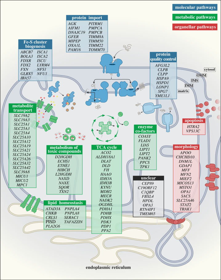
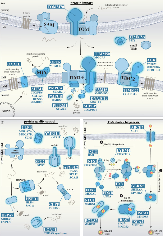
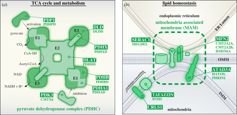
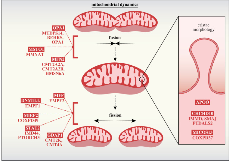
Mitochondrial diseases are a broad, genetically heterogeneous class of metabolic disorders characterized by deficits in oxidative phosphorylation (OXPHOS). Primary mitochondrial disease (PMD) defines pathologies resulting from mutation of mitochondrial DNA (mtDNA) or nuclear genes affecting either mtDNA expression or the biogenesis and function of the respiratory chain. Secondary mitochondrial disease (SMD) arises due to mutation of nuclear-encoded genes independent of, or indirectly influencing OXPHOS assembly and operation. Despite instances of novel SMD increasing year-on-year, PMD is much more widely discussed in the literature. Indeed, since the implementation of next generation sequencing (NGS) techniques in 2010, many novel mitochondrial disease genes have been identified, approximately half of which are linked to SMD. This review will consolidate existing knowledge of SMDs and outline discrete categories within which to better understand the diversity of SMD phenotypes. By providing context to the biochemical and molecular pathways perturbed in SMD, we hope to further demonstrate the intricacies of SMD pathologies outside of their indirect contribution to mitochondrial energy generation.

Keywords: mitochondria; mitochondrial protein import; mitochondrial quality control; secondary mitochondrial disease.


Conflict of interest statement

We declare we have no competing interests.

Figures

Figure 1.
Figure 1.
Categorical organization of mitochondrial genes associated with secondary mitochondrial disease (SMD). Genes with deleterious mutations impacting mitochondrial functions secondary to OXPHOS have been broadly classified into three main categories: (1) Molecular pathways related to protein biogenesis, including protein import, protein quality control and Fe-S cluster biogenesis (indicated in blue); (2) Metabolic pathways involving metabolite transport, metabolism of toxic compounds, enzymatic cofactors, TCA cycle metabolism and lipid homeostasis (indicated in green); and (3) Organellar pathways linked to mitochondrial health, including mitochondrial morphology and apoptosis (indicated in red). Genes linked to SMD with currently unclear functions are listed in the Unclear category (indicated in grey). OMM, outer mitochondrial membrane; IMS, intermembrane space; IMM, inner mitochondrial membrane.
Figure 2.
Figure 2.
Overview of protein biogenesis pathways linked to secondary mitochondrial disease. (a) Schematic depicting mitochondrial import pathways and genes associated with SMD. The majority of nuclear-encoded mitochondrial precursor proteins are targeted to mitochondria and imported via the translocase of the outer mitochondrial membrane (TOMM) complex. From here, precursor import pathways diverge to one of four key routes: (1) β-barrel insertion and assembly into the outer membrane via the sorting and assembly machinery (SAM); (2) cysteine-rich precursors of the intermembrane space are oxidized by CHCHD4 (MIA40) at the mitochondrial intermembrane space and assembly complex (MIA); (3) the N-terminal pre-sequence pathway via the translocase of the inner mitochondrial membrane 23 (TIMM23) complex, for import into the mitochondrial matrix, or lateral insertion of proteins into the inner membrane; and (4) the carrier pathway, where proteins with multi-spanning transmembrane domains are chaperoned by members of the small TIM family and delivered to the TIMM22 complex for lateral insertion into the inner membrane. The OXA1L insertase is responsible for the biogenesis of a number of inner membrane proteins, including components of the respiratory chain encoded by the mitochondrial genome. (b) Protein quality control systems within the mitochondrial intermembrane space and matrix. Within the IMS, the i-AAA protease (YME1L1 hexamer) and CLPB disaggregase clear misfolded and aggregated proteins, respectively. In the matrix, the HSP60/10 chaperone complex (HSPD1/HSPE1, respectively) facilitates protein folding while the m-AAA (AFG3L2/SPG7 hetero-hexamer or AFG3L2 homo-hexamer), CLPXP (CLPP and CLPX oligomer), and LONP1 proteases cooperate to degrade misfolded protein precursors. (c) Fe-S cluster biogenesis occurs through three major steps 1) [2Fe-2S] biosynthesis: NFS1, in complex with LYRM4, catalyses the release of sulfane (-SSH) from cysteine. FXN likely chaperones imported iron to the ISCU scaffold. FDX2 and FDXR reduce sulfane to sulfide and finalize [2Fe-2S] assembly at ISCU. 2) [2Fe-2S] trafficking: chaperone HSPA9 reacts with and detaches [2Fe-2S] from the ISCU. GLRX5 binds to and transfers the mature [2Fe-2S] cluster to apoproteins or shuttles the cluster for export. 3) [4Fe-4S] biosynthesis: IBA57, ISCA1 and ISCA2 facilitate the maturation of [4Fe-4S] clusters and can either deliver them to apoproteins directly or pass clusters on to other proteins (such as NFU1 or BOLA3) to target more specific downstream [4Fe-4S]-containing proteins. Gene names are boxed, and associated diseases are listed below or indicated here: (a) AGK (Sengers syndrome (MIM #212350) and CTRCT38 (MIM #614691)); AIFM1 (COXPD6 (MIM #300816), CMTX4 (MIM #310490), DFNX5 (MIM #300614) and SEMDHL (MIM #300232)); DNACJ19 (MGCA5 (MIM #610198)); GFER (MPMCD (MIM #613076)); MIPEP (COXPD31 (MIM #617228)); OXA1L (-); PAM16 (SMDMDM (MIM #613320)); PITRM1 (SCAR30 (MIM #619405)); PMPCA (SCAR2 (MIM #213200)); PMPCB (MMDS6 (MIM #617954)); TIMM22 (COXPD43 (MIM #618851)); TIMM50 (MGCA9 (MIM #617698)); TIMM8A (MTS (MIM #304700)); TOMM70 (-). (b) AFG3L2 (SPAX5 (MIM #614487)), OPA12 (MIM #618977) and SCA28 (MIM #610246)); CLPB (MGCA7A (MIM #619835), MGCA7B (MIM #616271) and SCN9 (MIM #619813)); CLPP (PRLTS3 (MIM #614129)); HSPA9 (SIDBA4 (MIM #182170) and EVPLS (MIM #616854)); HSPD1 (HLD4 (MIM #612233) and SPG13 (MIM #605280)); LONP1 (CODAS syndrome (MIM #600373)); SPG7 (SPG7 (MIM #607259)); YME1L1 (OPA11 (MIM #617302)). (c) ABCB7 (ASAT (MIM #301310)); BOLA3 (MMDS2 (MIM #614299)); FDX2 (MEOAL (MIM #251900)); FDXR (ANOA (MIM #617717)); FXN (FRDA (MIM #229300)); GLRX5 (SIDBA3 (MIM #616860) and SPAHGC (MIM #616859)); IBA57 (SPG74 (MIM# 616451) and MMDS3 (MIM #615330)); ISCA1 (MMDS5 (MIM #617613)); ISCA2 (MMDS4 (MIM #616370)); ISCU (HML (MIM #255125)); LYRM4 (COXPD19 (MIM #615595)), NFS1 (COXPD52 (MIM #619386)); NFU1 (MIM #MMDS1 (605711)).
Figure 3.
Figure 3.
TCA and lipid metabolic pathways compromised in secondary mitochondrial disease. (a) At the core of the PDHC complex is dihydrolipoyl transacetylase (DLAT; E2). The E2 core is anchored to dihydrolipoamide dehydrogenase (DLD; E3) via PDHX. Pyruvate dehydrogenase a/β heterotetrametric subunits (PDHA1/PDHB; E1), associate with E2. The PDHC catalyses the production of acetyl-CoA from pyruvate in a three-step process: (1) E1, in conjunction with thiamine pyrophosphate (TPP) cofactor, catalyses the decarboxylation of pyruvate, releasing CO2 and forming a hydroxyethyl-TPP intermediate. (2) E2 transfers the hydroxyethyl group from TPP to an oxidized lipoamide cofactor, releasing an acetyl group which is then transferred to coenzyme A (CoA-SH) to form acetyl-CoA and a reduced dihydrolipoamide-E2 core. (3) E3 then oxidizes the lipoyl group of dihydrolipoamide-E2 to form lipoamide-E2 and NADH. Activity of the PDHC can be tightly modulated by associated kinases and phosphatases; phosphorylation of E1 by pyruvate dehydrogenase kinase 3 (PDK3) inactivates PDC, and dephosphorylation of E1 by pyruvate dehydrogenase phosphatase 1 (PDP1) reactivates PDC. (b)Mitochondria-associated membranes (MAMs) are direct points of contact between mitochondria and endoplasmic reticulum (ER). MAMs support phospholipid exchange between the ER and mitochondria, with links to SERAC1 in this process. In addition, MAMs serve as complex signalling platforms, as selective enrichment of proteins at these intraorganellar contact points enables robust coordination of intracellular events, such as apoptosis, autophagy and calcium homeostasis. ER-mitochondrial contacts also regulate mitochondrial dynamics, recruiting proteins such as MFF and MFN2 to coordinate mitochondrial fission and fusion, respectively. Proteins such as TAFAZZIN and CRLS1 coordinate cardiolipin remodelling at the inner membrane to preserve correct lipid composition. ATAD3A is proposed to tether mitochondrial membranes at MAM sites and is therefore broadly implicated in the retention of mitochondrial network structure and cholesterol homeostasis, in addition to mtDNA nucleoid regulation. Gene names are boxed, and associated diseases are listed below or indicated here: (a) DLAT (PDHDD (MIM #245348)); DLD (DLDD (MIM #246900)); PDHA1 (PDHAD (MIM #312170)); PDHB (PDHBD (MIM #614111)); PDHX (PDHXD (MIM #245349)); PDK3 (CMTX6 (MIM #300905)); PDP1 (PDHPD (MIM #608782)). (b) ATAD3A (HAYOS (MIM #617183) and PHRINL (MIM #618810)); CRLS1 (-); MFN2 (CMT2A2A (MIM #609260), CMTA2A2B (MIM #617087) and HMSN6A (MIM #601152)); SERAC1 (MEGDEL (MIM #614739)); TAFAZZIN (BTHS (MIM #302060)).
Figure 4.
Figure 4.
Regulators of mitochondrial fission and fusion and secondary mitochondrial disease. Mitochondrial fusion events occur by successive fusion of outer and inner membranes. Mitochondrial fusion GTPases MFN1 and MFN2 mediate OM fusion in conjunction with MSTO1, a cytosolic accessory protein recruited to the OM via an unknown mechanism. Mitochondrial IM fusion is coordinated by balanced processing of the OPA1 GTPase from long-form (L-OPA1) into short-form (S-OPA1). L-OPA1 can form oligomers and promote fusion upon GTP hydrolysis. Excessive L-OPA1 processing into S-OPA1 can disrupt L-OPA1 fusion events and tip the balance towards mitochondrial network fission. Mitochondrial fission is an essential component in cellular proliferation and is also used to clear terminally damaged or toxic nodes from the network via mitophagy. Fission factors such as MFF, MIEF1 and MIEF2 recruit cytosolic GTPase DNM1L to the OM, where it assembles in a spiral formation to restrict mitochondria and sever the double membrane upon GTP hydrolysis. DNM1L GTPase activity is dynamically controlled via a number of post translational modifications, including phosphorylation as mediated by kinases such as STAT2. GDAP1 is another fission factor localized to the OM. While loss of GDAP1 prevents efficient mitochondrial fission, the exact role of GDAP1 in cooperation with other OM fission mediators is yet to be uncovered. Within the mitochondrial contact site and cristae organizing system (MICOS) only two components have been connected to secondary mitochondrial disease: CHCHD10 and MICOS13. MICOS13 is an IM scaffolding protein required for the integration of other MICOS members into the mature complex. CHCHD10 is an IMS protein peripherally associated with the MICOS and is believed to maintain complex stability. Gene names are boxed, and associated diseases are listed below or indicated here: CHCHD10 (IMMD (MIM #616209), SMAJ (MIM #615048) and FTDALS2 (MIM #615911)); DNM1L (EMPF1 (MIM #614388)); GDAP1 (CMT2K (MIM #607831) and CMT4A (MIM #214400)); MFF (EMPF2 (MIM #617086)); MFN2 (CMT2A2A (MIM #609260), CMT2A2B (MIM #617087) and HMSN6A (MIM #601152)); MICOS13 (COXPD37 (MIM #618329)); MIEF2 (COXPD49 (MIM #619024)); MSTO1 (MMYAT (MIM #617675)); OPA1 (MTDPS14 (MIM #616896), BEHRS (MIM #210000) and OPA1 (MIM #165500)); STAT2 (IMD44 (MIM #616636) and PTORCH3 (MIM #618886)).

References

    1. Gabaldón T, Huynen MA. 2004. Shaping the mitochondrial proteome. Biochim. Biophys. Acta (BBA) - Bioenerg. 1659, 212-220. (10.1016/j.bbabio.2004.07.011) - DOI - PubMed
    1. Gabaldón T, Huynen MA. 2003. Reconstruction of the proto-mitochondrial metabolism. Science 301, 609. (10.1126/science.1085463) - DOI - PubMed
    1. Friedman JR, Nunnari J. 2014. Mitochondrial form and function. Nature 505, 335-343. (10.1038/nature12985) - DOI - PMC - PubMed
    1. Pfanner N, Warscheid B, Wiedemann N. 2019. Mitochondrial proteins: from biogenesis to functional networks. Nat. Rev. Mol. Cell Biol. 20, 267-284. (10.1038/s41580-018-0092-0) - DOI - PMC - PubMed
    1. Burté F, Carelli V, Chinnery PF, Yu-Wai-Man P. 2015. Disturbed mitochondrial dynamics and neurodegenerative disorders. Nat. Rev. Neurol. 11, 11-24. (10.1038/nrneurol.2014.228) - DOI - PubMed