Review
. 2022 Dec 20;55(24):3572-3580.
doi: 10.1021/acs.accounts.2c00520. Epub 2022 Dec 7.

Relaxometry with Nitrogen Vacancy (NV) Centers in Diamond

Affiliations
Review

Relaxometry with Nitrogen Vacancy (NV) Centers in Diamond

Aldona Mzyk et al. Acc Chem Res. .

Abstract

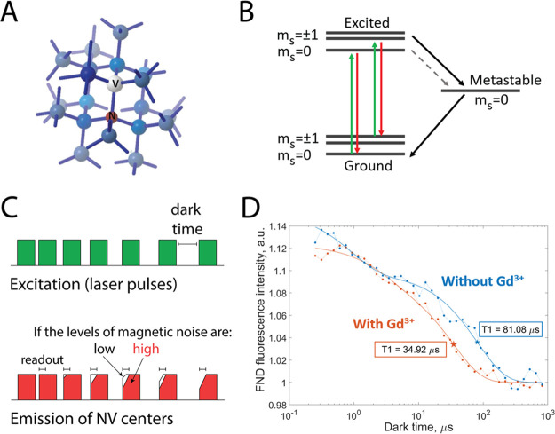
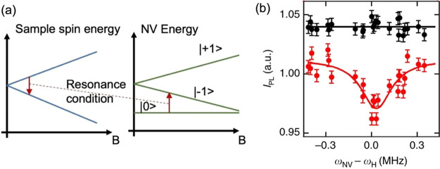
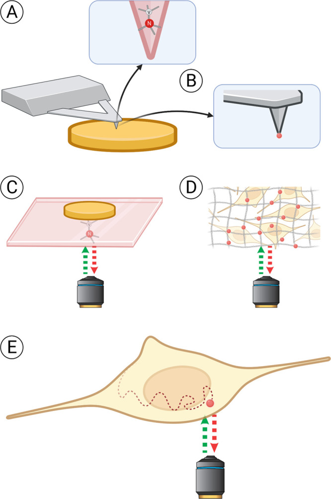
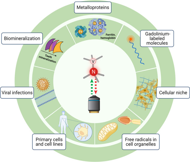
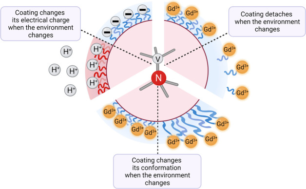
Relaxometry is a technique which makes use of a specific crystal lattice defect in diamond, the so-called NV center. This defect consists of a nitrogen atom, which replaces a carbon atom in the diamond lattice, and an adjacent vacancy. NV centers allow converting magnetic noise into optical signals, which dramatically increases the sensitivity of the readout, allowing for nanoscale resolution. Analogously to T1 measurements in conventional magnetic resonance imaging (MRI), relaxometry allows the detection of different concentrations of paramagnetic species. However, since relaxometry allows very local measurements, the detected signals are from nanoscale voxels around the NV centers. As a result, it is possible to achieve subcellular resolutions and organelle specific measurements.A relaxometry experiment starts with polarizing the spins of NV centers in the diamond lattice, using a strong laser pulse. Afterward the laser is switched off and the NV centers are allowed to stochastically decay into the equilibrium mix of different magnetic states. The polarized configuration exhibits stronger fluorescence than the equilibrium state, allowing one to optically monitor this transition and determine its rate. This process happens faster at higher levels of magnetic noise. Alternatively, it is possible to conduct T1 relaxation measurements from the dark to the bright equilibrium by applying a microwave pulse which brings NV centers into the -1 state instead of the 0 state. One can record a spectrum of T1 at varying strengths of the applied magnetic field. This technique is called cross-relaxometry. Apart from detecting magnetic signals, responsive coatings can be applied which render T1 sensitive to other parameters as pH, temperature, or electric field. Depending on the application there are three different ways to conduct relaxometry experiments: relaxometry in moving or stationary nanodiamonds, scanning magnetometry, and relaxometry in a stationary bulk diamond with a stationary sample on top.In this Account, we present examples for various relaxometry modes as well as their advantages and limitations. Due to the simplicity and low cost of the approach, relaxometry has been implemented in many different instruments and for a wide range of applications. Herein we review the progress that has been achieved in physics, chemistry, and biology. Many articles in this field have a proof-of-principle character, and the full potential of the technology still waits to be unfolded. With this Account, we would like to stimulate discourse on the future of relaxometry.


Conflict of interest statement

The authors declare no competing financial interest.

Figures

Figure 1
Figure 1
Foundation of NV center based relaxometry. (A) Nitrogen vacancy centers are defects of the diamond crystal structure: a nitrogen atom is present instead of one of the carbon atoms, and one of the neighboring positions in the lattice is vacant. (B) A diagram of the NV center’s energy levels. (C) The NV centers are pumped into a brighter ground state with a green laser pulse. The laser is switched off for a predefined period of time (dark time), and the NV centers are allowed to relax back to the darker equilibrium state. The observed fluorescence becomes less bright as the dark times increase. The transition from the bright polarized state to the darker equilibrium state happens faster at higher levels of magnetic noise (e.g., due to paramagnetic species). (D) The NV center’s fluorescence intensity plotted against the corresponding dark times can be used to calculate the T1 relaxation constant. If the relaxation is slower, one observes longer T1 values (blue; diamond without Gd3+). Faster relaxation can happen due to the changes in the NV center’s environment, such as the presence of paramagnetic species. In this case, the T1 curves are shifted to the left, producing shorter T1 values (orange; after adding paramagnetic Gd3+ ions).
Figure 2
Figure 2
Cross-relaxometry principle. (a) Cross-relaxometry offers spectral information. If T1 experiments are done in the presence of a varying magnetic field B, there is a condition where the energy difference between the NV center energy levels equals the difference in the energy levels of the spins in the sample. When the magnetic field is swept, there is a peak at this energy which is characteristic for the sample spin. (b) Example spectrum taken with the cross-relaxometry method. The scan over the Larmor frequency of 1H is shown in red. Data from ref (13).
Figure 3
Figure 3
Different ways to conduct relaxometry experiments. The sample is shown in yellow, the (nano)diamond hosting NV centers in red. Three main approaches can be defined. In the first case (A, B), the sample is scanned with a NV-containing probe, similarly to atomic force microscopy. The scanning tip can either be made of diamond hosting NV centers (A) or have a nanodiamond attached to it (B). The second approach (C, D) relies on obtaining information from a stationary diamond placed close to the sample. Most commonly, bulk diamond plates are used for that (C). Nanodiamonds immobilized on a supporting surface or embedded in polymer matrices offer a cheaper alternative. They can be measured through a cover glass, in a microfluidic chip, or diluted in solution in high concentrations. Also, they offer shorter measurement times than bulk diamond plates due to shorter T1 time. However, with nanodiamonds there is less control over the orientation of the NV centers and the defect properties are usually worse (D). In the third approach (E), individual nanodiamonds diffusing in the sample (chemical solution or a live cell) are tracked during the experiment, allowing one to map the spatial differences or to choose a specific position for the measurements. Nevertheless, direct measurement of single nanodiamonds in solution without immobilization is challenging. In cells, FNDs are transported/diffuse relatively slowly or can be anchored to specific locations (e.g., mitochondrial membrane, cell membrane).,
Figure 4
Figure 4
Applications of relaxometry for biomedical samples. Naturally occurring magnetic crystals, as well as metal ions incorporated in metalloproteins, are a source of magnetic signal that can be detected with NV-based sensing, often with bulk diamond plates. Paramagnetic species, such as gadolinium, can be used to label molecules and structures of interest for relaxometry measurements. Nanodiamonds hosting NV centers can be introduced in the cellular niche or even inside the cell for highly localized detection of magnetic signals, such as those produced by free radicals.
Figure 5
Figure 5
Coatings that respond to the changes in the environment can broaden the range of applications for relaxometry measurements. Coatings that change their charge in a specific pH range (e.g., poly(l-cysteine) with pKa = 8–10) will make the NV centers in the diamond sensitive to that range of pH changes. Gadolinium-labeled coatings that detach or expand upon stimulus bring the paramagnetic label closer to or further away from the diamond surface, directly affecting T1 values.

References

    1. Nie L.; Nusantara A. C.; Damle V. G.; Sharmin R.; Evans E. P. P.; Hemelaar S. R.; Van der Laan K.; Perona Martinez F. P.; Vedelaar T.; Chipaux M.; Schirhagl R.; Li R. Quantum monitoring of cellular metabolic activities in single mitochondria. Science advances 2021, 7, eabf057310.1126/sciadv.abf0573. - DOI - PMC - PubMed
    1. Nie L.; Nusantara A. C.; Damle V. G.; Baranov M. V.; Chipaux M.; Reyes-San-Martin C.; Hamoh T.; Epperla C. P.; Guricova M.; Cigler P.; Van den Bogaart G.; Schirhagl R. Quantum sensing of free radicals in primary human dendritic cells. Nano Lett. 2022, 22, 1818–1825. 10.1021/acs.nanolett.1c03021. - DOI - PMC - PubMed
    1. Reyes-San-Martin C.; Hamoh T.; Zhang Y.; Berendse L.; Klijn C.; Li R.; Kawalko J.; Mzyk A.; Schirhagl R.; et al. Nanoscale MRI for Selective Labeling and Localized Free Radical Measurements in the Acrosomes of Single Sperm Cells. ACS Nano 2022, 16 (7), 10701–10710. 10.1021/acsnano.2c02511. - DOI - PMC - PubMed
    1. Wu K.; Vedelaar T. A.; Damle V. G.; Morita A.; Mougnaud J.; San Martin C. R.; Zhang Y.; Van der Pol D. P. I.; Ende-Metselaar H.; Rodenhuis-Zybert I.; Schirhagl R. Applying NV center-based quantum sensing to study intracellular free radical response upon viral infections. Redox biology 2022, 52, 102279.10.1016/j.redox.2022.102279. - DOI - PMC - PubMed
    1. Boretti A.; Rosa L.; Blackledge J.; Castelletto S. Nitrogen-vacancy centers in diamond for nanoscale magnetic resonance imaging applications. Beilstein J. Nanotechnol. 2019, 10, 2128–2151. 10.3762/bjnano.10.207. - DOI - PMC - PubMed

Publication types