. 2023 Jan 31;97(1):e0133122.
doi: 10.1128/jvi.01331-22. Epub 2022 Dec 8.

Oropouche Virus Glycoprotein Topology and Cellular Requirements for Glycoprotein Secretion

Affiliations

Oropouche Virus Glycoprotein Topology and Cellular Requirements for Glycoprotein Secretion

Natalia S Barbosa et al. J Virol. .

Abstract

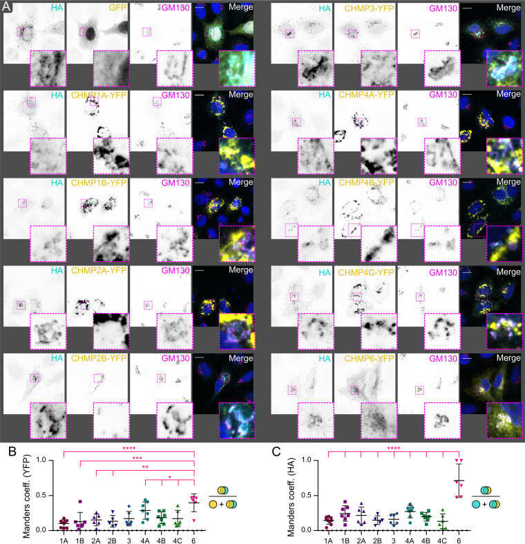
Oropouche virus (OROV; genus Orthobunyavirus) is the etiological agent of Oropouche fever, a debilitating febrile illness common in South America. We used recombinant expression of the OROV M polyprotein, which encodes the surface glycoproteins Gn and Gc plus the nonstructural protein NSm, to probe the cellular determinants for OROV assembly and budding. Gn and Gc self-assemble and are secreted independently of NSm. Mature OROV Gn has two predicted transmembrane domains that are crucial for glycoprotein translocation to the Golgi complex and glycoprotein secretion, and unlike related orthobunyaviruses, both transmembrane domains are retained during Gn maturation. Disruption of Golgi function using the drugs brefeldin A and monensin inhibits glycoprotein secretion. Infection studies have previously shown that the cellular endosomal sorting complexes required for transport (ESCRT) machinery is recruited to Golgi membranes during OROV assembly and that ESCRT activity is required for virus secretion. A dominant-negative form of the ESCRT-associated ATPase VPS4 significantly reduces recombinant OROV glycoprotein secretion and blocks virus release from infected cells, and VPS4 partly colocalizes with OROV glycoproteins and membranes costained with Golgi markers. Furthermore, immunoprecipitation and fluorescence microscopy experiments demonstrate that OROV glycoproteins interact with the ESCRT-III component CHMP6, with overexpression of a dominant-negative form of CHMP6 significantly reducing OROV glycoprotein secretion. Taken together, our data highlight differences in M polyprotein processing across orthobunyaviruses, indicate that Golgi and ESCRT function are required for glycoprotein secretion, and identify CHMP6 as an ESCRT-III component that interacts with OROV glycoproteins. IMPORTANCE Oropouche virus causes Oropouche fever, a debilitating illness common in South America that is characterized by high fever, headache, myalgia, and vomiting. The tripartite genome of this zoonotic virus is capable of reassortment, and there have been multiple epidemics of Oropouche fever in South America over the last 50 years, making Oropouche virus infection a significant threat to public health. However, the molecular characteristics of this arbovirus are poorly understood. We developed a recombinant protein expression system to investigate the cellular determinants of OROV glycoprotein maturation and secretion. We show that the proteolytic processing of the M polypeptide, which encodes the surface glycoproteins (Gn and Gc) plus a nonstructural protein (NSm), differs between OROV and its close relative Bunyamwera virus. Furthermore, we demonstrate that OROV M glycoprotein secretion requires the cellular endosomal sorting complexes required for transport (ESCRT) membrane-remodeling machinery and identify that the OROV glycoproteins interact with the ESCRT protein CHMP6.

Keywords: Bunyamwera virus; ESCRT; Oropouche fever; Oropouche virus; arbovirus; bunyavirus; polyprotein processing; virus budding.


Conflict of interest statement

The authors declare no conflict of interest.

Figures

FIG 1
FIG 1
Recombinant production and secretion of Oropouche virus (OROV) glycoproteins. (A) Schematic representation of the recombinant constructs used to express the OROV M polyprotein, individual glycoproteins, or domains thereof. Expression constructs were preceded by the OROV M secretion signal sequence (SS; unshaded) and an hemagglutinin epitope tag (HA; red shading). Regions corresponding to Gn, NSm, and Gc are shaded in orange, yellow, and green, respectively, with cytosolic tails denoted (CT). Transmembrane regions (black) as predicted by TMHMM (31) and polyprotein cleavage sites (arrows) are shown. (B, C) HEK293T cells were transfected with HA-tagged OROV M (poly)protein constructs. After 48 h, the cell lysates and supernatants were harvested, subjected to SDS-PAGE, and immunoblotted using anti-HA and anti-OROV antibodies to detect secreted proteins, the latter polyclonal antibody detecting Gc but not Gn, and with anti-EEA1 (B) or anti-tubulin (C) antibodies used as loading controls. The data are representative of two (B) or three (C) independent experiments. Nonspecific bands are marked (*). (D) HEK293T cells were transfected with OROV M (poly)proteins lacking an N-terminal epitope tag or infected with OROV (MOI = 1). After 24 h, the cells were harvested and subjected to SDS-PAGE and immunoblotting using antibodies shown, with anti-actin serving as a loading control. Nonspecific bands are marked (*). The data are representative of four independent experiments. (E) Schematic representation of OROV M polyprotein processing. TM, transmembrane; EEA1, early endosome antigen 1.
FIG 2
FIG 2
Intracellular localization of OROV glycoproteins. (A to F) HeLa cells were transfected with plasmids expressing the HA-tagged OROV glycoproteins or the M polyprotein (as indicated). The cells were fixed and costained with rabbit anti-HA (Alexa Fluor [AF]488) and either mouse anti-GM130 (AF568) or mouse anti-CNX2 (AF568) antibodies to identify Golgi and ER compartments, respectively, before analysis using wide-field fluorescence microscopy. CNX2, calnexin 2. Bars = 10 μm.
FIG 3
FIG 3
Disruption of the Golgi apparatus prevents OROV glycoprotein secretion. (A) HEK293T cells were transfected with HA-tagged OROV M polyprotein, and after 18 h the cell supernatant was harvested and reserved. The culture medium was replenished and supplemented with 5 μg/mL brefeldin A, 1 μM monensin, or dimethyl sulfoxide (DMSO) as a control. After 6 h, the cells and supernatant were harvested. All samples were then subjected to SDS-PAGE and immunoblotted using the antibodies shown, with anti-tubulin acting as a loading control. (B) Ratio of Gn (anti-HA) and Gc (anti-OROV) protein present in supernatants collected before or after drug treatment. The mean ± SD is shown for four independent experiments. *, P ≤ 0.05; **, P ≤ 0.01 (One-way analysis of variance (ANOVA) using Dunnett’s test for multiple comparisons to DMSO control cells.) (C) HeLa cells transfected and drug treated as in panel A before fixation, costaining with rabbit anti-HA (AF568) plus mouse anti-GM130 (AF488) to identify Golgi membranes and analysis using wide-field fluorescence microscopy. Bars = 10 μm.
FIG 4
FIG 4
OROV protein secretion depends on VPS4 activity. (A) HEK293 cells stably expressing green fluorescent protein (GFP)-tagged wild-type VPS4 (VPS4wt) or a dominant-negative mutant (VPS4E/Q) under the control of the ecdysone response element or parental cells as a control were transfected with HA-tagged M polyprotein and treated with either 1 μM ponasterone A (Pon A) or with DMSO as a control. After 48 h, the cells (C) and supernatant (S) were harvested, subjected to SDS-PAGE, and immunoblotted using antibodies shown. (B) Quantitation of the ratio of OROV Gn (anti-HA; left) or Gc (anti-OROV; right) that was secreted following treatment with Pon A versus DMSO. The means ± SD from three independent experiments are shown. ***, P ≤ 0.001 (one-way ANOVA using Dunnett’s test for multiple comparisons to parental control cells). (C) HEK293 cells stably expressing VPS4wt (wt) or VPS4E/Q (E/Q) under the control of the ecdysone response element or parental cells (P) as a control were infected with OROV (MOI = 1) and, after 4 h, were treated with 1 μM Pon A or DMSO as a control. After 24 h cells and supernatant were harvested, subjected to SDS-PAGE and immunoblotted using antibodies shown, with anti-OROV detecting both N and Gc proteins. (D) Quantitation of the ratio of OROV Gc (left) and N (right) secretion in cells treated with Pon A versus DMSO. The means ± SD from three independent experiments are shown. *, P ≤ 0.05 (one-way ANOVA using Dunnett’s test for multiple comparisons to parental control cells). (E) HEK293 cells expressing GFP-tagged VPS4wt or VPS4E/Q, or parental cells, were transfected with HA-tagged OROV M polyprotein and treated with 1 μM Pon A for 18 h before being fixed and costained with rabbit anti-HA (AF568) and mouse anti-GM130 (AF647) antibodies and then analyzed by wide-field fluorescence microscopy. Bars = 10 μm. (F) HeLa cells were cotransfected with plasmids expressing HA-tagged OROV M polyprotein and YFP-VPS4wt. After 24 h, the cells were fixed and costained with rabbit anti-HA (AF568) and (top) mouse anti-GM130 (AF647) or (bottom) sheep anti-TGN46 (AF647) and then analyzed by confocal microscopy. GAPDH, glyceraldehyde-3-phosphate dehydrogenase; ns, not significant; YFP, yellow fluorescent protein. Bars = 10 μm.
FIG 5
FIG 5
OROV glycoproteins colocalize with CHMP6 but not with other endosomal sorting complexes required for transport (ESCRT) III components. (A) HeLa cells were cotransfected with plasmids expressing HA-tagged OROV M polyprotein and YFP-tagged CHMP proteins (as indicated). After 24 h, the cells were fixed and costained with rabbit anti-HA (AF568) and mouse anti-GM130 (AF647) and analyzed by confocal microscopy. Bars = 10 μm. (B) Manders correlation coefficients were measured for each YFP-tagged CHMP protein and represent the amount of YFP fluorescence that colocalizes with HA signal as a proportion of all YFP signal. The data points represent individual cells (n = 5 to 9), and means ± SD are shown. Correlation is significantly higher for CHMP6-YFP than for all other CHMP proteins (one-way ANOVA with Tukey’s multiple-comparison test; *, P < 0.05; **, P < 0.01; ***, P < 0.001; ****, P < 0.0001). (C) As for panel B but measuring the colocalization of HA-tagged OROV glycoproteins with YFP-tagged CHMP proteins as a proportion of all HA signal.
FIG 6
FIG 6
OROV glycoproteins interact with CHMP6. (A) HeLa cells were transfected with HA-tagged OROV M polyprotein. After 18 h, the cells were treated with 1 μM monensin or mock-treated for 6 h before being fixed and costained with mouse anti-HA (AF568), rabbit anti-CHMP6 (AF488) and sheep anti-TGN46 (AF647) before analysis by confocal microscopy. Bars = 10 μm. (B) HeLa cells were cotransfected with HA-tagged OROV M polyprotein and YFP-tagged CHMP6. The cells were drug treated and fixed as in panel A before staining with rabbit anti-HA (AF568) and sheep anti-TGN46 (AF647) before analysis by confocal microscopy. Bars = 10 μm. (C) Manders correlation coefficients were measured and represent CHMP fluorescence (AF488 or YFP) that colocalizes with HA-tagged OROV glycoprotein signal as a proportion of all AF488/YFP signal (left) or HA fluorescence that colocalizes with A488/YFP signal as a proportion of all HA signal (right). The data points represent individual cells (n = 6 to 7), and means ± SD are shown. There is no significant difference in colocalization between endogenous and YFP-tagged CHMP6, nor is there a significant change in colocalization when monensin is added to stall glycoprotein trafficking at the Golgi (one-way ANOVA with Tukey’s multiple-comparison test). (D) HEK293T cells were cotransfected with HA-tagged M polyprotein and GFP or CHMP6-YFP. After 48 h cells, the supernatants were harvested, subjected to SDS-PAGE, and immunoblotted using the antibodies listed. (E) Ratio of Gn (anti-HA) signal in the culture supernatant versus cell lysate. The data are from five independent experiments. *, P ≤ 0.05 (paired ratio t test). (F, G) HEK293T cells were cotransfected with HA-M polyprotein and either GFP, CHMP6-YFP, or CHMP5-YFP. After 18 h, the cells were treated with 1 μM monensin or mock-treated for 6 h. The cells were harvested, and the lysates were subjected to affinity capture (IP) using either GFP (F) or HA (G) affinity matrices before SDS-PAGE and immunoblotting using the antibodies shown. Representative blots from two (F) or three (G) independent experiments are shown. Nonspecific bands arising from partial proteolysis of bait proteins (F) (YFP-tagged CHMP5/6 to YFP, or GFP following removal of the additional residues encoded by the multiple cloning site of the “empty” pEGFP-C1 vector used in this experiment) or the light chain of the anti-HA antibody resin used for affinity capture (G) are marked (*).

Similar articles

Cited by

References

    1. Sakkas H, Bozidis P, Franks A, Papadopoulou C. 2018. Oropouche fever: a review. Viruses 10:175. 10.3390/v10040175. - DOI - PMC - PubMed
    1. Romero-Alvarez D, Escobar LE. 2018. Oropouche fever, an emergent disease from the Americas. Microbes Infect 20:135–146. 10.1016/j.micinf.2017.11.013. - DOI - PubMed
    1. Gutierrez B, Wise EL, Pullan ST, Logue CH, Bowden TA, Escalera-Zamudio M, Trueba G, Nunes MRT, Faria NR, Pybus OG. 2020. Evolutionary dynamics of Oropouche virus in South America. J Virol 94:e01127-19. 10.1128/JVI.01127-19. - DOI - PMC - PubMed
    1. Hughes HR, Adkins S, Alkhovskiy S, Beer M, Blair C, Calisher CH, Drebot M, Lambert AJ, de Souza WM, Marklewitz M, Nunes MRT, Shí X, ICTV Report Consortium . 2020. ICTV virus taxonomy profile: peribunyaviridae. J Gen Virol 101:1–2. 10.1099/jgv.0.001365. - DOI - PMC - PubMed
    1. Elliott RM. 2014. Orthobunyaviruses: recent genetic and structural insights. Nat Rev Microbiol 12:673–685. 10.1038/nrmicro3332. - DOI - PubMed

Publication types

MeSH terms

Substances

Supplementary concepts

LinkOut - more resources