Review
. 2023 Mar 23;36(1):e0006019.
doi: 10.1128/cmr.00060-19. Epub 2022 Dec 8.

Human Listeriosis

Affiliations
Review

Human Listeriosis

Merel M Koopmans et al. Clin Microbiol Rev. .

Abstract

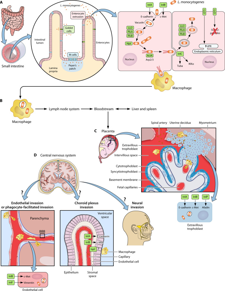
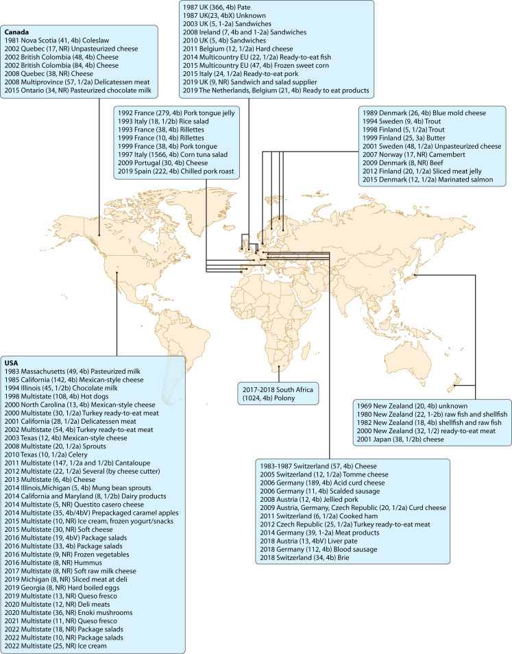
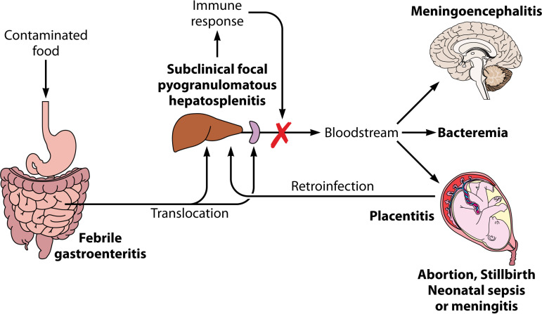
Listeria monocytogenes is a Gram-positive facultative intracellular pathogen that can cause severe invasive infections upon ingestion with contaminated food. Clinically, listerial disease, or listeriosis, most often presents as bacteremia, meningitis or meningoencephalitis, and pregnancy-associated infections manifesting as miscarriage or neonatal sepsis. Invasive listeriosis is life-threatening and a main cause of foodborne illness leading to hospital admissions in Western countries. Sources of contamination can be identified through international surveillance systems for foodborne bacteria and strains' genetic data sharing. Large-scale whole genome studies have increased our knowledge on the diversity and evolution of L. monocytogenes, while recent pathophysiological investigations have improved our mechanistic understanding of listeriosis. In this article, we present an overview of human listeriosis with particular focus on relevant features of the causative bacterium, epidemiology, risk groups, pathogenesis, clinical manifestations, and treatment and prevention.

Keywords: Listeria monocytogenes; bacterial genetics; epidemiology; histopathology; listeriosis; neurolisteriosis; pathophysiology; pregnancy-related listeriosis.


Conflict of interest statement

The authors declare no conflict of interest.

Figures

FIG 1
FIG 1
Pathogenesis of L. monocytogenes infection. (A) Invasion of the intestine through intestinal villi enterocytes, goblet cells and M cells. Entry into non-phagocytic cells is mediated by expression of bacterial surface-associated internalins A and B (InlA and InlB), which use as host ligands the adherens junction protein E-cadherin and the Met tyrosine kinase receptor, respectively. After host cell internalization, the listerial pore-forming toxin listeriolysin O (LLO) and phospholipases A and B (PlcA and PlcB) lyse the phagocytic vacuole membrane. The released bacteria replicate in the cytosol aided by the listerial virulence factor Hpt, which promotes rapid intracellular proliferation by allowing utilization of host-cell hexose phosphates. Then, the listerial actin-polymerizing protein ActA recruits host cell Arp2/3 complexes and induces actin-based motility, which propels the bacteria through the cytosol and into neighbouring cells, where the infection cycle starts again. InlC, another listerial virulence factor, assists in the process of cell-to-cell spread by targeting the cytoskeletal protein Tuba, and also interacts with IκB kinase (IKKα) dampening the innate immune response. (B) L. monocytogenes is taken up by macrophages which transport the bacteria to the lymph node system, and via the bloodstream to the primary target organs (liver and spleen), and from there to the secondary target organs (placenta or brain). See Fig. 3. (C) L. monocytogenes can colonize the placenta via cell-to-cell spread from infected macrophages to extravillous cytotrophoblasts, or via direct invasion of the trophoblast through InlA and InlB. Another internalin family protein, InlP, has been reported to facilitate placental invasion involving interaction with the cell junction-associated host protein afadin. (D) L. monocytogenes can gain access into the central nervous system in different ways: via cell-to-cell spread from infected phagocytes, or via direct (InlA/B-mediated) invasion of endothelial cells of brain microcapillaries, the basolateral side of the choroid plexus, or nerve cells of trigeminal nerve terminals (followed by intra-axonal ascension to the rhombencephalon). Invasion of brain endothelial cells is further facilitated by interaction of the listerial internalin family protein InlF with the host intermediate filament protein vimentin. See text for details.
FIG 2
FIG 2
Outbreaks of Listeria between 1969 and 2022. Described by year, location, number of patients, serotype and source of infection. NR = not reported.
FIG 3
FIG 3
Pathophysiology of foodborne listeriosis. L. monocytogenes bacteria cross the epithelial barrier of the intestine, translocate to the mesenteric lymph nodes, and reach their primary target organs, i.e., liver and spleen. There they establish infectious foci that in an immunocompetent individual are efficiently cleared by cell-mediated immunity. In adult people with no predisposing conditions, the process is largely subclinical. In this population, exposure to larger infective doses may cause febrile gastroenteritis and, in rare cases, invasive disease. In immunocompromised adults and elderly people who are unable to mount an efficient T-cell-mediated immune response, the primary infectious foci are inadequately resolved and Listeria bacteria may be released to the bloodstream. This results in febrile bacteremia and, eventually, invasive infection of the brain. In pregnant women, L. monocytogenes colonizes the uterus in addition to the liver and spleen. While the infection is controlled in the latter organs, the placental immune tolerance mechanisms provide a permissive niche for the proliferation of L. monocytogenes. Bacteria from the placental reservoir released to the bloodstream may reinfect the mother’s liver and spleen, contributing to infection maintenance and amplification (395). Transplacental dissemination to the fetus results in abortion, stillbirth, or neonatal sepsis. A late-onset congenital form is also observed in neonates, often accompanied by meningitis. Reproduced from reference 410, based on an earlier depiction in reference 61.

Similar articles

Cited by

References

    1. Vivant AL, Garmyn D, Piveteau P. 2013. Listeria monocytogenes, a down-to-earth pathogen. Front Cell Infect Microbiol 3:87. 10.3389/fcimb.2013.00087. - DOI - PMC - PubMed
    1. Stea EC, Purdue LM, Jamieson RC, Yost CK, Truelstrup Hansen L. 2015. Comparison of the prevalences and diversities of Listeria species and Listeria monocytogenes in an urban and a rural agricultural watershed. Appl Environ Microbiol 81:3812–3822. 10.1128/AEM.00416-15. - DOI - PMC - PubMed
    1. Kathariou S. 2002. Listeria monocytogenes virulence and pathogenicity, a food safety perspective. J Food Prot 65:1811–1829. 10.4315/0362-028x-65.11.1811. - DOI - PubMed
    1. Taege AJ. 1999. Listeriosis: recognizing it, treating it, preventing it. Cleve Clin J Med 66:375–380. 10.3949/ccjm.66.6.375. - DOI - PubMed
    1. Wemekamp-Kamphuis HH, Karatzas AK, Wouters JA, Abee T. 2002. Enhanced levels of cold shock proteins in Listeria monocytogenes LO28 upon exposure to low temperature and high hydrostatic pressure. Appl Environ Microbiol 68:456–463. 10.1128/AEM.68.2.456-463.2002. - DOI - PMC - PubMed

Publication types