. 2022 Dec 7;72(1):32.
doi: 10.1186/s12576-022-00857-x.

Critical role of Rho proteins in myosin light chain di-phosphorylation during early phase of endothelial barrier disruption

Affiliations

Critical role of Rho proteins in myosin light chain di-phosphorylation during early phase of endothelial barrier disruption

Mayumi Hirano et al. J Physiol Sci. .

Abstract

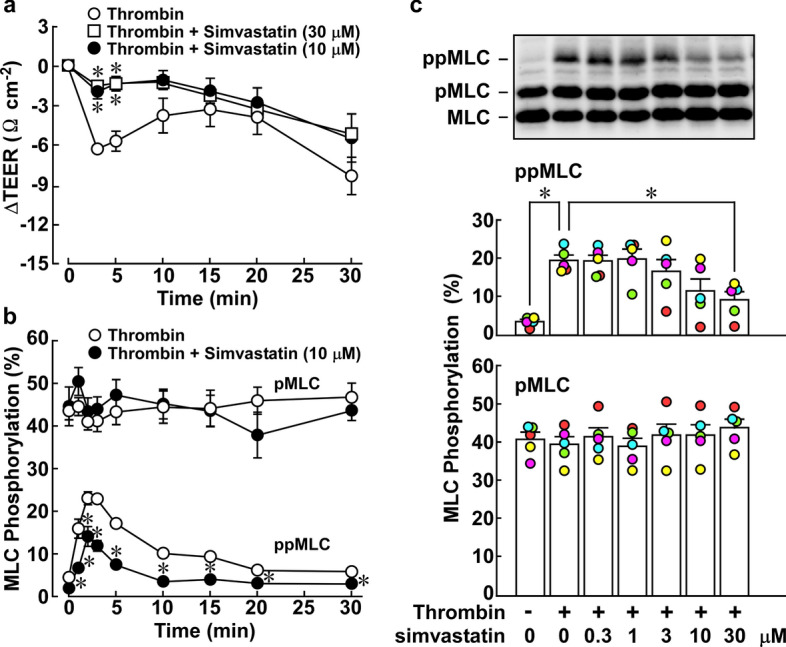
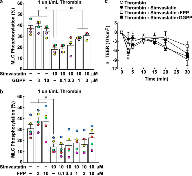
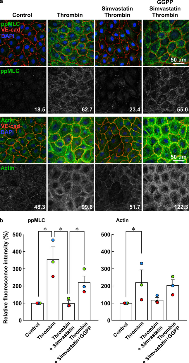
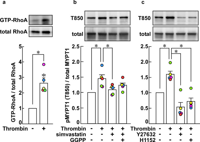
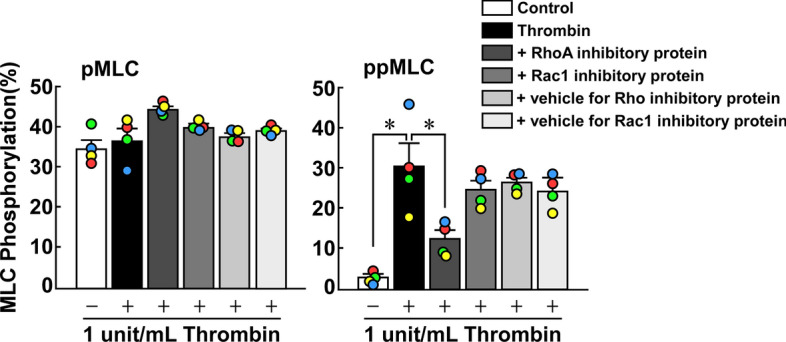
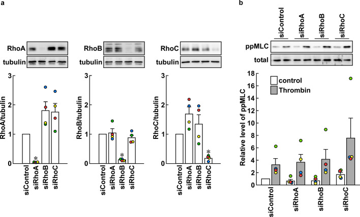
We previously reported the Rho-associated coiled-coil containing protein kinase (ROCK)-mediated di-phosphorylation of myosin light chain (MLC) and actin bundle formation at the cell periphery as early events of the endothelial barrier disruption. We herein examined the role of RhoA during early events of barrier disruption. Treatment of cultured porcine aortic endothelial cells with simvastatin prevented the decrease in trans-endothelial electrical resistance, MLC di-phosphorylation and peripheral actin bundle formation seen 3 min after thrombin stimulation. Co-treatment with geranylgeranyl pyrophosphate rescued the thrombin-induced events. Thrombin increased a GTP-bound form of RhoA and phosphorylation of myosin phosphatase target subunit 1 (MYPT1) at the ROCK site. The intracellular introduction of the inhibitory protein of RhoA inhibited the thrombin-induced di-phosphorylation of MLC. However, knockdown of either one of RhoA, RhoB or RhoC failed to inhibit thrombin-induced MLC di-phosphorylation. The findings suggest that Rho proteins play a critical role during early events of thrombin-induced barrier disruption.

Keywords: Actin filaments; Barrier function; Myosin light chain; Phosphorylation; Small G protein; Vascular endothelial cells.


Conflict of interest statement

The authors declare no competing interests.

Figures

Fig. 1
Fig. 1
Effects of simvastatin pretreatment on the thrombin-induced decrease in trans-endothelial electric resistance (TEER) and mono- and di-phosphorylation of myosin light chain (pMLC, ppMLC) in porcine aortic endothelial cells (PAECs). a, b The time courses of thrombin-induced changes in TEER (a, n = 6), pMLC and ppMLC (b, n = 4) are summarized. c A representative immunoblot and the summary for the concentration-dependent effects of simvastatin on pMLC and ppMLC at 3 min after thrombin stimulation are shown (n = 5). PAECs at confluence were pretreated or left untreated with simvastatin at the indicated concentrations for 16 h. The cells were then stimulated with 1 unit/mL thrombin, in the presence of simvastatin, when pretreated. All original images of the immunoblot analyses are shown in Additional file 1: Figure S1. The data are expressed as the mean ± SEM. *P < 0.05 vs. thrombin, according to an ANOVA followed by Dunnett’s post hoc test
Fig. 2
Fig. 2
Effects of geranylgeranyl pyrophosphate (GGPP) and farnesyl pyrophosphate (FPP) on the inhibition by simvastatin of the thrombin-induced di-phosphorylation of myosin light chain (ppMLC) and decrease in the trans-endothelial electrical resistance (TEER) in porcine aortic endothelial cells (PAECs). a, b The effects of simvastatin and GGPP (a; n = 4) or FPP (b; n = 5) on the thrombin-induced ppMLC were summarized. PAECs were pretreated or left untreated with simvastatin, GGPP or FPP, either alone or their combination, at the indicated concentrations for 16 h. The cells were then stimulated with 1 unit/mL thrombin, in the presence of simvastatin, GGPP or FPP, when pretreated. The levels of ppMLC were evaluated 3 min after thrombin stimulation. All original images of the immunoblot analyses in panels a and b are shown in Additional file 1: Figure S2. c Shown are the time courses of the change in the TEER after stimulation with 1 unit/mL thrombin in PAECs with and without pretreatment with 10 µM simvastatin, 3 µM GGPP or 10 µM FPP, as indicated (n = 5). The data are expressed as the mean ± SEM. *P < .05 vs. simvastatin (a, b) and thrombin + simvastatin (c); #P < 0.05 vs. thrombin (c), according to an ANOVA followed by Dunnett’s post hoc test
Fig. 3
Fig. 3
Effects of simvastatin and geranylgeranyl pyrophosphate (GGPP) on localization of di-phosphorylation of myosin light chain (ppMLC) and actin bundle formation in porcine aortic endothelial cells (PAECs). a Representative merged images of confocal microphotographs show the triple fluorescence staining of either F-actin/VE-cadherin (VE-Cad)/nuclei (DAPI) or ppMLC/VE-Cad/DAPI in PAECs in the indicated colors. The extracted images of ppMLC and actin (both in a green channel) in a gray scale are shown below the corresponding merged images. The readings of the fluorescence intensity were indicated at a lower right corner of each image. b Summaries (n = 3) of quantitative analysis of fluorescence intensities of ppMLC and actin. The data are expressed as the mean ± SEM. *P < 0.05 according to an ANOVA followed by Fisher post hoc test. PAECs were either pretreated or left untreated with 10 µM simvastatin for 16 h with and without co-treatment with 3 µM GGPP (n = 3). The images were obtained 3 min after stimulation with 1 unit/mL thrombin. The cells were challenged to thrombin, in the presence of simvastatin and GGPP, when pretreated. Control indicates images of cells without any treatment. Thrombin induced peripheral accumulation of ppMLC and actin bundles in close proximity to VE-cadherin. Simvastatin inhibited the thrombin-induced peripheral accumulation of ppMLC and actin bundles, while co-treatment with GGPP prevented the simvastatin effects. The results obtained in the two additional sets of the experiments are shown in Additional file 1: Figure S3
Fig. 4
Fig. 4
Activation of RhoA and Rho-associated coiled-coil containing protein kinase (ROCK) by thrombin in porcine aortic endothelial cells (PAECs). a The levels of GTP-bound form of RhoA before and 3 min after stimulation with 1 unit/mL thrombin were summarized (n = 5). b, c Shown are the representative immunoblots and the summaries of the level of phosphorylation of MYPT1 at the ROCK site corresponding to T850 in human MYPT1 before and 3 min after stimulation with 1 unit/mL thrombin in the presence and absence of 10 µM simvastatin and 3 µM GGPP (b; n = 5), and 10 µM Y27632 and 1 µM H1152 (c; n = 5). PAECs were pretreated with simvastatin for 16 h in the presence or absence of GGPP, and with Y27632 or H1152 for 30 min, and then stimulated with thrombin in their presence, when pretreated (n = 5). The basal level of MYPT1 phosphorylation was assigned a value of 1. The data are expressed as the mean ± SEM. *P < 0.05 vs. thrombin, according to an ANOVA followed by Dunnett’s post hoc test. All original images of the immunoblot analyses are shown in Additional file 1: Figure S4
Fig. 5
Fig. 5
Effects of introduction of the inhibitory proteins of RhoA and Rac1/Cdc42 on the thrombin-induced mono- and di-phosphorylation of MLC (pMLC and ppMLC) in porcine aortic endothelial cells (PAECs). PAECs were pretreated with 5 μM inhibitory proteins of RhoA and Rac1/Cdc42 for 30 min prior to and during stimulation with 1 unit/mL thrombin. MLC phosphorylation was evaluated with Phos-tag SDS-PAGE followed by immunoblot detection of MLC. The data are expressed as the mean ± SEM (n = 4). *P < 0.05, according to an ANOVA followed by Dunnett’s post hoc test. All original images of the immunoblot analyses are shown in Additional file 1: Figure S5
Fig. 6
Fig. 6
Effect of knockdown of RhoA, RhoB and RhoC on thrombin-induced di-phosphorylation of myosin light chain (ppMMLC) in porcine aortic endothelial cells (PAECs). Representative immunoblot images and summaries of immunoblot detection of RhoA, RhoB and RhoC (a), and ppMLC before and 3 min after stimulation with 1 unit/mL thrombin (b). The samples obtained before thrombin stimulation were used to detect Rho proteins. The data are expressed as mean ± SEM (n = 4). *P < 0.05 vs. the other conditions according to an ANOVA followed by Fisher post hoc test. In b, the values of siRhoA, siRhoB and siRhoC before and after thrombin stimulation showed no significant difference from those of siControl, according to an ANOVA followed by Dunnett’s post hoc text. All original images of the immunoblot analyses are shown in Additional file 1: Figure S6

References

    1. Mehta D, Malik AB. Signaling mechanisms regulating endothelial permeability. Physiol Rev. 2006;86:279–367. doi: 10.1152/physrev.00012.2005. - DOI - PubMed
    1. Komarova YA, Mehta D, Malik AB. Dual regulation of endothelial junctional permeability. Sci STKE. 2007 doi: 10.1126/stke.4122007re8. - DOI - PubMed
    1. Goddard LM, Iruela-Arispe ML. Cellular and molecular regulation of vascular permeability. Thromb Haemost. 2013;109:407–415. doi: 10.1160/TH12-09-0678. - DOI - PMC - PubMed
    1. Vandenbroucke E, Mehta D, Minshall R, Malik AB. Regulation of endothelial junctional permeability. Ann N Y Acad Sci. 2008;1123:134–145. doi: 10.1196/annals.1420.016. - DOI - PubMed
    1. Schnittler H, Taha M, Schnittler MO, Taha AA, Lindemann N, Seebach J. Actin filament dynamics and endothelial cell junctions: the Ying and Yang between stabilization and motion. Cell Tissue Res. 2014;355:529–543. doi: 10.1007/s00441-014-1856-2. - DOI - PubMed