. 2022 Dec 7;17(1):40.
doi: 10.1186/s13062-022-00354-6.

Alpha-actnin-4 (ACTN4) selectively affects the DNA double-strand breaks repair in non-small lung carcinoma cells

Affiliations

Alpha-actnin-4 (ACTN4) selectively affects the DNA double-strand breaks repair in non-small lung carcinoma cells

Daria Kriger et al. Biol Direct. .

Abstract

Background: ACTN4 is an actin-binding protein involved in many cellular processes, including cancer development. High ACTN4 expression is often associated with a poor prognosis. However, it has been identified as a positive marker for platinum-based adjuvant chemotherapy for non-small cell lung cancer (NSCLC). The goal of our study was to investigate the involvement of ACTN4 in the NSCLC cells' response to the genotoxic drugs.

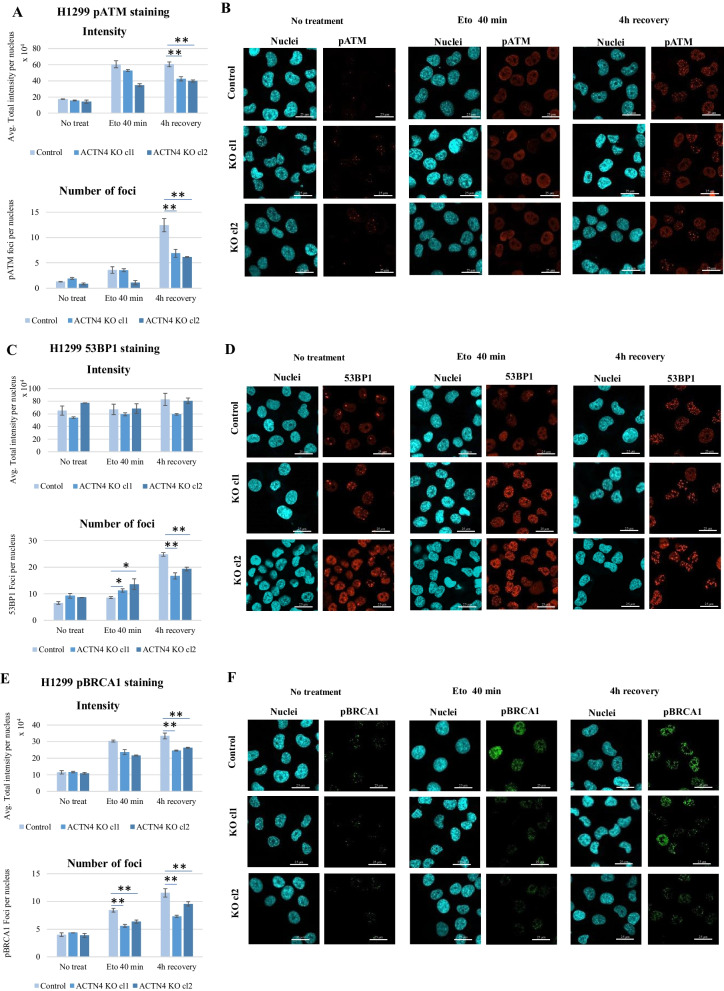
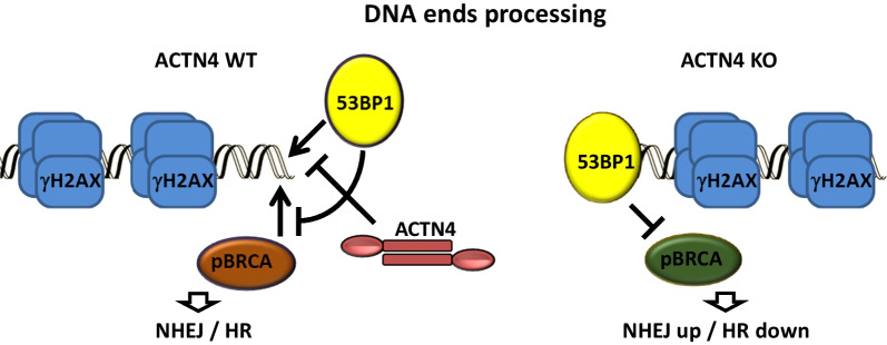
Results: We generated H1299 cells with the ACTN4 gene knock-out (ACTN4 KO), using the CRISPR/Cas9 system. The resistance of the cells to the cisplatin and etoposide was analyzed with the MTT assay. We were also able to estimate the efficiency of DNA repair through the DNA comet assay and gamma-H2AX staining. Possible ACTN4 effects on the non-homologous end joining (NHEJ) and homologous recombination (HR) were investigated using pathway-specific reporter plasmids and through the immunostaining of the key proteins. We found that the H1299 cells with the ACTN4 gene knock-out did not show cisplatin-resistance, but did display a higher resistance to the topoisomerase II inhibitors etoposide and doxorubicin, suggesting that ACTN4 might be somehow involved in the repair of DNA strand breaks. Indeed, the H1299 ACTN4 KO cells repaired etoposide- and doxorubicin-induced DNA breaks more effectively than the control cells. Moreover, the ACTN4 gene knock-out enhanced NHEJ and suppressed HR efficiency. Supporting the data, the depletion of ACTN4 resulted in the faster assembly of the 53BP1 foci with a lower number of the phospho-BRCA1 foci after the etoposide treatment.

Conclusions: Thus, we are the first to demonstrate that ACTN4 may influence the resistance of cancer cells to the topoisomerase II inhibitors, and affect the efficiency of the DNA double strand breaks repair. We hypothesize that ACTN4 interferes with the assembly of the NHEJ and HR complexes, and hence regulates balance between these DNA repair pathways.

Keywords: ACTN4; DNA repair; Etoposide resistance; Homologous recombination (HR); Non-homologous end joining (NHEJ); Non-small cell lung cancer (NSCLC).


Conflict of interest statement

The authors declare no conflict of interest.

Figures

Fig. 1
Fig. 1
The ACTN4 knock-out affects the viability of H1299 cells under genotoxic stress. A Western Blot analysis of ACTN4 expression levels in H1299 (left) and H460 (right) control and ACTN4 knock-out (ACTN4 KO) cells. Three H1299 and two H460 ACTN4 KO clones used in the study are presented. BD The MTT assay performed on H1299 (B, C) and H460 (D) control, ACTN4 KO and ACTN4 OE cells exposed by different concentrations of cisplatin (B) and etoposide (C, D) for 72 h. Results for three H1299 ACTN4 KO clones are presented in C to ensure the effect. E The H1299 (left) and H460 (right) cells were treated by 50 uM etoposide for 24 h to induce apoptosis. Annexin V (APC)/PI (PE) plots and percentages of cells in quadrants (means of three replicas) are presented. H1299 ACTN4 KO cells display significantly (p < 0.001) lower early apoptosis (AV) than the control cells while H460 cells show the opposite. The data are presented as the mean of at least three replicas ± SEM. ***p < 0.001, **p < 0.01, *p < 0.05 compared to untreated cells (multiple Student’s t-test). Each assay was performed at least three times
Fig. 2
Fig. 2
ACTN4 knock-out does not change cell cycle in H1299 cells but increases G0-G1 phase under genotoxic conditions. H1299 Control and ACTN4 KO cells were treated with 1.5 uM etoposide for 18 (A) or 48 (B) hours. Cell cycle distributions are presented as graphs (upper panels) and flow cytometry plots (lower panels). The data are presented as the mean of at least three replicas in each experiment ± SEM and repeated three times
Fig. 3
Fig. 3
Depletion of ACTN4 in H1299 and H460 cells leads to more efficient DNA break repairs induced by the topoisomerase II inhibitors. Control, ACTN4 KO and ACTN4 OE cells were treated with 50 uM etoposide (Eto) or 1.5 uM doxorubicin (Doxo) for 40 min. The drug was then replaced with fresh media and 4 or 8 h of cells recovery. A Analysis of gamma-H2AX immunostaining in H1299 control and ACTN4 KO cells. Images of the etoposide-induced foci formation are presented in the left panel. Mean numbers of foci per nucleus were calculated in Eto or Doxo treated cells. The graphs represent the mean gamma-H2AX foci numbers between three replicas (± SE) with at least 500 cells per replica analyzed. Scale bars in panel represent 25 μm. B An alkaline comet assay is performed on H1299 and H460 cells. Images of the DNA comets obtained from H1299 control, ACTN4 KO and ACTN4 OE cells are presented in the left panel. Scale bars in panel represents 250 μm. The graphs represent mean Tail DNA percentages of at least 100 analyzed cells ± SE. ***p < 0.001, *p < 0.05 compared to untreated cells (multiple Student’s t-test). Each assay was performed at least three times. The experiments were performed in triplicate
Fig. 4
Fig. 4
ACTN4 cells show enhanced NHEJ and suppressed HR. H1299 cells were transfected with NHEJ and HR specific reporter constructs along with a plasmid, encoding dsRed-fused I-SceI endonuclease that performs site-specific cleavage of the reporters. Representative FACS plots display NHEJ and HR specific repair of the reporter GFP as an increase of GFP-positive cells (upper quadrants). The graph represents DNA repair efficiency estimated as the ratio of GFP + /dsRed + cells. Data are presented as mean of three replicas ± SD. ***p < 0.0001compared to untreated cells (Student’s t-test). The assay was performed three times
Fig. 5
Fig. 5
ACTN4 expression affects the distribution of the key DSBs repair proteins in H1299 cells after the etoposide-induced DNA mage. The H1299 Control and ACTN4 KO cells were treated with 50 uM etoposide for 40 min. The media was then replaced with a fresh one, and cells were incubated for 4 h to recover from the damage. Changes in total intensity and foci number of pATM (AB), 53BP1 (CD), pBRCA1 (EF) proteins were estimated by immunofluorescence. Representative images are presented on B, D and F. Scale bars in panels represent 25 μm. Graphs A, C, E present the mean of three replicas ± SE. Over 500 cells were analyzed per replica. **p < 0.0001, **p < 0.001, *p < 0.05 compared to untreated cells (Student’s t-test)
Fig. 6
Fig. 6
Possible mechanism of ACTN4 influence on the DSBs repair pathways. ACTN4 may possibly interfere with 53BP1 binding to unprocessed DNA ends, hence suppressing the end processing suitable for NHEJ and promoting assembly of the pBRCA-positive HR complexes. In H1299 ACTN4 KO cells however, DNA binding of 53BP1 is increased, which results in enhancing the NHEJ pathway and the suppression of pBRCA complexes and HR pathways

Similar articles

Cited by

References

    1. Honda K, Yamada T, Endo R, Ino Y, Gotoh M, Tsuda H, et al. Actinin-4, a novel actin-bundling protein associated with cell motility and cancer invasion. J Cell Biol. 1998;140(6):1383–1393. doi: 10.1083/jcb.140.6.1383. - DOI - PMC - PubMed
    1. Agarwal N, Adhikari AS, Iyer SV, Hekmatdoost K, Welch DR, Iwakuma T. MTBP suppresses cell migration and filopodia formation by inhibiting ACTN4. Oncogene. 2013;32(4):462–470. doi: 10.1038/onc.2012.69. - DOI - PMC - PubMed
    1. Kumeta M, Yoshimura SH, Harata M, Takeyasu K. Molecular mechanisms underlying nucleocytoplasmic shuttling of actinin-4. J Cell Sci. 2010;123(7):1020–1030. doi: 10.1242/jcs.059568. - DOI - PubMed
    1. Li J, Wang H. H3K27ac-activated EGFR-AS1 promotes cell growth in cervical cancer through ACTN4-mediated WNT pathway. Biol Direct. 2022;17(1):3. doi: 10.1186/s13062-021-00315-5. - DOI - PMC - PubMed
    1. An HT, Yoo S, Ko J. α-Actinin-4 induces the epithelial-to-mesenchymal transition and tumorigenesis via regulation of Snail expression and β-catenin stabilization in cervical cancer. Oncogene. 2016;35(45):5893–5904. doi: 10.1038/onc.2016.117. - DOI - PubMed