

Combined application of dinitrofluorobenzene and ovalbumin induced AD-like dermatitis with an increase in helper T-cell cytokines and a prolonged Th2 response
- PMID: 36476273
- PMCID: PMC9730685
- DOI: 10.1186/s12865-022-00531-2
Combined application of dinitrofluorobenzene and ovalbumin induced AD-like dermatitis with an increase in helper T-cell cytokines and a prolonged Th2 response
Abstract
Background: The progression of acute-to-chronic atopic dermatitis is accompanied by multiple helper T-cell cytokine responses, but the mechanisms and relative importance of these changes remain unclear. There is no animal model for atopic dermatitis that recapitulates these cytokine responses.
Objective: We sought to build a novel mouse model for atopic dermatitis (AD) that recapitulates these helper T-cell responses and some dynamic changes in cytokine responses in the progression of AD.
Methods: Female BALB/c mice were subjected to the application of dinitrofluorobenzene (DNFB) and ovalbumin (OVA) to induce AD-like dermatitis. Skin lesions and serum were collected from mice in the acute and chronic phases to detect changes in cytokine responses and other features of AD.
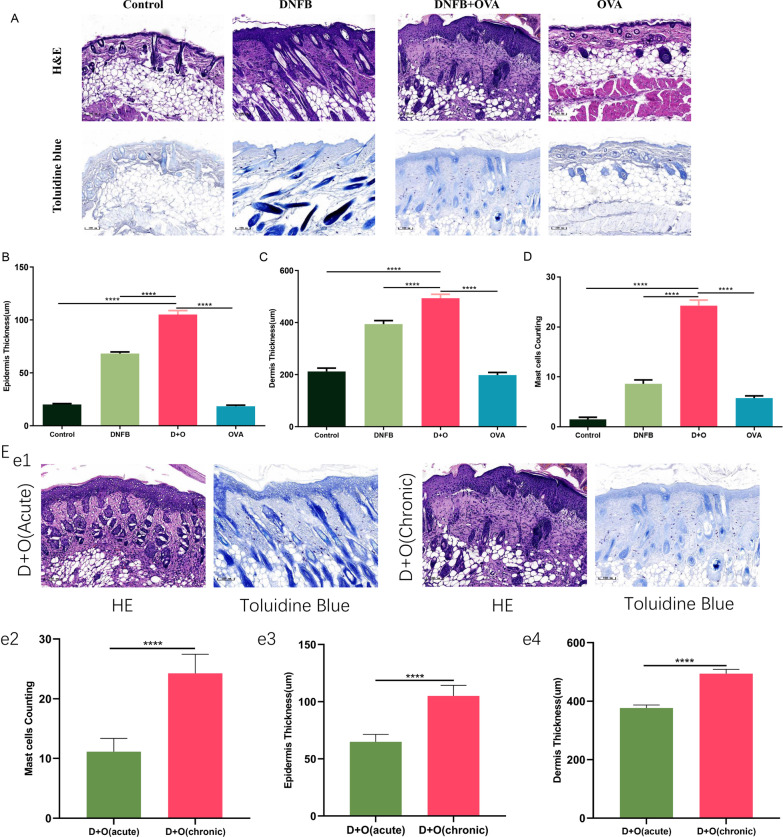
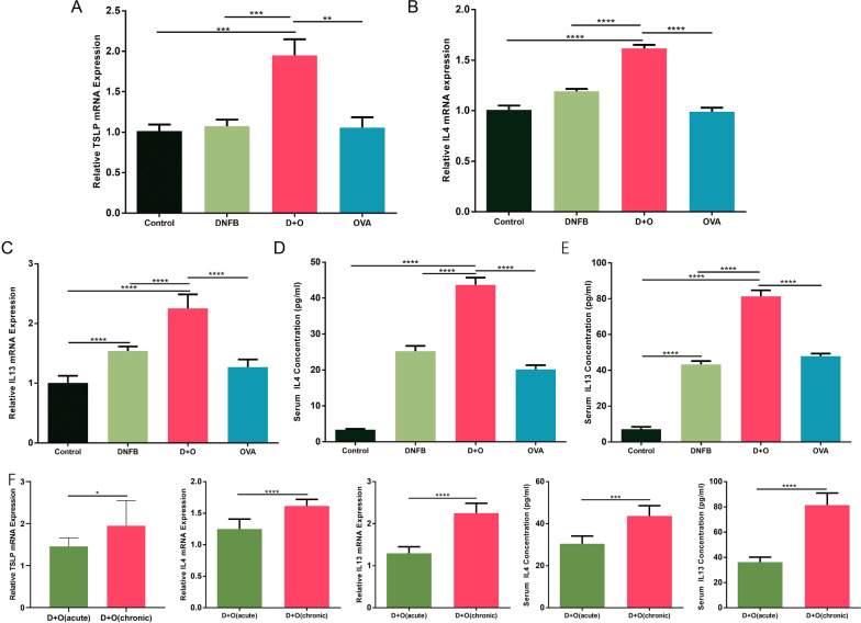
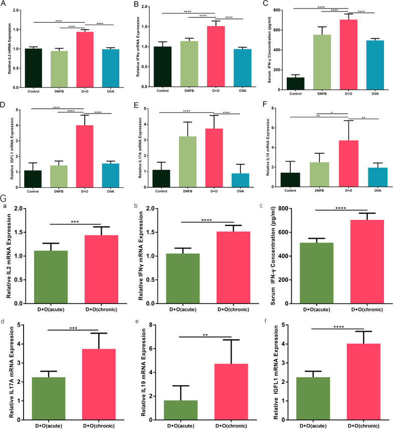
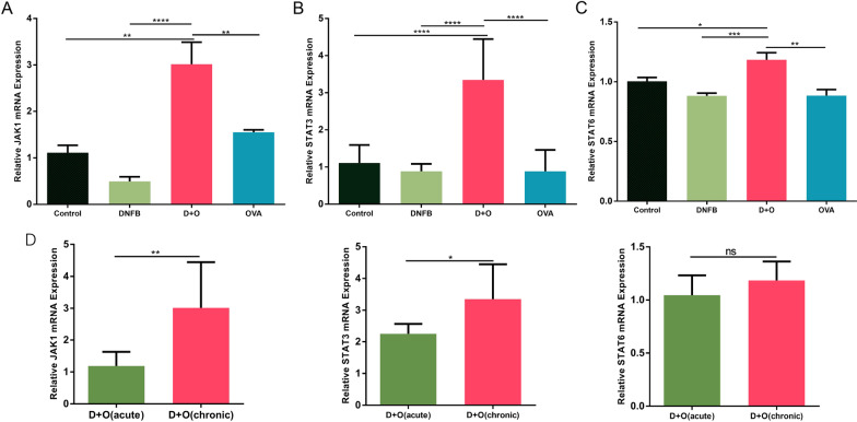
Results: Combined application of DNFB and OVA successfully induced AD-like dermatitis and histological changes as well as epidermal barrier dysfunction. In the acute phase of AD-like dermatitis, Th2-associated cytokines were mainly increased in serum and skin lesions. In the chronic phase of AD-like dermatitis, Th2-associated cytokines were still highly expressed, while Th1- and Th17-associated cytokines were also gradually increased. Compared with the acute phase, the JAK-STAT signaling pathway was highly expressed in the chronic phase of AD-like dermatitis.
Conclusion: The combined application of DNFB and OVA could be used to build a new mouse model for atopic dermatitis. This mouse model recapitulates the helper T-cell responses and some dynamic changes in cytokine responses in the progression of acute-to-chronic in human AD. The JAK-STAT signaling pathway plays a pivotal role in the chronicity of AD.
Keywords: Atopic dermatitis; BALB/c mice; Dinitrofluorobenzene; Ovalbumin.
© 2022. The Author(s).
Conflict of interest statement
The authors declare that they have no competing interests.
Figures


Similar articles
-
Comprehensive analysis of phenotypes and transcriptome characteristics reveal the best atopic dermatitis mouse model induced by MC903.J Dermatol Sci. 2024 Jun;114(3):104-114. doi: 10.1016/j.jdermsci.2024.05.003. Epub 2024 May 18. J Dermatol Sci. 2024. PMID: 38806322
-
Calcium-Based Antimicrobial Peptide Compounds Attenuate DNFB-Induced Atopic Dermatitis-Like Skin Lesions via Th-Cells in BALB/c Mice.Int J Mol Sci. 2022 Sep 26;23(19):11371. doi: 10.3390/ijms231911371. Int J Mol Sci. 2022. PMID: 36232673 Free PMC article.
-
Sophoricoside from Styphnolobium japonicum improves experimental atopic dermatitis in mice.Phytomedicine. 2021 Feb;82:153463. doi: 10.1016/j.phymed.2021.153463. Epub 2021 Jan 14. Phytomedicine. 2021. PMID: 33545490
-
Potential Natural Biomolecules Targeting JAK/STAT/SOCS Signaling in the Management of Atopic Dermatitis.Molecules. 2022 Jul 21;27(14):4660. doi: 10.3390/molecules27144660. Molecules. 2022. PMID: 35889539 Free PMC article. Review.
-
The role of cytokines/chemokines in the pathogenesis of atopic dermatitis.Curr Probl Dermatol. 2011;41:80-92. doi: 10.1159/000323299. Epub 2011 May 12. Curr Probl Dermatol. 2011. PMID: 21576949 Review.
Cited by
-
A refined comparative mouse model of acute and chronic atopic dermatitis.J Anim Sci Technol. 2025 May;67(3):636-650. doi: 10.5187/jast.2024.e93. Epub 2025 May 31. J Anim Sci Technol. 2025. PMID: 40519610 Free PMC article.
-
Anti-Atopic Dermatitis Effect of Azalomycin F on 2,4-Dinitrofluorobenzene-Induced Mice and Potential Mechanism.Int J Mol Sci. 2024 Nov 29;25(23):12846. doi: 10.3390/ijms252312846. Int J Mol Sci. 2024. PMID: 39684557 Free PMC article.
References
-
- Odhiambo JA, Williams HC, Clayton TO, Robertson CF, Asher MI. ISAAC Phase Three Study Group. Global variations in prevalence of eczema symptoms in children from ISAAC Phase Three. J Allergy Clin Immunol. 2009;124(6):1251-8.e23. doi: 10.1016/j.jaci.2009.10.009. PMID: 20004783. - PubMed
Publication types
MeSH terms
Substances
LinkOut - more resources
Full Text Sources
