

Decoding the molecular cascade of embryonic-uterine modulators in pregnancy loss of PCOS mother- an "in vivo" study
- PMID: 36476384
- PMCID: PMC9727897
- DOI: 10.1186/s12958-022-01041-x
Decoding the molecular cascade of embryonic-uterine modulators in pregnancy loss of PCOS mother- an "in vivo" study
Abstract
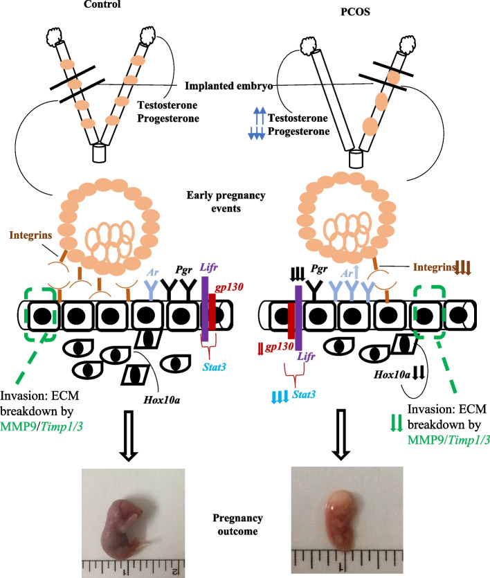
Background: Polycystic ovary syndrome is associated with an increased rate of spontaneous abortion/early pregnancy loss and pups delivered to PCOS animals were abnormal. Currently, assisted reproductive technology has been used to help numerous infertile couples to have their babies. However, there is a low implantation rate after the transfer of embryos. Till now, it could not be concluded whether the reduced pregnancy rates observed were due to abnormal embryos or endometrial modification. Further, transgenic mouse models have been used to find out the molecular deficits behind early pregnancy complications. But, the deletion of crucial genes could lead to systemic deficiencies/embryonic lethality. Also, pregnancy is a complex process with overlapping expression patterns making it challenging to mimic their stage-specific role. Therefore, the motive of the current study was to investigate the probable molecular cascade to decipher the early pregnancy loss in the letrozole-induced PCOS mouse model.
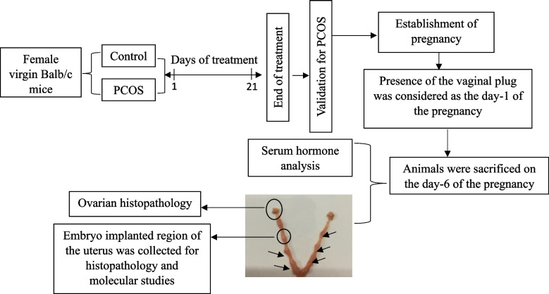
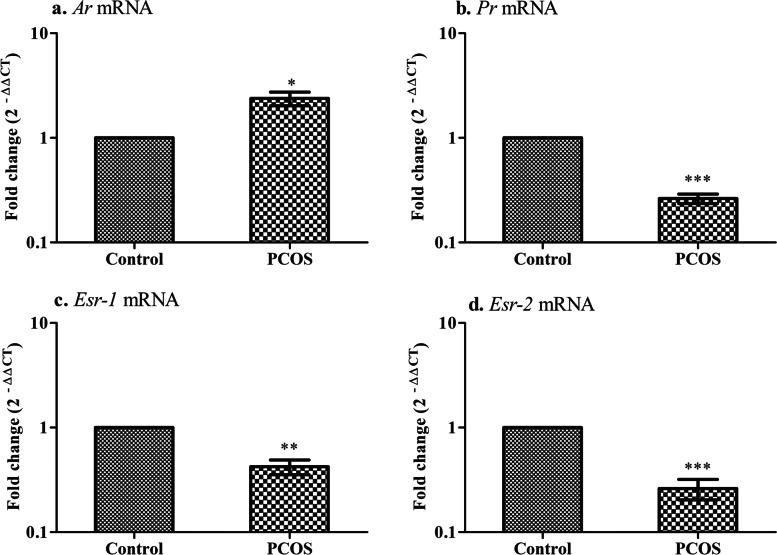
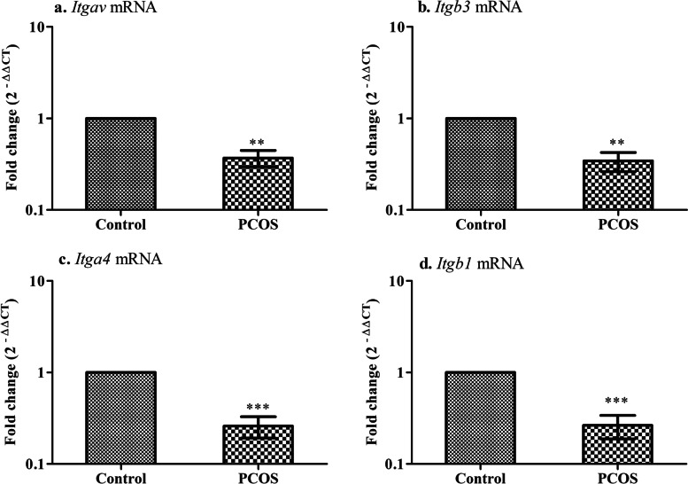
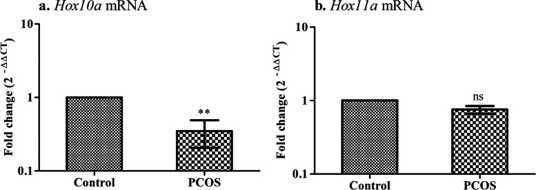
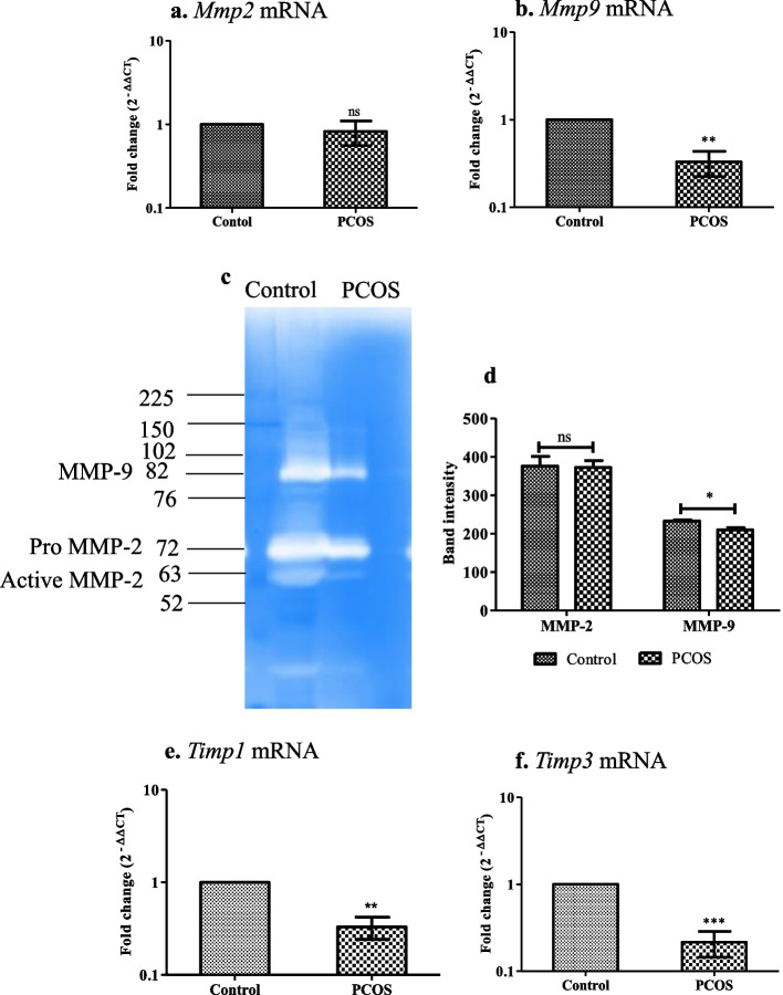
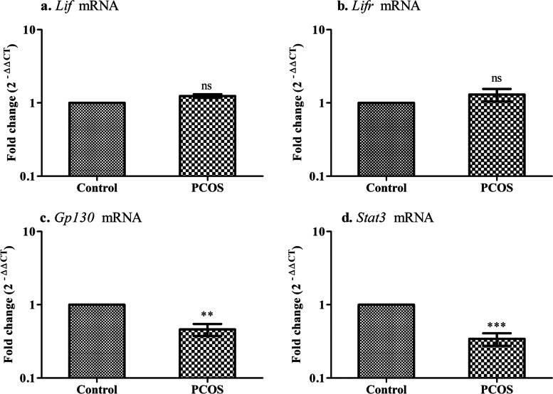
Methods: PCOS was induced in mice by oral administration of letrozole daily for 21 days. Following, the pregnancy was established and animals were sacrificed on the day 6th of pregnancy. Animals were assessed for early pregnancy loss, hormonal profile, mRNA expression of steroid receptors (Ar, Pr, Esr1/2), decidualization markers (Hox10/11a), adhesion markers (Itgavb3, Itga4b1), matrix metalloproteinases and their endogenous inhibitor (Mmp2/9, Timp1/2) and key mediators of LIF/STAT pathway (Lif, Lifr, gp130, stat3) were analyzed in the embryo implanted region of the uterus. Morphological changes in ovaries and implanted regions of the uterus were assessed.
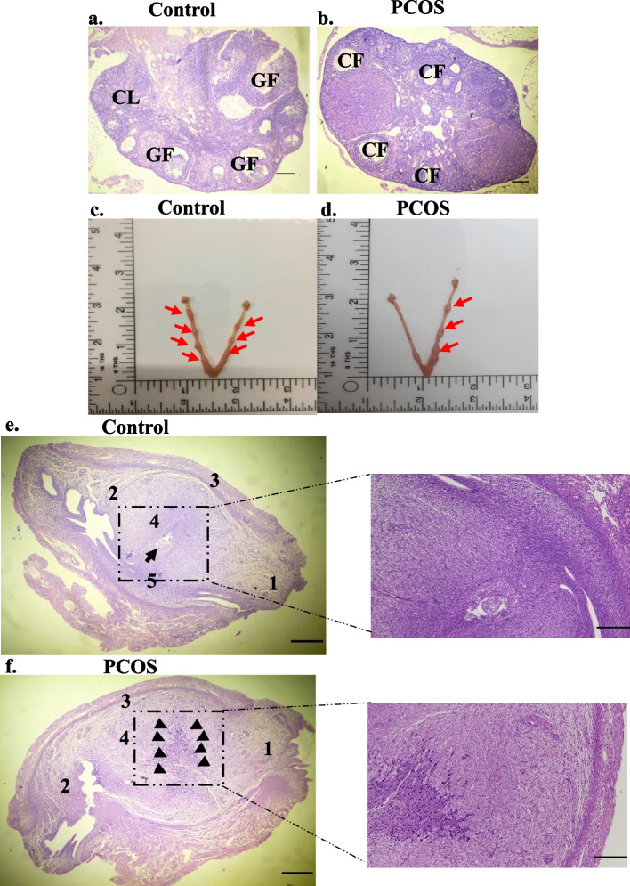
Results: Mice treated with letrozole demonstrated significant increases in testosterone levels along with a decline in progesterone levels as compared to control animals. PCOS animals also exhibited decreased fertility index and disrupted ovarian and embryo-containing uterus histopathology. Altered gene expression of the steroid receptors and reduced expression of Hox10a, integrins, Mmp9, Timp1/3, Gp130 & Stat3 was observed in the implanted region of the uterus of PCOS animals.
Conclusion: Our results reveal that majority of the molecular markers alteration in the establishment of early pregnancy could be due to the aberrant progesterone signaling in the embryonic-uterine tissue of PCOS animals, which further translates into poor fetal outcomes as observed in the current study and in several IVF patients.
Keywords: Letrozole; Mice; Polycystic ovary syndrome; Pregnancy loss; Progesterone.
© 2022. The Author(s).
Conflict of interest statement
The authors declare no competing interests.
Figures
Similar articles
-
Partially purified non-polar phytocomponents from Aloe barbadensis Mill. gel restores metabolic and reproductive comorbidities in letrozole-induced polycystic ovary syndrome rodent model- an "in-vivo" study.J Ethnopharmacol. 2022 Jun 12;291:115161. doi: 10.1016/j.jep.2022.115161. Epub 2022 Mar 7. J Ethnopharmacol. 2022. PMID: 35271948
-
Altered expression of leukemia inhibitory factor (LIF), LIFR, gp130, and IL11 in the embryo implantation site of rat model with prenatal androgen-induced polycystic ovary syndrome.Biochem Biophys Res Commun. 2022 May 21;605:24-30. doi: 10.1016/j.bbrc.2022.03.053. Epub 2022 Mar 11. Biochem Biophys Res Commun. 2022. PMID: 35306361
-
Increased uterine androgen receptor protein abundance results in implantation and mitochondrial defects in pregnant rats with hyperandrogenism and insulin resistance.J Mol Med (Berl). 2021 Oct;99(10):1427-1446. doi: 10.1007/s00109-021-02104-z. Epub 2021 Jun 28. J Mol Med (Berl). 2021. PMID: 34180022 Free PMC article.
-
Uterine Function: From Normal to Polycystic Ovarian Syndrome Alterations.Curr Med Chem. 2018;25(15):1792-1804. doi: 10.2174/0929867325666171205144119. Curr Med Chem. 2018. PMID: 29210631 Review.
-
Polycystic ovary syndrome: Endometrial markers.Best Pract Res Clin Obstet Gynaecol. 2016 Nov;37:66-79. doi: 10.1016/j.bpobgyn.2016.03.008. Epub 2016 Apr 1. Best Pract Res Clin Obstet Gynaecol. 2016. PMID: 27156350 Review.
Cited by
-
Higher Cumulative Live Birth Rate but Also Higher Late Miscarriage Risk in Non-Obese Women with Polycystic Ovary Syndrome Undergoing the First IVF/ICSI Cycle.Int J Womens Health. 2024 Feb 23;16:289-298. doi: 10.2147/IJWH.S445021. eCollection 2024. Int J Womens Health. 2024. PMID: 38415060 Free PMC article.
-
Identification of crucial pathways and genes linked to endoplasmic reticulum stress in PCOS through combined bioinformatic analysis.Front Mol Biosci. 2025 Jan 9;11:1504015. doi: 10.3389/fmolb.2024.1504015. eCollection 2024. Front Mol Biosci. 2025. PMID: 39850756 Free PMC article.
References
MeSH terms
Substances
Grants and funding
LinkOut - more resources
Full Text Sources
Medical
Research Materials
Miscellaneous
