. 2023 Jan 31;43(1):BSR20220369.
doi: 10.1042/BSR20220369.

Remodeling of algal photosystem I through phosphorylation

Affiliations

Remodeling of algal photosystem I through phosphorylation

Muhammad Younas et al. Biosci Rep. .

Abstract

Photosystem I (PSI) with its associated light-harvesting system is the most important generator of reducing power in photosynthesis. The PSI core complex is highly conserved, whereas peripheral subunits as well as light-harvesting proteins (LHCI) reveal a dynamic plasticity. Moreover, in green alga, PSI-LHCI complexes are found as monomers, dimers, and state transition complexes, where two LHCII trimers are associated. Herein, we show light-dependent phosphorylation of PSI subunits PsaG and PsaH as well as Lhca6. Potential consequences of the dynamic phosphorylation of PsaG and PsaH are structurally analyzed and discussed in regard to the formation of the monomeric, dimeric, and LHCII-associated PSI-LHCI complexes.

Keywords: Light harvesting proteins; green algae; phosphorylation/dephosphorylation; photosystems.


Conflict of interest statement

The authors declare that there are no competing interests associated with the manuscript.

Figures
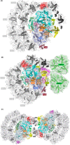
Figure 1
Figure 1. An overview of PsaG and/or PsaH subunits in monomeric, dimeric, and state transition complex of PSI
(A) Cartoon representation of the Chlamydomonas PSI with ten Lhca (Protein Data Bank (PDB) ID: 6JO5). Subunits are color-coded. PsaG subunit is highlighted in surface. (B) Cartoon representation of the Chlamydomonas PSI coupled with additional LHCII trimers during state transition (PDB ID: 7DZ7). Subunits are color-coded. PsaG and PsaH subunits are highlighted in surface. (C) Cartoon representation of the Chlamydomonas PSI dimer (PDB ID: 7ZQD). Subunits are color-coded. PsaG subunits are marked in red circles.
Figure 2
Figure 2. Influence of different light conditions on the abundance and phosphorylation of photosynthetic thylakoid membrane proteins
Volcano plots of FDR (-log10) against log2 fold changes (HL/NL), illustrating differences in phosphorylation ( A) and protein (B) levels. Differential expression analysis was performed using Limma. Color code: PSII (blue), PSI (red), LHC (green), others (black). $, doubly phosphorylated peptides (multiplicity = 2); *, nonproteotypic peptide (LhcbM4/6/8); **, nonproteotypic peptide (LHCSR1/3). Detailed data is available in Supplementary Table S1. For each light condition, abundance data of four replicates of each of the strains CC-124, CC-125, and CC-4375 IFT46::YFP were treated as equally weighted, independent replicates (n=12).
Figure 3
Figure 3. Phosphorylated sites in PsaG and PsaH subunits
(A) An overview of PsaG phosphorylated sites using PSI dimer as a reference structure. For convenience, only one side of the PSI dimer is shown. The phosphorylated residues (pSer65, pThr66, pThr71, and pThr72) are represented in sticks. (B) A zoomed-in view of the phosphorylated residues (pSer65, pThr66, pThr71, and pThr72) in PsaG, as shown in sticks representation. (C) An overview of the phosphoresidue pSer50 in PsaH, as shown in stick representation, using the reference structure PSI–LHCI–LHCII (PDB ID: 7DZ7).
Figure 4
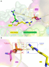
Figure 4. Potential functional role of PsaH phosphorylation at position 50 (pSer50)
(A) Superposition of the dimeric PSI structure (PDB ID: 7ZQD) with PSI–LHCI–LHCII (PDB ID: 7DZ7) shows a tilting of R158 in PsaL of PSI–LHCI–LHCII toward the phosphorylated serine residue (pSer50) in PsaH. The PsaL subunits are shown in cartoon while the residues are displayed as sticks. (B) Distance between the atoms in the phosphate group of pSer50 and the guanidinium group of R158 within 4.0 Å limit.
Figure 5
Figure 5. Stabilization of the loop in PsaL, which is involved in the interaction with pS50 in PsaH as well as pT27 in LhcbM1
(A) Superposition of the PsaL subunits from state transition complex (yellow) and the PSI monomer (blue). Red arrow indicates the missing residues (142–159) from monomeric PSI. The interacting partners that lie in this loop of PsaL in state transition complex are shown in gray sticks. (B) Superposition of the PsaL subunits from state transition complex (7DZ7) and PSI dimer (7ZQD). The missing residues (143–156) from PSI dimer are highlighted by the red arrow. Note that the R158 (unresolved in the monomer) is available in the dimeric PSI.
Figure 6
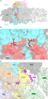
Figure 6. Interactions of residues from the stabilized loop in PsaL with the residues from neighboring subunits (PsaH, PsaI, PsaD, and LhcbM1) during state transitions (also summarized in Table 1)
For convenience, only the residues involved in the interaction are shown.
Figure 7
Figure 7. An overview of the distorted loop in PsaG that also contains the phosphorylated residues
(A) A look into the missing loop region (indicated with the red arrow) in PSI monomer (PDB ID: 6JO5). (B) The loop (including encircled small helix; residues 63–85) is stabilized in the PSI dimer (PDB ID: 7ZQD). The phosphorylated residues are marked with asterisks. (C) Superposition of PsaG from PSI monomer (PDB ID: 6IJO) and state transition complex (PDB ID: 7DZ7) reveals the stabilization of the loop in the state transition complex as well.
Figure 8
Figure 8. Interactions of PsaG residues (63–85) with the nearby residues from PsaB and Lhca9, using the PSI dimer as a reference structure (also summarized in Table 2)

References

    1. Ben-Shem A., Frolow F. and Nelson N. (2003) Crystal structure of plant photosystem I. Nature 426, 630–635 10.1038/nature02200 - DOI - PubMed
    1. Amunts A., Drory O. and Nelson N. (2007) The structure of a plant photosystem I supercomplex at 3.4 A resolution. Nature 447, 58–63 10.1038/nature05687 - DOI - PubMed
    1. Jordan P., Fromme P., Witt H.T., Klukas O., Saenger W. and Krauss N. (2001) Three-dimensional structure of cyanobacterial photosystem I at 2.5 A resolution. Nature 411, 909–917 10.1038/35082000 - DOI - PubMed
    1. Nelson N. and Ben-Shem A. (2004) The complex architecture of oxygenic photosynthesis. Nat. Rev. Mol. Cell Biol. 5, 971–982 10.1038/nrm1525 - DOI - PubMed
    1. Hippler M. and Nelson N. (2021) The plasticity of photosystem I. Plant Cell Physiol. 62, 1073–1081 10.1093/pcp/pcab046 - DOI - PubMed

MeSH terms

Substances